
Abstract
An immunohistochemical method using biotinyl tyramine was recently introduced to amplify weak staining signals. Despite its high sensitivity, however, tyramine-based immunostaining has been limited by its increased background staining. In this study, to develop an improved protocol of biotinyl tyramine-based immunohistochemistry minimizing the background staining, we determined which staining steps lead to the nonspecific reaction and the most appropriate blocking agents for background-provoking steps. Trypton casein peptone and distilled water with Tween-20 were shown to be most effective as a blocking agent and a rinsing solution, respectively. In conclusion, we developed an optimized protocol for biotinyl tyramine-based immunohistochemistry with minimal background staining.
T
Tissue specimens were collected from surgically dissected specimens submitted to the Department of Pathology at Seoul National University Hospital. All archival materials were routinely fixed in 10% neutral buffered formalin and embedded in paraffin. Sections (5-μm) were prepared on silane-coated slides (Sigma; St Louis, MO).
The reagents used to prepare the blocking solutions were as follows: nonfat dried (ND) milk (skim milk) from Beckton Dickinson (Sparks, MD); casein sodium salt, bovine serum albumin (BSA), and goat globulin from Sigma; and trypton casein peptone (TCP) from Amresco (Solon, OH). The immunostaining kit and all of antibodies (Abs), including anti-CD20 (L26), anti-ER (6F11), anti-CD3 (PS1), anti-vimentin (V9), anti-desmin (DE-R-11), anti-GFAP (GF-01), and anti-CD99 (DN16), were obtained from DiNonA (Seoul, Korea).
Tissue sections on microslides were deparaffinized with xylene, hydrated in serially diluted alcohol, and then immersed in 3% H2O2 to remove the endogenous peroxidase (HRP) activity. AR was done by heating the slides in 10 mM sodium citrate buffer (pH 6.0) in a microwave oven. Then the sections were incubated with primary Abs for 60 min and, after three successive rinsings with washing buffer, further incubated with biotinylated goat anti-mouse Abs (DiNonA) for 20 min. After rinsing, the tissue sections were incubated with HRP-conjugated streptavidin (SA-HRP) (DiNonA) for 20 min at room temperature (RT). The slides were washed and the chromogen was developed for 5 min with liquid 3,3′-diaminobenzidine (DAB) (DiNonA). The slides were then counterstained with Meyer's hematoxylin, dehydrated, and mounted with Canada balsam for examination.
Biotinyl tyramine was prepared as previously described elsewhere (Kerstens et al. 1995). The procedures for the biotinyl tyramine-based immunostaining can be summarized as follows: Step I, primary Ab incubation; Step II, secondary Ab incubation; Step III, SA-HRP incubation; Step IV, biotinyl tyramine incubation; Step V, SA-HRP incubation; Step VI, visualization and mounting. The staining procedure of Steps I, II, V, and VI was carried out according to the conventional immunostaining procedure as described above. The Step III is exactly the same as step V. At Step IV, biotinyl tyramine solution was applied for 10 min at RT.
To determine which step was responsible for the background staining, we added three steps (Steps IV, III, and II) one after another to the conventional immunostaining method with only SA-HRP incubation (Steps V and VI) without primary Ab incubation and compared their background levels. Whereas the sole application of Step V and the addition of Step IV produced no and minimal background in lymph nodes, respectively, the successive addition of Steps III and IV induced intense nonspecific reactivity. Moreover, the additional application of Step II as the first step also yielded highly intense background staining. This nonspecific reactivity in lymph nodes was notably marked on lymphocyte cytoplasm, high endothelial venules, and attached adipose tissue.
To develop an optimized blocking agent, we tested which reagents block the background staining induced by the secondary Ab incubation step (Step II) and the SA-HRP incubation step (Step III), among 3 mg/ml goat globulin, 4% BSA, 4% casein sodium salt, 2% ND milk, or 8% TCP. When the various kinds of blocking agents were applied at Step III by diluting SA-HRP in PBS containing them, the use of 8% TCP almost eliminated the background staining in lymph nodes with adipose tissue. Moreover, 2% ND milk was also effective to a lesser extent, but the other reagents were ineffective. For Step II, a blocking test was performed, and the use of TCP, BSA, and ND milk showed notable if not complete elimination of background staining, with no difference among these agents. However, goat globulin and 4% casein sodium salt were revealed to be less effective. Therefore, to avoid misinterpretation due to Step III-related background staining, 8% TCP was used as the Step III blocking agent.
Various kinds of solutions, such as PBS (pH 7.3), Tris buffer (pH 7.0), citrate buffer (pH 5.0), imidazole buffer (pH 7.0), borate buffer (pH 8.0), and distilled water (DW), were tested to determine which rinsing solution is the most effective for reduction of background staining. Tween-20 was added to all these solutions as a detergent. Unexpectedly, DW with 0.03% Tween-20 showed remarkable reduction of background staining, wheras PBS, citrate buffer, and imidazole buffer were minimally effective, and borate buffer and Tris buffer were mildly effective.
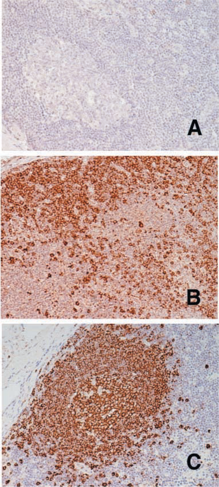
Immunostaining of human lymph node tissues with anti-CD20 Ab under various immuostaining conditions. Anti-CD20 Ab was diluted at 1:10,000. Conventional immunostaining without tyramine-based signal amplification did not elicit positive staining (
On the basis of these results, we can suggest a modified protocol for biotinyl tyramine-based immunostaining, employing TCP as Step II and III blocking agent and DW with Tween-20 for the rinsing solution. To confirm the effectiveness of this modified protocol, we performed immunostaining of human lymph nodes with anti-CD20 Ab. With the conventional immunostaining method, the lymph nodes were stained with moderate intensity at 1:200 dilution, but no staining was observed at 1:10,000 dilution (Figure 1A). Biotinyl tyramine-based immunostaining without modification showed that lymph nodes were stained with the same intensity at 1:10,000, but with significant background staining (Figure 1B). However, our modified protocol for biotinyl tyramine-based immunostaining produced not only the same degree of staining intensity as that of the unmodified tyramine-based signal amplification method but also the drastically decreased background staining (Figure 1C). Furthermore, applied to other tissues and other primary Abs, this improved protocol showed remarkably decreased background staining without impairing the high sensitivity (Table 1).
In this study, we determined the cause of background staining in biotinylated tyramine-based IHC and exploited a novel modified protocol of the standard staining technique. The biotinylated Ab incubation step and the SA-HRP incubation step were critical for nonspecific background staining. The primary cause of this background staining may be the binding of SA-HRP to macromolecules through nonspecific ionic or hydrophobic interactions. The binding of SA-HRP to endogenous biotin or biotin-like proteins may be the second source of nonspecificity, because lymphoid tissue is known to be free of endogenous biotin (Kim et al. 2002). It is evident that the background staining is not induced by the biotinyl tyramine itself. Rather, the background staining by the secondary Ab and SA-HRP appears to be gradually amplified by biotinyl tyramine. On the basis of our results, we suggest that this mechanism be generally applied to nonspecific background staining in the various multi-step signal amplification systems. Complete elimination of nonspecific reaction of the secondary Ab and SA-HRP is critical in minimizing background staining. Several blocking agents to suppress nonspecific reaction have been developed (Duhamel and Johnson 1985; Vogt et al. 1987). In immunofluorescence staining, ND milk was reported to be superior to BSA in reducing nonspecific nuclear staining of kidney (Duhamel and Johnson 1985). In an ELISA study, among various blocking agents, instant dry milk was reported to be most effective for blocking nonspecific binding of HRP-conjugated secondary Ab (Vogt et al. 1987). In accordance with these previous results, ND milk and TCP were the most effective blocking agent for both secondary Ab and SA-HRP in our study. In addition, we showed that DW with Tween-20 was the most effective rinsing solution for background staining. The conventional blocking method, in which blocking agents are applied before incubation of the primary Ab, turned out to be ineffective for reduction of background staining in biotinyl tyramine-based IHC (data not shown). Although some methods, including the addition of Tween-20 to biotinylated tyramine and lowering of the biotinylated tyramine concentration have been suggested to reduce the background staining (Freedman and Maddox 2001), none of them was effective in eliminating the background staining (data not shown). In contrast, the application of our modified protocol with various Abs and tissues resulted in an apparent reduction of nonspecific background staining without impairing the sensitivity.
Immunostaining of tissues with primary Abs using the modified protocol for biotinylated tyramine-based IHC minimizing background staining; data are expressed in terms of dilution ratio at which the same intensity of staining was obtained and symbols in parentheses indicate the intensity of background staining
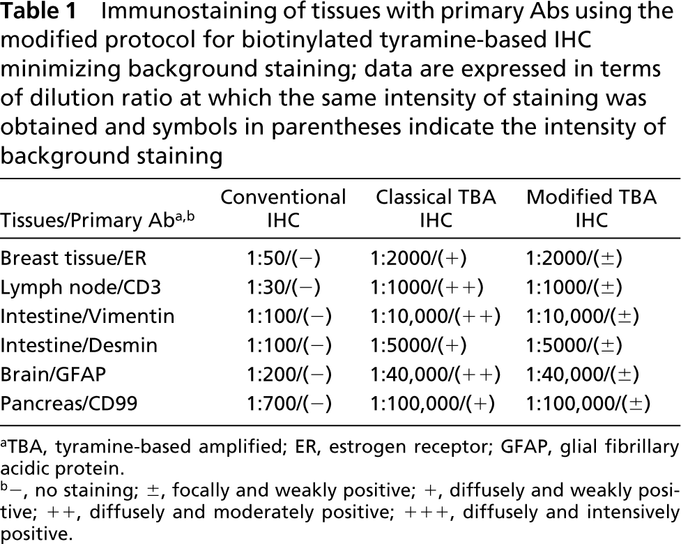
aTBA, tyramine-based amplified; ER, estrogen receptor; GFAP, glial fibrillary acidic protein.
b-, no staining; ±, focally and weakly positive; +, diffusely and weakly positive; ++, diffusely and moderately positive; +++, diffusely and intensively positive.
In conclusion, our results show that a modified protocol of biotinyl tyramine-based IHC can minimize nonspecific background staining without compromising high sensitivity.
Footnotes
Acknowledgements
Supported by a grant (2002) for Technical Renovation Development Enterprise from the Korea Small and Medium Business Administration.
We wish to thank Hee Jin Kim for helpful comments during the preparation of the manuscript.