
Abstract
Specific enzymes play key roles in many pathophysiological processes and therefore are targets for therapeutic strategies. The activity of most enzymes is largely determined by many factors at the post-translational level. Therefore, it is essential to study the activity of target enzymes in living cells and tissues in a quantitative manner in relation to pathophysiological processes to understand its relevance and the potential impact of its targeting by drugs. Proteases, in particular, are crucial in every aspect of life and death of an organism and are therefore important targets. Enzyme activity in living cells can be studied with various tools. These can be endogenous fluorescent metabolites or synthetic chromogenic or fluorogenic substrates. The use of endogenous metabolites is rather limited and nonspecific because they are involved in many biological processes, but novel chromogenic and fluorogenic substrates have been developed to monitor activity of enzymes, and particularly proteases, in living cells and tissues. This review discusses these substrates and the methods in which they are applied, as well as their advantages and disadvantages for metabolic mapping in living cells.
Keywords
S
Many enzymes, and particularly proteases, are present in cells and tissue compartments in an inactive form because they are either synthesized as precursors that have little if any catalytic activity and need post-translational activation or they are bound to an endogenous inhibitor. The inactive enzyme can be converted to its active form by proteolytic processing by specific proteases, autocatalysis, or by binding of co-factors or removal of inhibitors. Hence, large amounts of inactive and therefore not functional enzyme can be accumulated in a tissue compartment. However, the enzyme can become activated rapidly on demand. This can be achieved, for example, by an amplification loop, in which a small amount of the active protease can directly or indirectly activate its inactive precursor in a defined cell or tissue compartment, resulting in an exponential rate of activation to ensure that the protease can accomplish its function locally when required. Endogenous inhibitors are present in tissues to establish a threshold that regulates the concentration of active proteases in cells and tissues, thus keeping proteolysis under control (Thornberry and Lazebnik 1998). It is the balance between post-translational activation and inhibition that determines the activity of a (proteolytic) enzyme. Therefore, histochemical or cytochemical localization of the activity of an enzyme (also called catalytic histochemistry and cytochemistry, or metabolic mapping) is a powerful approach to study whether an enzyme is functionally involved in a pathophysiological process because it links the enzyme activity to cell and tissue structure.
Localization of the activity of an enzyme is traditionally performed at substrate concentrations that produce maximal amounts of colored or fluorescent final reaction product. These concentrations are usually high so that the maximal velocity of the enzyme (Vmax) is obtained. However, these high substrate concentrations are seldom present in vivo. Moreover, the affinity of an enzyme for its substrate(s) can also be under post-translational control (Jonges et al. 1992; Swezey and Epel 1986; Van Noorden et al. 1997a), e.g., due to interactions of the enzyme with other macromolecular structures. Variations in Vmax and Km greatly affect substrate conversion by an enzyme at physiological concentrations (Van Noorden and Jonges 1995a). Because of these considerations, metabolic mapping becomes more and more focused on the visualization of enzyme reactions in living cells and tissues at substrate concentrations that are physiological. When metabolic mapping is performed quantitatively with digital microscopy (Chieco et al. 2001), substrate concentrations can be varied and local Vmax and Km values can be determined easily in distinct tissue compartments or cell populations on the basis of the amount of colored or fluorescent final reaction product generated (Jonker et al. 1995). In this way, metabolic mapping provides many possibilities to link the actual functioning of an enzyme with pathophysiological alterations so that, for example, specific inhibitors can be tested for their therapeutic use.
Localization and quantification of the activity of enzymes in living cells and tissues can be performed by analysis of either the production or the consumption of fluorescent endogenous molecules, such as NADPH and NADH, or the formation of colored or fluorescent products generated from synthetic chromogenic or fluorogenic substrates using digital microscopy or flow cytometry. This setup allows quantitative monitoring of enzyme reactions in cells and tissues in time and space while the reaction proceeds. Although endogenous fluorescent molecules such as NAD(P)H, flavins, and porphyrins can be useful indicators of enzyme activity, such endogenous metabolites are limited in number and are not specific for a single enzyme. These molecules are both produced and consumed permanently in various enzymatic processes. Therefore, monitoring of these molecules in cells and tissues is not a good parameter of the activity of a specific enzyme. Consequently, the design, synthesis, and application of synthetic chromogenic and fluorogenic substrates are indispensable to identify specific enzymatic reactions in in vivo experiments. These substrates must meet a series of criteria for the application to living cells and tissues, such as the following.
First, substrates must have access to the enzyme under study such that the physiological and structural integrity of cells or tissue compartments is maintained. Preferably, these substrates diffuse freely into intact cells without the aid of loading procedures. In general, lipophilic substrates can be introduced into cells by simply adding a small amount of a concentrated solution of substrate, dissolved in a solvent such as dimethylformamide or dimethylsulfoxide, to the incubation medium. The concentration of the solvent should be less than 1% in the final incubation medium to avoid adverse effects of the solvent on the living cells or tissues. However, for quantitative purposes an important question is the exact (intracellular) concentration of the substrate at the site of the active enzyme.
Second, the substrate must be selectively converted by the target enzyme.
Third, fluorescence or absorption of the enzyme product must be sufficiently strong to permit detection at physiological substrate concentrations. When high concentrations of substrates are used, activity can be visualized and increases in absorbance or fluorescence can be readily detected, but then the enzyme is active at non-physiological concentrations of the substrate.
Fourth, in the case of synthetic substrates, conversion by the target enzyme should be similar to that of the natural substrate(s) of the enzyme, so that increase in absorbance or fluorescence in time is a proper reflection of physiological substrate fluxes. In other words, the energy differences between synthetic substrate and product should be similar to that of the natural substrate(s) and product(s) of that enzyme.
Fifth, products of synthetic substrates must accumulate at the site of enzyme activity and the enzyme product should not diffuse away from the site of enzyme activity. Chemical characteristics of the enzyme product are often the cause of a higher affinity for cellular compartments other than those of the active target.
Finally, synthetic substrates should be nontoxic for cells and tissues.
When enzyme reactions are analyzed quantitatively, one should realize that kinetic parameters of enzymes can be seriously affected by various properties of the synthetic chromogenic or fluorogenic substrates. A number of these pitfalls are listed below.
Affinity of the enzyme for its synthetic substrate is too low. However, affinity can be manipulated by chemical substitution of side groups in synthetic substrates.
Synthetic substrates contain more than one site of action for an enzyme. For example, fluorogenic protease substrates often contain two groups that can be cleaved off. Enzymatic hydrolysis of the first group yields a fluorescent half-product and fluorescence increases after hydrolysis of the second group.
More than one enzyme can convert the substrate.
Permeability barriers for the substrate in intact cells limit the availability of the synthetic substrate for the enzyme and are rate-limiting, rather than the enzyme reaction itself.
The product inhibits the enzyme reaction or the product diffuses away from the site of the active enzyme.
All these phenomena can affect the kinetics of enzymes and can therefore complicate the analysis of the activity of enzymes by metabolic mapping. For example, for accurate determination of enzyme activity with substrates containing two sites of action for an enzyme, the two-step catalysis, the channeling effect of intermediate products and the intracellular substrate concentrations should be considered, as described by Huang (1991).
This review discusses the advantages and disadvantages of fluorescent metabolites and chromogenic and fluorogenic substrates for their use in metabolic mapping in living cells and tissues on the basis of these criteria and considerations.
Metabolic Mapping in Living Cells by Use of Endogenous Fluorescent Metabolites
A number of cellular metabolites are fluorescent. These fluorescent endogenous metabolites can be used to monitor enzyme reactions in living cells and tissues (Table 1). However, most of these metabolites must be excited with ultraviolet (UV) light, which rapidly damages living cells and tissues (König 2000). Furthermore, autofluorescence from molecules other than those under study can mask fluorescence of the specific metabolites. We discuss here characteristics of autofluorescence produced by endogenous metabolites, measurements of concentrations of endogenous fluorescent metabolites, and possible damage that can occur in cells that are irradiated with UV light.
The characteristics of autofluorescence are as follows:
Autofluorescence of cells is composed of at least four distinct excitation and emission maxima: the tryptophan peak (290-nm excitation, 330-nm emission), the NAD(P)H peak (350-nm excitation, 450-nm emission), the riboflavin (FAD) peak (450-nm excitation, 530-nm emission), and a yet unidentified peak (500-nm excitation, 530-nm emission) (Heintzelman et al. 2000).
Autofluorescence produced by fluorescent metabolites is mainly found in discrete cytoplasmic vesicles.
Autofluorescence varies strongly in living cells. For example, it is low in freshly prepared cells and increases with time during culture until a plateau is reached.
Comparison of spectra of intact cells with spectra of known cell metabolites indicates that autofluorescence in cells arises mainly from intracellular NADH and riboflavin, flavin co-enzymes, and flavoproteins present in mitochondria. Co-enzymes fluoresce when in the reduced state (NAD(P)H) and do not fluoresce in the oxidized state (NAD(P)), whereas flavins fluoresce when in the oxidized state (FAD) and fluorescence disappears during reduction (FADH2). It is not known why autofluorescence in living cells varies so widely but, to a certain extent, intensities of autofluorescence reflect intracellular concentrations of NADH and FAD (Aubin 1979).
The spectra of the components NADH and riboflavin compare well with the spectra of autofluorescent cells and the metabolic activity in these cells. Ramanujam et al. (1994) demonstrated that NADH fluorescence increases when tissue progresses from normal to dysplastic, which might be explained by the fact that abnormal tissues have an increased metabolic rate and, therefore, increased concentrations of the fluorescent electron carriers NADH and FAD and decreased concentrations of the nonfluorescent NAD and FADH2. A complicating factor involved in the autofluorescence of NADH and FAD is that the protein-bound and therefore also enzyme-bound forms of these compounds exhibit fluorescence emission maxima that are shifted to shorter wavelengths by 20 nm and 5 nm, respectively. Furthermore, nonspecific binding of NADH to serum components such as albumin has also been shown to enhance its fluorescence, with a shift of the emission spectrum to shorter wavelengths (Canessa-Fischer and Davis 1966). Such nonspecific binding may contribute to autofluorescence when serum proteins in culture media remain bound to the surface of cells after their removal from culture. These different excitation and emission spectra of components such as NADH and FAD lead to wavelength shifts and band broadening in spectra, which makes (quantitative) analysis difficult. The use of fluorescent properties of NAD(P)H and FAD to monitor enzyme reactions in living cells was performed in the 1960s and 1970s by pioneers of living cell cytochemistry, such as Kohen and Thorell and co-workers (Table 1; for review see Van Noorden and Butcher 1991), because they were not able to use tools of modern cytochemistry such as synthetic substrates. However, as stated above, NAD(P)H is produced by a series of enzymes and is used by many others involved in various metabolic processes such as the respiratory chain, biosynthesis, and detoxification. Therefore, the approach to analyzing autofluorescence for metabolic mapping in living cells and tissues has become obsolete.
Endogenous metabolites for metabolic mapping in living cells
Experiments with HeLa cells transfected with GFP-tagged histone 2B clearly demonstrated the phototoxic effects of excitation light. It was expected that a beta sheet surrounding the GFP fluorochrome would prevent energy transfer to surrounding molecules to enhance the quantum efficiency and thus would restrict phototoxic effects. However, transfected cells appeared to be much more vulnerable to phototoxic effects than untransfected cells, which resulted in cell-cycle arrest or cell death. These living cells can be imaged only during the entire cell cycle, when the total amount of excitation light is kept to an absolute minimum of approximately 10 J cm-2 (E. Manders, personal communication). Manders' experiments demonstrated the dose-dependent relationship between the amount of excitation light and cell damage. However, the type of fluorophore also plays an important role.
Exposure of cells to stressful conditions, such as excitation, triggers stress responses. Solar UV light is a major source of environmental stress for mammalian cells (Tyrrell 1996a, b; König 2000). UV-C (200–290 nm) is strongly absorbed by nucleic acids and causes DNA damage by inducing the formation of pyrimidine dimers, resulting in mutations and eventually carcinogenesis (Hall et al. 1988). UV-A (320–380 nm) is less strongly absorbed by nucleic acids but causes the production of a variety of reactive oxygen species (ROS), including superoxide and lipid peroxides (Jurkiewicz and Buettner 1996; Scharffetter-Kochanek et al. 1997). UV-A irradiation can produce lipid peroxidation when superoxide anion and hydrogen peroxide form hydroxyl radicals in the presence of iron. Hydroxyl radicals can then react with polyunsaturated fatty acids to abstract hydrogen, forming lipid hydroperoxyl radicals with a half-life of seconds, which are then able to diffuse over significant distances before detoxification. Propagation and diffusion allow a peroxidative chain reaction to spread through membranes, generating new radical species as the reaction proceeds and greatly amplifying the damage produced (Petkau 1986; Vladimirov 1986; Nishi et al. 1990). Intermediate UV-B (290–320 nm) is absorbed by nucleic acids but also contains an oxidative component (Tyrrell 1996a). UV irradiation also triggers the activation of surface death receptors, such as epidermal growth factor (EGF) receptor, CD95/Fas, or certain members of the tumor necrosis factor (TNF-α) receptor family (Rosette and Karin 1996), the src family of tyrosine kinases (Devary et al. 1992), and phosphatidyl-inositol 3-kinase (PI-3) (Kabuyama et al. 1998). These are believed to activate protein phosphorylation cascades, resulting in mitogen-activated protein (MAP) kinase activation. The various MAP kinases are activated in a wavelength-specific manner by UV light (Kabuyama et al. 1998). Furthermore, UV irradiation results in the activation of various genes that are involved in the regulation of cell proliferation, such as tumor suppressor p53, which acts as a cell-cycle checkpoint. UV irradiation causes cells to be arrested in G1-phase, as was observed in experiments that followed cells in time under UV exposure (Maltzman and Czyzyk 1984; Hall et al. 1993; Di Leonardo et al. 1994). Expression of bax is enhanced by p53 after transcriptional activation (Miyashita et al. 1994; Selvakumaran et al. 1994; Miyashita and Reed 1995; Hansen and Braithwaite 1996). Bax induces cytochrome c release from mitochondria and activation of caspase-9, which activates caspase-3 and induces apoptosis. Vitamin E, a lipid-soluble antioxidant that scavanges ROS and singlet oxygen, inhibits activation of various MAP kinases. In conclusion, metabolic mapping in living cells and tissues on the basis of endogenous fluorescent metabolites has only limited applications. Concentrations of these metabolites do not reflect the activity of a specific enzyme and these metabolites must be excited with UV light, which is detrimental to living cells and tissues. Excitation light of short wavelengths is far more damaging to cells because the energy content is higher than that of light of longer wavelengths. Moreover, UV light can induce activation of signal transduction pathways within minutes, leading to profound alterations in cell metabolism.
Synthetic Chromogenic Substrates for Metabolic Mapping in Living Cells
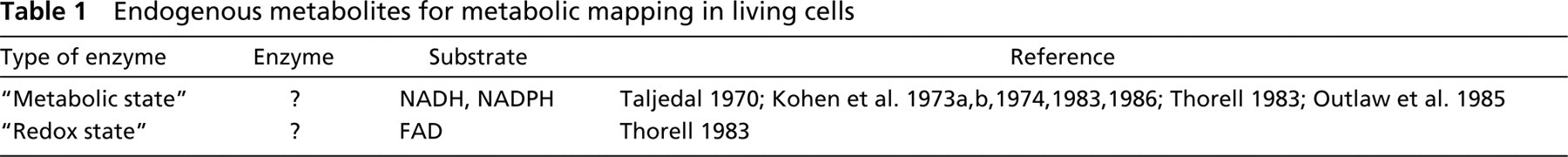
Tetrazolium salt methods are established methods for the localization of the activity of dehydrogenases, reductases, and oxidases. Enzyme-catalyzed oxidation of the substrate liberates protons that are then transferred to a tetrazolium salt such as (tetra)nitro BT as final electron acceptor. In this way, a water-insoluble formazan is produced (Lojda et al. 1976; Seidler 1991; Van Noorden and Frederiks 1992). This method has been applied to quantify glucose-6-phosphate dehydrogenase (G6PDH) activity in living hepatocytes of marine fish (Winzer et al. 2001a; Table 2). Formazan was localized exclusively in the cytoplasm of cells, leaving the nucleus unstained (Figure 1). Polyvinyl alcohol was used in the incubation medium as stabilizer in a concentration of 2% rather than 18%. To prevent precipitation of the tetranitro BT in the incubation medium due to the low amounts of polyvinyl alcohol present, a final concentration of 1 mM instead of 5 mM was used. Under these conditions, capturing of electrons is just as efficient as at high tetrazolium salt concentrations (Van Noorden 1988). The intact plasma membrane did not appear to be a barrier to substrate, co-enzyme and dye molecules necessary to detect intracellular G6PDH activity. Nevertheless, the authors mention that quantitative histochemical data obtained with the use of cryostat sections were higher than those obtained with intact hepatocytes, possibly caused by the process of penetration of components into the living cells.
D, T-diaphorase that can use either NADH or NADPH as substrate was demonstrated in living fish hepatocytes as well (Winzer et al. 2001b). Activity in hepatocytes appeared to be lower when NADPH was used as substrate. The method was also adopted for the demonstration of activity of aldehyde dehydrogenase in living fish hepatocytes (Table 2).
Reduction of tetrazolium salts has been used as test for viability of eukaryotic and prokaryotic cells. Although this approach is not specific for a particular dehydrogenase or reductase, it has been used successfully for subcellular localization of reducing enzyme systems in intact human hepatoma cells (Bernas and Dobrucki 1999).
In conclusion, chromogenic substrates thus far have been only rarely exploited for metabolic mapping in living cells and tissues, but this approach is promising, especially for high-throughput screening of effects of potential drugs on living cells, as absorbance measurements are simpler than fluorescence measurements.
Synthetic Fluorogenic Substrates for Metabolic Mapping in Living Cells
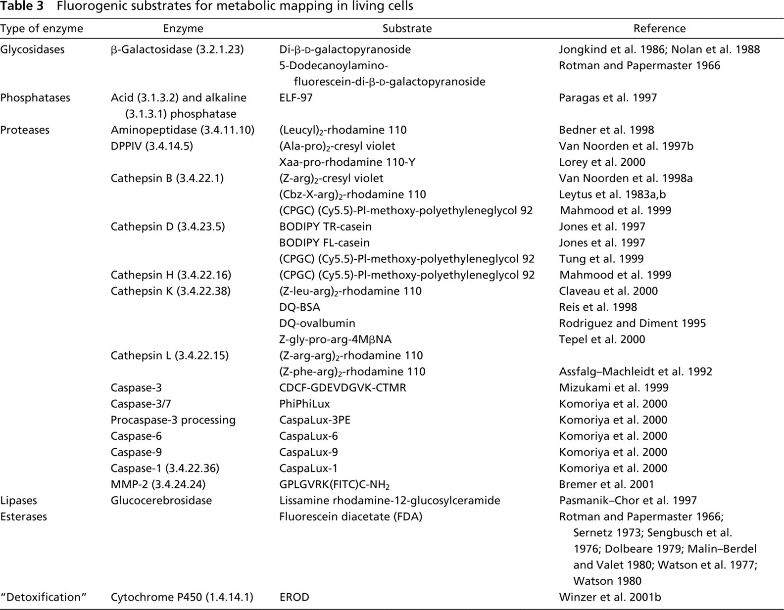
Synthetic fluorogenic substrates can be used for determination of viability of cells, but also for metabolic mapping. Various fluorogenic substrates have been developed, especially for hydrolytic enzymes such as proteases. Here we discuss synthetic fluorogenic substrates that have been applied to living cells and tissues (Table 3).
Fluorochromes Used in Synthetic Fluorogenic Substrates
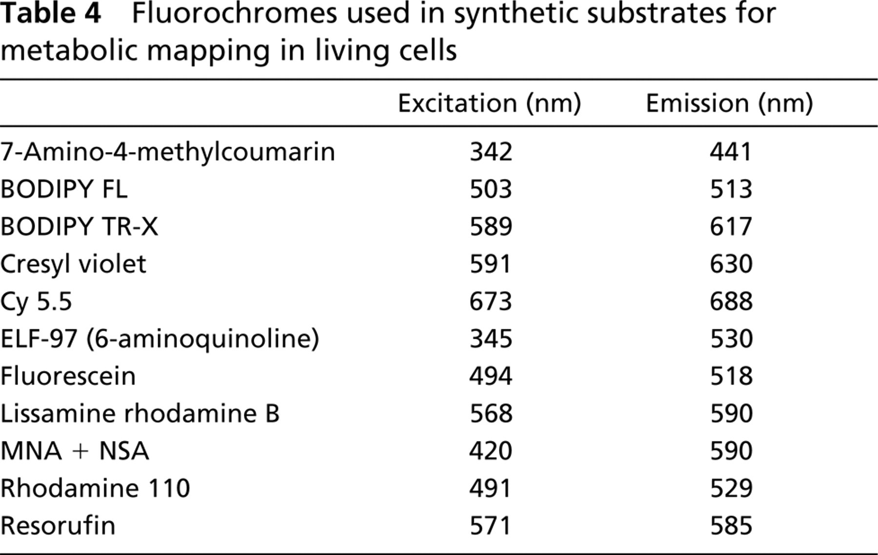
Fluorogenic substrates are usually esters or ethers of phenolic fluorophores; 7-hydroxy-4-methylcoumarin (β-methylumbelliferone) and its analogues fluorescein and resorufin are the most common examples of fluorophores used in fluorogenic substrates (Haugland 1995). Other fluorophores for fluorogenic substrates of hydrolytic enzymes are rhodamine 110 (Leytus et al. 1983a, b) and cresyl violet (Van Noorden et al. 1997b). These synthetic substrates are small molecular substrates, consisting of amino acids attached to a fluorochrome. Internally quenched fluorogenic substrates (Matayoshi et al. 1990) and, in the future, fluorescence resonance energy transfer (FRET)-based substrates will become increasingly available. These types of substrates are macromolecular substrates, consisting of fluorochromes attached to proteins. Spectral characteristics of the various fluorochromes used in fluorogenic substrates are presented in Table 4.
Formation of esters or ethers of phenolic fluorophores results in a shift to shorter wavelengths of absorbance and either partial or total quenching of the long-wavelength fluorescence of the fluorophore. Fluorescein-based substrates are not fluorescent because quenching is complete as the dye is converted into a non-fluorescent colorless lactone by the formation of two ether or ester bonds. Therefore, these substrates do not cause background fluorescence and are among the most sensitive fluorogenic substrates known. Fluorescein is water-soluble, and free fluorescein can be retained in living cells for at least a few minutes at room temperature (Rotman and Papermaster 1966; Sengbusch et al. 1976; Watson et al. 1977; Dolbeare and Phares 1979; Dolbeare 1990). Fluorescein accumulation was claimed to be linear up to 80 min of the reaction to detect β-galactosidase activity in living fibroblasts (Jongkind et al. 1986). Afterwards, intracellular accumulation of fluorescence levels off as a result of both product inhibition and fluorescein diffusion out of the cells (Kohen et al. 1973a, b; Watson et al. 1977). Cooling of cells on ice after loading with substrate slows leakage of fluorescein (Nolan et al. 1988), but this prevents metabolic mapping under physiological conditions for most organisms.
Chromogenic substrates for metabolic mapping in living cells
Cytochrome P450 activity in living cells can be analyzed using the 7-ethoxyresorufin-O-deethylase (EROD) assay based on the formation of fluorescence of resorufin (Burke and Mayer 1974). The assay has been modified by Behrens et al. (1998) for study of living trout hepatocytes. Enzyme activity can also be assessed by using 7-ethoxycoumarin-O-deethylase (ECOD) as substrate (Scholz and Segner 1999).
Activity of cathepsin K was localized by Tepel et al. (2000) using the substrate benzyloxycarbonyl-glycyl-prolyl-arginine-4-methoxy-β-naphthylamide (Bz-Gly-Pro-Arg-4MβNA) in combination with nitrosalicylaldehyde, which couples directly with the proteolytically released 4MβNA to produce a yellow fluorescent final reaction product that can then be visualized by fluorescence microscopy (Van Noorden et al. 1987). Living thyroid epithelial cells showed the precipitate to be mainly present at the cell surface, indicating the inability of these living cells to internalize either the substrate or the coupling reagent.
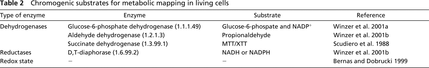
A unique fluorogenic substrate for phosphatases, 2-(5′-chloro-2-phosphoryloxyphenyl)-6-chloro-4(3H)-quinazolinone or ELF-97-phosphate, has recently been developed (Paragas et al. 1997). The ELF-97-alcohol that results after liberation of the phosphate group by phosphatase activity forms a bright yellow-green fluorescent precipitate at the site of the activity (Figure 2). Fluorescence of ELF-97 product is very stable; there is minimal diffusion of the reaction product from the site of enzymatic activity because it is water-insoluble and it provides high spatial resolution (Breininger and Baskin 2000). The ELF-97 phosphatase substrate contains only a single phosphate group, which makes it suitable for simple kinetic analysis of phosphatase activity. Another advantage is its large Stoke's shift. It is maximally excited at 345 nm and emits at 530 nm (yellow; Figure 2). The substrate itself is weakly blue fluorescent, unfortunately in the region of autofluorescence. Therefore, it is not possible to use the blue fluorescence of this synthetic substrate to determine its intracellular concentration. In that case, ratio images of both substrate and product could be made and concentration of both substrate and product determined. A major disadvantage of the ELF-97 product is the short excitation wavelength in the range of UV-A, which makes it largely unsuitable for metabolic mapping in living cells. Moreover, the substrate can, in principle, be cleaved by every phosphatase in a living cell, thus providing little specificity. In conventional enzyme histochemistry, the composition of the incubation medium determines which phosphatase is detected (e.g., acid pH for acid phosphatase and alkaline pH for alkaline phosphatase), but in a living cell each phosphatase is present in its own natural environment.
Rhodamine-based fluorogenic dipeptidide substrates have been synthesized by Leytus et al. (1983a, b). The non-fluorescent substrate diffuses readily into living cells, in which it can be hydrolyzed into a strongly fluorescent product that is entrapped within cells because of its positive charge (Assfalg-Machleidt et al. 1992). Activity of cysteine proteinases has been detected in living human monocytes and rat macrophages with the use of flow cytometry. Rhodamine production was completely inhibited by specific inhibitors of cys-teine proteinases such as E-64 and Z-Phe-Ala-CHN2. Rhodamine 110 is a diamino analogue of fluorescein that exhibits properties similar to those of fluorescein (Table 4). When amino groups of rhodamine 110 are blocked by acetylation, the intensely colored dye is converted into a colorless and non-fluorescent compound, implying that the conjugation of the system of the chromophore is interrupted (Drexhage 1976). Therefore, rhodamine-based substrates are highly reactive because loss of acetylation is accompanied by a large increase in the degree of conjugation and hence a large increase in stability. (Cbz-Arg-NH)2-rhodamine 110 is a stable compound that does not exhibit spontaneous hydrolysis in aqueous solutions during assays. Like fluorescein-based substrates, rhodamine 110-based bisubstituted substrates have low fluorescence coefficients that increase when the bisubstituted derivates are converted into their corresponding monosubstituted compounds and increase again after conversion of the monosubstituted compounds into their corresponding unsubstituted compounds. Nevertheless, this does not complicate the interpretation of kinetic data providing that less than 15% of the substrate is hydrolyzed during an assay (Leytus et al. 1983b). Under these conditions, the increase in fluorescence is solely due to the production of the monosubstituted (Cbz-Arg-NH)-rhodamine 110 compound. Bisubstitution of (Cbz-Arg-NH)2-rhodamine 110 also has certain advantages. The effective concentration of susceptible amide bonds is twice the substrate concentration, thus lowering the Km. Furthermore, the presence of two amino acid residues per rhodamine moiety enhances the water-solubility of the substrate.
Localization of glucose-6-phosphate dehydrogenase activity in living isolated hepatocytes of European flounder. The colored final reaction product, formazan, is localized in the cytoplasm, leaving the nucleus unstained. This approach is fast and simple and is therefore suitable for high-throughput screening of effects of drugs or for biomonitoring in environmental research. After Winzer et al. (2001a). Bar = 15 μm.
Gallery of images of a living human fibroblast incubated in a medium containing ELF-97 phosphate. First image is taken at 30 sec after the incubation was started and each subsequent image at intervals of 15 sec. Yellow fluorescence is produced by phosphatases, most likely acid phosphatases, in lysosomes, as most ELF-97 product is generated in intracellular granular form. Bar = 2 μm.
Localization of dipeptidyl peptidase IV (DPPIV) protein (also known as CD26) using an antibody coupled with FITC (
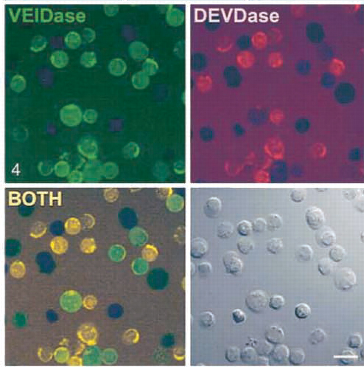
Caspase activity in living but apoptotic thymocytes as demonstrated by incubation with CaspaLux-6 substrate for caspase 6 (VEIDase) and PhiPhiLux substrate for caspases 3 and/or 7 (DEVDase). Combination of these images (BOTH) shows distribution patterns of caspase 6 activity and activity of caspases 3 and/or 7 in the same cells. Images were taken after 140-min incubation. The Nomarski image is at lower right. The dark images of cells in each fluorescence image correspond to those cells in which intracellular caspase activation is not yet detectable. Bar = 20 μm. From Komoriya et al. (2000), with permission.
Fluorogenic substrates for metabolic mapping in living cells
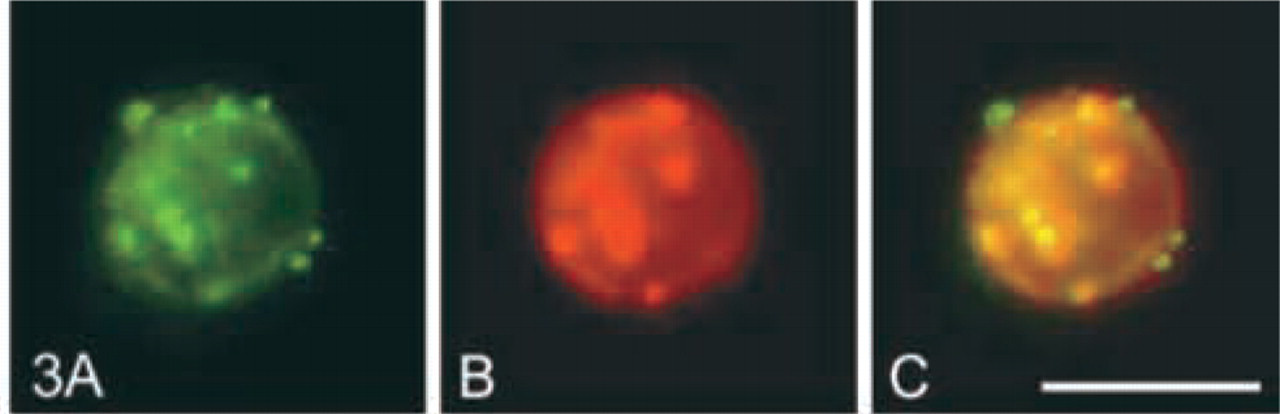
In contrast, coumarin-based substrates are less useful than rhodamine-based substrates because suboptimal conditions for detection of the fluorophore must be used to maximize spectral differences between substrate and product. Furthermore, a structural change in the aminocoumarin moiety does not take place when the substrate is cleaved and, therefore, coumarin-based substrates are less useful than rhodamine-based substrates (see also below). Recently, a new type of fluorogenic substrate for proteases has been synthesized based on the leaving group cresyl violet (Van Noorden et al. 1997b). Cresyl violet has been used in histology for over a century because of its metachromatic properties. In the early days it was employed for Nissl staining of neural tissue and is excellent for staining connective tissue. Furthermore, it stains nuclei violet, cytoplasm blue, and amyloid, mucin, and mast cell granules red. It has also been used for vital staining of white blood cells and for staining of cancer cells in biopsies. Cresyl violet is also a fluorescent molecule. It can be used in combination with other fluorescent dyes, such as fluorescein, or green fluorescent protein. Two amino-acid side chains can be bound by peptide bonds to the two amide groups in the fluorophore. At present, a bifunctional synthetic substrate for dipeptidyl peptidase IV (DPPIV) (Ala-Pro)2-cresyl violet (Van Noorden et al. 1997b), and one for cathepsin B (Z-Arg)2-cresyl violet (Van Noorden et al. 1998a, b), are available. In Figure 3, activity of DPPIV is demonstrated on the membrane of a living human T-helper-cell and is co-localized with the DPPIV protein (also known as CD26), showing the good localization properties of fluorescent cresyl violet.
Fluorochromes used in synthetic substrates for metabolic mapping in living cells
Casein conjugates of two BODIPY dyes, one named BODIPY fluorescein (FL) and the other BODIPY Texas red (TR), have been developed by Jones et al. (1997) as fluorogenic protease substrates. The basic structure of the BODIPY fluorophore is 4,4-difluoro-4-bora-3a,4a-diaza-s-indacene. Solutions of the alkyl-substituted derivates have a green fluorescein-like fluorescence, but when substituents that yield additional conjugation are added to the molecule, both absorption and emission shift to longer wavelengths. Conjugates are labeled to such an extent that the dyes are efficiently quenched in the protein, yielding virtually non-fluorescent substrate molecules. These fluorogenic substrates release highly fluorescent BODIPY-labeled peptides, and this release is proportional with protease activity. These quenched substrates are suitable for continuous assay of enzymatic activity in living cells and tissues, particularly in fluorescence microplate readers using either fluorescein excitation and emission wavelengths to measure BODIPY FL casein hydrolysis or Texas Red wavelengths to detect proteolysis of BODIPY TR casein. Compared with the FITC-casein assay, the BODIPY-labeled casein protease assay is simple and precise and has greater sensitivity and a broader dynamic range of detection. In this way, it is possible to detect the activity of a wide variety of proteolytic enzymes with high sensitivity (Jones et al. 1997).
DQ-BSA is also based on the BODIPY fluorochrome. BODIPY FL is conjugated to BSA at a high molar ratio. The resulting conjugate is self-quenched by fluorescence energy transfer between neighboring BODIPY molecules. Although this substrate is not membrane-permeable, it can be internalized by forming a complex with anti-BSA that is taken up by the Fc receptor of macrophages (Reis et al. 1998). DQ-ovalbumin is also nonpermeable to cell membranes but can be internalized by mannose receptor-mediated endocytosis by antigen-presenting cells (Rodriguez and Diment 1995).
The fluorogenic substrates for caspases in Table 3 are based on peptides of 18 amino acids containing caspase cleavage sequences, with two identical fluorophores covalently attached near their termini. Such substituted peptides are assumed to have an oval-shaped structure in solution due to the formation of intramolecular excitonic H-dimers between the fluorophores (Packard et al. 1996,1997). In such rhodamine-derivatized dimers, the fluorophore fluorescence is quenched for 90–99%. When a protease cleaves the peptide backbone of this complex, the cyclic structure incorporating the fluorophores is broken and two highly fluorescent substituted peptide fragments are generated (Figure 4). Whereas normal peptides of 18 amino acids do not enter cells without a specialized means of transport, the caspase substrates of this design are permeable to cells (Komoriya et al. 2000).
Substrates have been designed for specific enzymes, such as cathepsin D, which is overexpressed in a number of cancers (Rochefort and Liaudet-Coopman 1999). The Cy5.5 fluorochrome can be excited in the near-infrared region and has been attached to the amino terminal of a sequence of 11 amino acids that is specifically recognized by cathepsin D. The peptides were subsequently attached to a synthetic graft polymer for efficient tumor delivery (Tung et al. 1999). The spatial proximity of the fluorochromes results in quenching of fluorescence in the bound state. A 350-fold-higher fluorescence signal was observed after cleavage in vitro. Cell culture experiments using a rodent tumor cell line stably transfected with human cathepsin D confirmed enzyme-specific production of fluorescence. This sequence, but not a scrambled control sequence, resulted in specific production of fluorescence due to enzyme activity in vitro. The same type of substrate has also been used to demonstrate matrix metalloproteinase (MMP)-2 activity. The substrate consists of three elements: the quenched Cy5.5 fluorochrome, an MMP-2-sensitive peptide sequence, and a poly-
The small molecular substrates listed in Table 3 are not very fluorescent themselves, but the internally quenched macromolecular substrates in Table 3 may have the advantage that fluorescence is negligible and therefore sensitivity of the enzymatic assay is higher due to an increased signal-to-noise ratio. On the other hand, when the substrate is also fluorescent but with spectral characteristics that are different from those of the product, measurement of intracellular substrate concentrations is possible.
Enzyme products that can be excited in the red or near-infrared region of the spectrum are, in principle, the best for in vivo imaging of enzyme activity because the excitation light is less harmful than light of shorter wavelenghts.
In the near future, frequency resonance energy transfer (FRET)-based substrates will become available for analysis of enzyme activity in living cells. FRET-based substrates should be synthesized in such a way that two different fluorophores, of which the emission peak of one overlaps with the excitation peak of the other, are located in close proximity at opposite sides of a bond susceptible to enzymatic cleavage. Preferably, these two fluorophores have a large Stoke's shift. Excitation of the fluorochrome with excitation and emission peaks at shorter wavelengths can then, in theory, result in enhanced fluorescence of the second fluorochrome, with excitation and emission peaks at longer wavelengths when the substrate is not enzymatically processed. When the substrate is cleaved, the FRET phenomenon disappears and fluorescence of the fluorochrome with excitation and emission peaks at shorter wavelengths appears. For example, when the fluorophores Alexa Fluor 488 and rhodamine are combined in such a FRET-based substrate, excitation of the Alexa Fluor 488 fluorophore in the intact substrate results in enhanced emission of rhodamine fluorescence, which is a measure for the local (intracellular) substrate concentration. Alexa Fluor 488 fluorescence itself is a measure of the amount of substrate processed. Therefore, FRET-based substrates will enable measurements of both the amount of product generated by enzymatic activity and the intracellular concentration of the substrate, even in sub-compartments of cells. The use of this type of substrate would solve the problem of estimating local substrate concentrations in cells or cell compartments to calculate accurately Vmax and Km values for enzymes in living cells. Labeling peptide sequences with two different dyes has been described by Bark and Hahn (2000).
The FRET phenomenon can also be useful to demonstrate specificity of a substrate for an enzyme. When the enzyme of interest is tagged with, e.g., green fluorescent protein (GFP) by transfection, co-localization of the enzyme and the enzyme product containing a fluorophore that has an excitation peak that overlaps the emission peak of GFP may result in the FRET phenomenon.
In conclusion, fluorophores with high fluorescence quantum yield should be selected for incorporation into synthetic fluorogenic enzyme substrates to obtain sufficient sensitivity to analyze enzyme reactions at physiological substrate concentrations. Substrates that contain fluorophores with excitation peaks in the red or infrared region of the spectrum are the substrates of choice. Moreover, kinetic parameters of the enzyme for the synthetic fluorogenic substrate should resemble that for its natural substrate(s), as explained below. In addition to small molecular fluorogenic substrates, macromolecular substrates containing quenched fluorophores are useful for analysis of specific activity of enzymes in living cells and tissues, and the concept of FRET-based fluorogenic substrates is intriguing.
Reactivity of Synthetic Fluorogenic Substrates
The ability of an enzyme to discriminate among many potential substrates is an important factor in maintaining organization of most biological functions in the biocomplexity of cells and tissues. Although substrate selection can be regulated at many levels in a biological context, such as spatial and temporal localization of enzyme and substrate, concentrations of enzyme and substrate, and requirement of co-factors, substrate specificity at the enzyme active site is the overriding principle that determines the turnover of a substrate (Harris et al. 2000). The effectiveness of the conversion of a substrate by an enzyme can be defined as the product of the second-order rate constant Kcat/Km or specifity constant (Knight 1977; McRae et al. 1981) and the relative detectability of the fluorescent leaving group given by the molar fluorescence coefficient. On the basis of this criterion (Cbz-Arg-NH)2-rhodamine 110 is a substrate with higher effectiveness than 7-(N-Cbz-
Rhodamine-based substrates exhibit a wide range of specificity constants. Amino acids in the P2 position in dipeptide substrates determine specificity in a large part. Comparison of the kinetic constants of plasmin or thrombin for the best dipeptide substrates with those for (Cbz-Arg-NH)2-rhodamine 110 indicates that the large increases in Kcat/Km obtained by extending the single amino acid substrate with an appropriate P2 residue in a dipeptide substrate are primarily the result of a large increase in Kcat and a decrease in Km. Therefore, the specificity of proteinases for synthetic substrates depends to a great extent on interactions between amino acids in the active site of a protease and amino acid residues in the peptide substrate. Because occupation of the P2 position does not increase specificity of coumarin thiolester-based substrates as much as it increases specificity of rhodamine-based substrates, selectivity can be much greater with rhodamine-based substrates. Therefore, rhodamine-based substrates are in principle more useful to detect selectively protease activity in living cells and tissues.
In conclusion, characterization of substrate specificity of an enzyme provides useful information for the dissection of complex biological pathways and also provides the basis for the design of selective substrates and potent inhibitors to study enzyme activity.
Localization of Final Fluorescent Reaction Product
When a fluorescent final reaction product of an enzyme accumulates at a certain site, this does not automatically imply that it has been produced in that site. Chemical properties of the fluorophore have effects on its intracellular localization. The charge of a fluorophore, such as rhodamine, can lead to accumulation in mitochondria. However, the charge of a molecule depends on its pKa value. For example, rhodamine 110 has a pKa of 4.3 at physiological pH 7.2, and one in a thousand rhodamine molecules is in the protonated acidic state with a positive charge. In the protonated state, rhodamine 110 accumulates in mitochondria as a result of the intramitochondrial electric potential (Jeannot et al. 1997). Other factors, such as lipophilicity or hydrophobicity, are also of crucial importance, not only for the uptake of these substrates by living cells but also for final localization of the fluorescent product. Therefore, it appears attractive to develop fluorochromes with a chemical anchor (Lorey et al. 2000) or to use a trapping agent to stop fluorochromes from diffusing into cell compartments on the basis of their charge, lipophilicity, or hydrophobicity, or the pH of intracellular compartments. In this way, diffusion of the fluorescent enzyme product from the site of enzyme activity can be limited.
In conclusion, chemical properties of fluorogenic substrate and fluorescent product must be taken into account for proper localization of enzyme activity in living cells and tissues.
Conclusions
The present overview of methods for detection of enzyme activity in living cells and tissues indicates our limited knowledge of molecular interactions that take place during incubation and recording of the formation of a colored or fluorescent reaction product. Metabolic mapping in living cells and tissues on the basis of endogenous fluorescent metabolites has only limited applications. Concentrations of these metabolites do not reflect the activity of a specific enzyme and these metabolites must be excited with UV light, which is detrimental to living cells and tissues. Excitation light of short wavelengths is far more damaging to cells because the energy content is higher than that of longer wavelengths. Moreover, UV light can induce activation of signal transduction pathways within minutes, leading to profound alterations in cellular metabolism.
Chromogenic substrates thus far have rarely been exploited for metabolic mapping in living cells and tissues, but this approach is promising, especially for high-throughput screening of effects of potential drugs on living cells, because absorbance measurements are far more simple than fluorescence measurements.
Important criteria for the selection of fluorophores to incorporate into synthetic fluorogenic substrates are a high-fluorescence quantum yield to obtain sufficient sensitivity to analyze enzyme reactions at physiological substrate concentrations. Substrates that contain fluorophores with excitation peaks in the red or infrared region of the spectrum are the substrates of choice. Moreover, kinetic parameters of the enzyme for the synthetic fluorogenic substrate should resemble those for its natural substrate(s). In addition to small molecular fluorogenic substrates, macromolecular substrates containing quenched fluorophores are useful for analysis of specific activity of enzymes in living cells and tissues, and the concept of FRET-based fluorogenic substrates is promising.
Intrinsic chemical properties of fluorophores in synthetic substrates have a strong effect on their detection and also on the reactivity of the substrate. These phenomena may be due to many factors, such as sterical hindrance or different chemical properties of the fluorophores used. The specificity exhibited by many enzymes depends, to a large extent, on the interaction of subsite amino acids in the active site of the enzyme. Specificity can be characterized with the use of synthetic substrates by studying variations in the specificity constant on substitution or alteration of single amino acid residues in the substrates. Furthermore, localization of fluorophores in living cells at the site of enzyme activity is a major issue. Addition of chemical anchors to a fluorophore may improve localization properties.