
Abstract
The mineral phase in calcified tissues represents an additional factor to be considered during their preservation for ultrastructural analyses. Microwave (MW) irradiation has been shown to facilitate fixative penetration and to improve structural preservation and immunolabeling in a variety of soft tissues. The aim of the present study was to determine whether MW processing could offer similar advantages for hard tissues. Rat hemimandibles were immersed in 4% formaldehyde + 0.1% glutaraldehyde buffered with 0.1 M sodium cacodylate, pH 7.2, and exposed to MWs for three periods of 5 min at temperatures not exceeding 37C. They were then decalcified in 4.13% EDTA, pH 7.2, for 15 hr, also under MW irradiation. Osmicated and non-osmicated samples were dehydrated in graded concentrations of ethanol and embedded in LR White resin. Sections of incisor, molars, and alveolar bone were processed for postembedding colloidal gold immunolabeling using antibodies against ameloblastin, amelogenin, bone sialoprotein, or osteopontin. Ultrastructural preservation of tissues was in most cases comparable to that obtained by perfusion-fixation, and there was no difference in distribution of labeling with those previously reported for the antibodies used. However, the immunoreactivities obtained were generally more intense, particularly at early stages of tooth formation. Amelogenin was abundant between differentiating ameloblasts and labeling for osteopontin appeared over the Golgi apparatus of odontoblasts after initiation of dentine mineralization. We conclude that MW irradiation represents a simple method that can accelerate the processing of calcified tissues while yielding good structural preservation and antigen retention. (
Keywords
T
Microwave (MW) processing has been shown to improve tissue preservation during immersion-fixation in a variety of soft tissues (Kok and Boon 1990; Login and Dvorak 1994; reviewed in Hayat 2000) but there are only few reports for hard tissues. MW has been used for the ultrastructural preservation of rat molar tooth germs (Massa and Arana–Chavez 2000) and to shorten EDTA decalcification time (Madden and Henson 1997; Faltin et al. 2001). This is an important aspect for the study of calcified tissues, such as human teeth, which require prolonged exposure to decalcifying solutions that may may cause damage to cell structure. Furthermore, a number of studies have applied MW irradiation to enhance immunolabeling at both the light and electron microscopic levels (Cuevas et al. 1995; Jamur et al. 1995; Chicoine and Webster 1998; Rangell and Keller 2000). Although the mechanism for preservation and antigenicity improvement by MWs is still not fully understood, it clearly offers an alternative for tissue processing for immunocytochemistry. The aim of the present study, therefore, was to verify whether MW processing can yield good ultrastructural preservation and antigenicity retention in mineralized tissues, and to determine whether it has an effect on the distribution of typical noncollagenous matrix proteins in bone and teeth. These representative proteins were selected on the basis of their proposed roles and importance in cell- and matrix-mediated events during mineralization (reviewed in Nanci and Smith 2000).
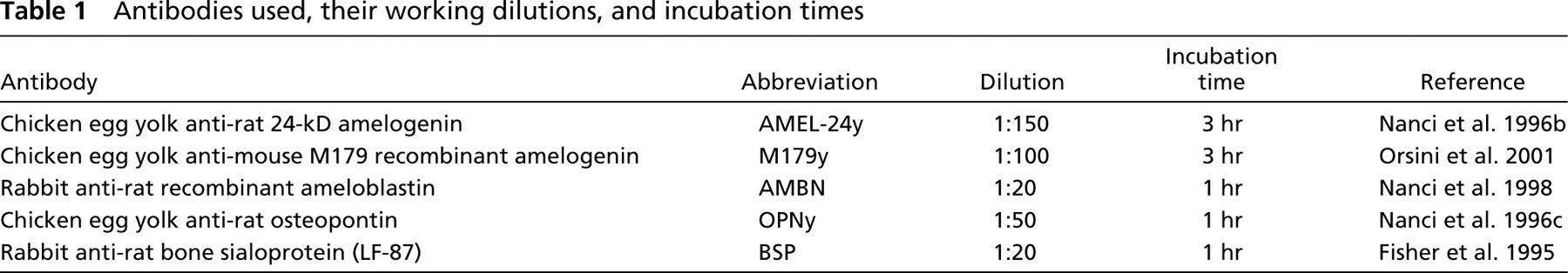
Antibodies used, their working dilutions, and incubation times
Materials and Methods
All animal procedures were in accordance with guidelines of the Comité de déontologie de l'expérimentation sur les animaux of Université de Montréal.
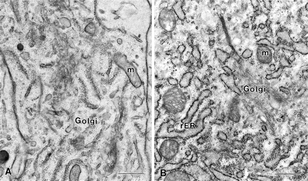
Electron micrographs illustrating the ultrastructural appearance of the Golgi region of (
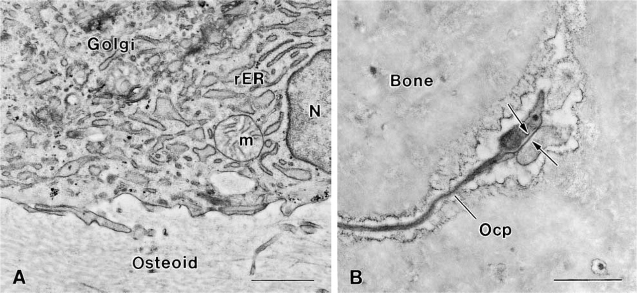
Electron micrographs illustrating the ultrastructural appearance of alveolar bone obtained with MW irradiation. (
Microwave Processing of Tissues
Male Wistar rats (Charles River Canada; St-Constant, PQ, Canada) weighing approximately 100 g were anesthetized with Metofane (methoxyfluorane; Janssen Pharmaceutica, North York, ON, Canada) and decapitated. The hemimandibles were dissected out and quickly immersed in 4% paraformaldehyde + 0.1% glutaraldehyde buffered at pH 7.2 with 0.1 M sodium cacodylate. All soft tissues covering the bone were gently removed. In addition, the alveolar bone overlying the buccal aspect at the apical end of the incisor was partially broken and the tip of the incisor was fractured using a bone-cutting forceps. Specimens were immersed in a beaker containing 40 ml of fixative at room temperature (RT), which was subsequently placed in a 20 × 20-cm glass recipient filled with ice and placed in a Pelco 3440 laboratory MW oven (Ted Pella; Redding, CA). The temperature probe of the oven was submersed in the fixative and the specimens were immediately exposed to MW irradiation at a 100% setting for three periods of 5 min with the temperature programmed to a maximum of 37C (Massa and Arana–Chavez 2000). After MW irradiation, specimens were transferred into fresh fixative and left in it overnight at 4C. They then were washed in 0.1 M sodium cacodylate buffer, pH 7.2, for 1 hr. In some cases, a 3-mm segment from the apical end of the incisor was left undecalcified. Decalcification of mandibles was carried out in an aqueous solution of 4.13% EDTA (Warshawsky and Moore 1967) under MW irradiation for a cumulative time of 15 hr in a Pelco 3440 laboratory MW oven. Specimens were placed in a beaker containing 25 ml of the decalcifying solution, which was placed in a larger glass recipient filled with ice. The temperature probe was submersed in the EDTA solution and the specimens were immediately exposed to MW irradiation at a 100% setting for periods of 15 min with the temperature programmed to a maximum of 37C. The decalcifying solution was changed every hour and the ice replaced as required. After decalcification, rat mandibles were divided into segments using a molar reference line (Smith and Nanci 1989) and washed extensively in 0.1 M sodium cacodylate buffer, pH 7.2. Some specimens were postfixed in potassium ferrocyanide-reduced osmium tetroxide (Neiss 1984) for 1 hr at RT, whereas others were left unosmicated. All samples were dehydrated in graded concentrations of ethanol and embedded in hard grade LR White resin (London Resin Company; London, UK). Toluidine blue-stained 1-μm-thick sections were examined in a light microscope and regions containing the incisor or the molars with their surrounding alveolar bone were selected for ultrathin sectioning. Eightynm-thick sections were cut with a diamond knife on a Reichert Ultracut E ultramicrotome and collected on Formvar–carbon-coated 200-mesh nickel grids.
Postembedding Colloidal Gold Immunocytochemistry
Ultrathin sections of osmicated samples were pretreated with an aqueous solution of 5% sodium metaperiodate for 60 min (Bendayan and Zollinger 1983) and washed with distilled water. Unosmicated sections were directly processed for immunolabeling using antibodies and conditions summarized inTable 1. Grids incubated with chicken egg yolk antibodies (AMEL-24y, M179y, OPNy) were incubated with a rabbit anti-chicken IgG secondary antibody (Cappel Research Products; Scarborough, ON, Canada) diluted 1:2000 for 1 hr. The sites of antibody–antigen binding were then revealed by floating the grids on a drop of protein A–gold complex for 30 min. The complex was prepared as described in Bendayan (1995) using colloidal gold particles of 10–12 nm (Frens 1973). Grids were washed with PBS between incubation steps and sections blocked by placing the grids on a drop of PBS–1% ovalbumin for 15 min. After the protein A–gold, the grids were jet-washed with PBS followed by distilled water. All steps were carried out at RT. Sections were then stained with uranyl acetate and lead citrate and examined in a JEOL 1200 EX II transmission electron microscope operated at 60 kV. Controls for the specificity of the labeling consisted of incubating the sections with antibodies to unrelated proteins, with the secondary antibody followed by protein A–gold, or with protein A–gold alone.
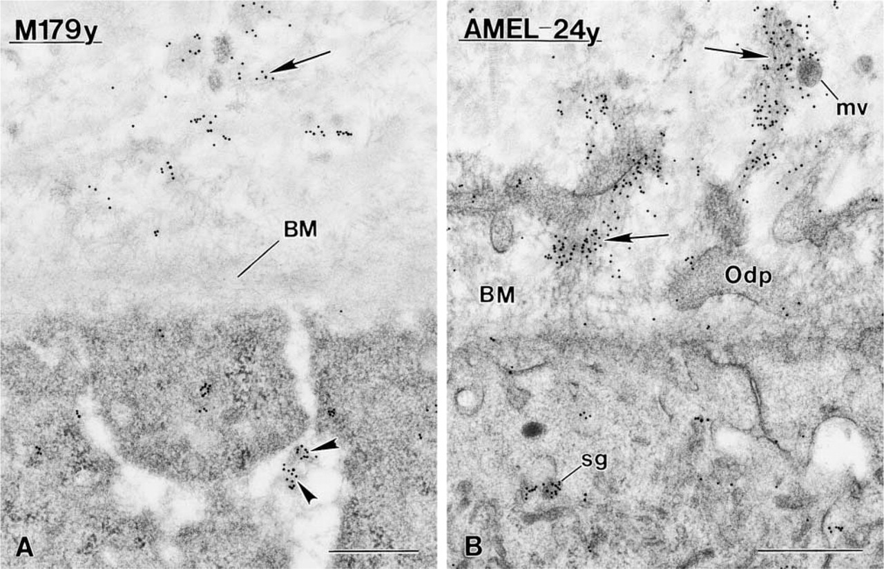
Immunocytochemical preparations of early presecretory stage ameloblasts incubated with anti-amelogenin antibodies. (
Results
Preservation of Tissues
Cells of incisor and molar tooth organs, as well as of alveolar bone, exhibited good ultrastructural preservation (Figures 1 and 2). The plasma membrane was well delineated and the distinctive structural details of organelles were clearly apparent. Mitochondria were not swollen and showed tightly packed cristae. There was no space between cells and the extracellular matrix they produce. In particular, there was no “stippled material” interposed between ameloblasts and enamel, as is usually seen in immersion-fixed teeth (see Nanci and Warshawsky 1984).
Postembedding Colloidal Gold Immunocytochemistry
As immunolabelings of enamel, dentine, bone and cementum with the antibodies used in the present study have been extensively described for conventionally fixed tissues, only those results that highlight issues pertinent to MW fixation and to the discussion will be presented.
Labeling for amelogenin was detected between differentiating ameloblasts before they fully reversed polarity. At this early time point, there were some gold particles in the unmineralized mantle dentine associated with either the basement membrane at the apical surface of ameloblasts or nearby patches of organic matrix (Figure 3A). At a slightly later time, labeled secretory granules were found at the distal pole of ameloblasts. The patches in the forming dentine matrix were larger and more intensely immunoreactive. These were frequently associated with odontoblast cell processes and occasionally with matrix vesicles (Figure 3B). When the basement membrane was no longer present, abundant patches of matrix, intensely immunoreactive for amelogenin, were present among the superficial collagen fibrils (Figure 4A). The ameloblasts associated with these patches exhibited immunoreactivity over the Golgi apparatus, secretory granules, and multivesicular bodies (Figures 4B and 4C). Differentiating ameloblasts showed immunoreactivity for ameloblastin, but there was no readily apparent labeling over the unmineralized mantle dentine. During the secretory stage, the Golgi apparatus, secretory granules, and enamel matrix were intensely labeled (Figures 5A and 5B). Gold particles over enamel were concentrated at growth sites (Figure 5A). No significant labeling for either ameloblastin or amelogenin was observed in differentiating odontoblasts.
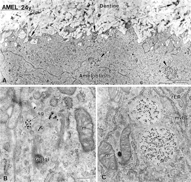
Electron micrographs of late presecretory stage ameloblasts incubated with chicken anti-24-kD rat amelogenin (AMEL-24y). (
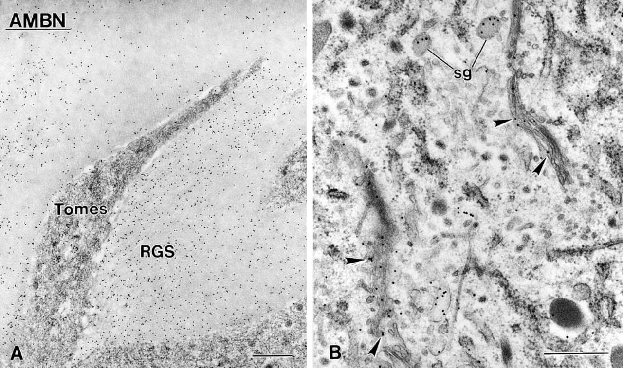
Immunocytochemical preparations for ameloblastin (AMBN). (
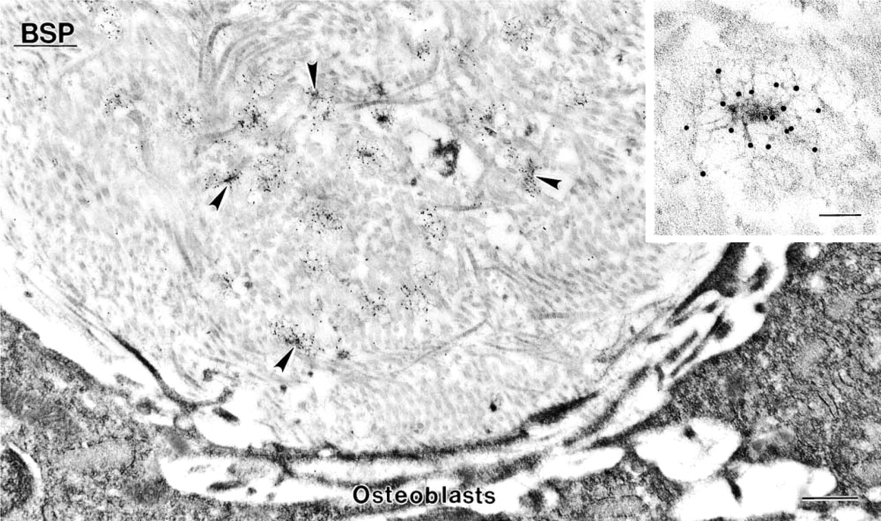
Electron micrograph showing a region of bone formation in which mineralization foci (arrowheads) are found. Immunoreactivity for bone sialoprotein (BSP) is present over the foci.
Electron micrographs illustrating labeling for (
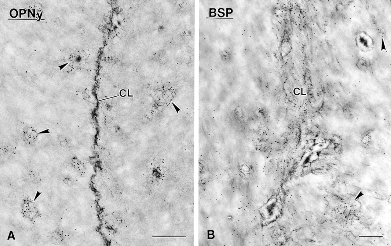
In regions of bone development, labeling for bone sialoprotein (Figure 6) and for osteopontin was observed in close relation to mineralization foci. Cement lines in bone as well as interfibrilar matrix in both bone and cementum were also immunoreactive for both noncollagenous proteins (Figures 7A, 7B, and 8). The lamina limitans outlining osteocyte lacunae and canaliculi lodging their processes occasionally showed labeling for osteopontin (not usually observed with this antibody). Distinctively, at early stages of tooth development, the Golgi apparatus of odontoblasts was labeled for osteopontin (Figures 9D–9F). The density of labeling over this organelle increased from the time when mineral crystals appeared in matrix vesicles to when a uniform layer of mantle dentine was evident (Figures 9A–9C). However, no significant labeling was observed over dentine matrix at any stage of dentinogenesis.
Discussion
The present study shows that immersion-fixation with MW irradiation can be applied to the immunocytochemical characterization of calcified tissues. The procedure is simple, yields ultrastructural preservation comparable to that obtained with perfusion-fixation, and retains antigenicity of the various noncollagenous matrix proteins examined. Although the exact mechanism of the MW effects is still not fully defined, it has been postulated that irradiation induces oscillation of water molecules (Moran et al. 1988). Therefore, fixative agents in aqueous solutions may take advantage of this physicochemical condition to increase their rate of penetration into tissues. Microwaves may also have an effect on chemical reactions during crosslinking by aldehydes because good ultrastructural preservation was obtained despite the use of relatively weak fixative solutions. However, MWs may also thermally “coagulate” proteins during irradiation (Horobin and Flemming 1990), potentially slowing the penetration rate ultimately impacting on the quality of preservation. This problem was avoided by maintaining the temperature of the specimens near the physiological level during fixation.
Decalcification of large mineralized samples, e.g., as human teeth, requires exposure to EDTA for long periods of time, introducing alterations in their ultrastructure. Although irradiation with MWs has been previously used to shorten decalcification time (Madden and Henson 1997; Faltin et al. 2001), our results additionally show that MW decalcification does not interfere with preservation of antigenicity. Shortening of decalcification time may represent an additional advantage for immunocytochemical studies because weak fixatives are generally used for preservation, making tissues more susceptible to alterations by decalcifying agents.
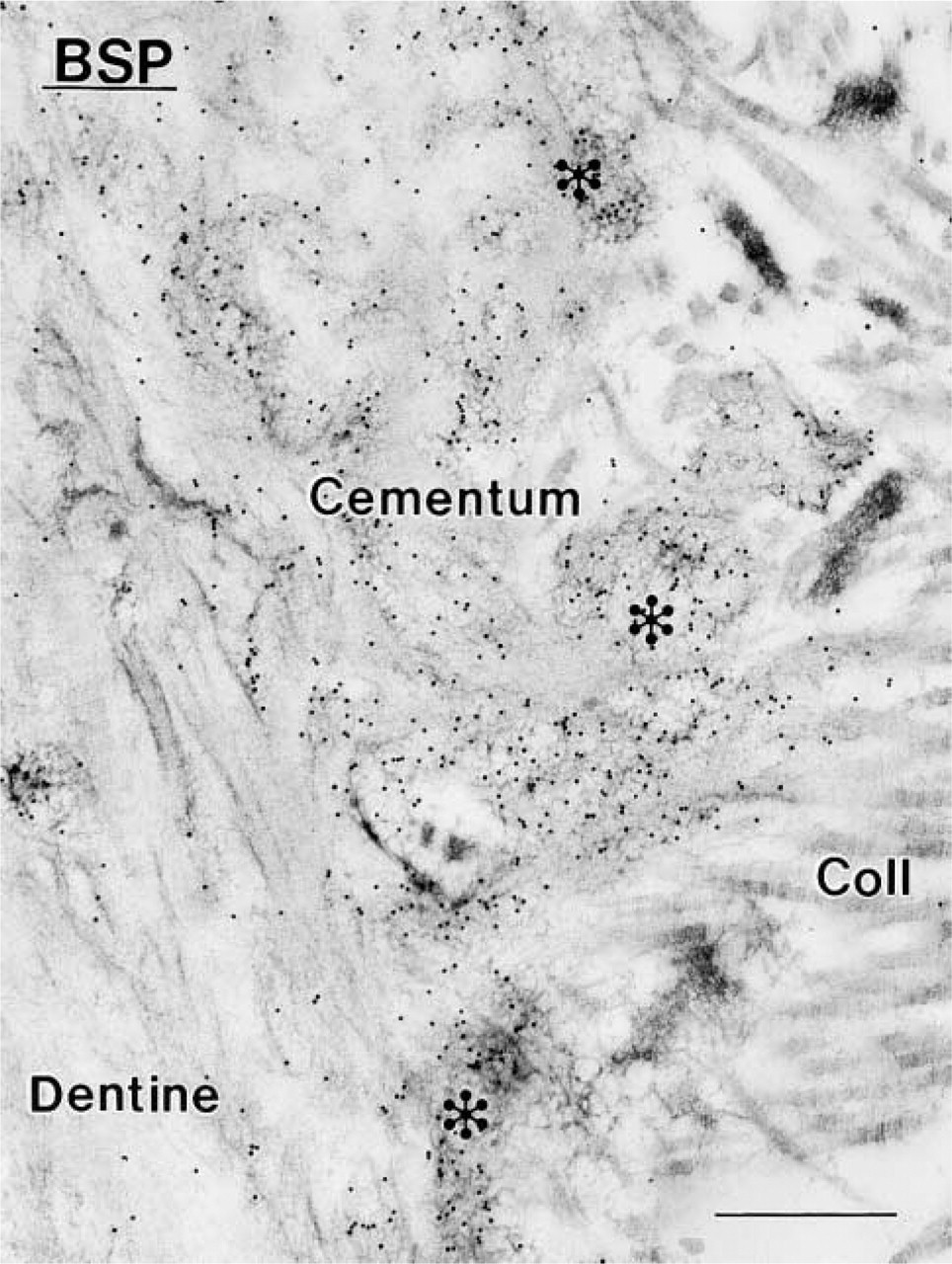
Electron micrograph illustrating portion of acellular cementum from the root of a third molar. There is intense immunoreactivity for bone sialoprotein (BSP) over granular organic matrix (asterisks) occupying the space between the inserted portions of extrinsic collagen fibers (Coll). Note the absence of any interfacial cementing line between the dentine and cementum. Bar = 0.5 μm.
After establishing that MW irradiation yielded good ultrastructural preservation, our aim was to determine its impact on the immunodetection of major noncollagenous matrix proteins present in calcified tissues. In general, the immunoreactivity obtained was at least as intense as in previous reports (see references below) and, in most cases, it appeared stronger. This is consistent with the findings that MW irradiation can be used for antigen retrieval (reviewed in Hayat 2000). Aspects of the immunolabelings that validate the observed benefits of MW irradiation and highlight important aspects of calcified tissue biology are discussed below.
Enamel Proteins
It has long been believed that initiation of dentinogenesis precedes amelogenesis. However, a number of immunocytochemical studies have shown that differentiating ameloblasts already secrete amelogenin during the presecretory stage of amelogenesis (discussed in Nanci and Smith 1992; Nanci et al. 1998). In the present study, amelogenin was immunodetected early during odontogenesis, along the lateral surfaces of differentiating ameloblasts that had not yet fully reversed polarity. When differentiation was more advanced, characteristic patches of enamel matrix appeared between collagen fibrils of the unmineralized mantle dentine, near the ameloblast surface. In general, these patches showed a somewhat stronger immunoreactivity for amelogenin than is usually observed in perfused material (see Nanci and Smith 1992; Nanci et al. 1998 for comparative images). In contrast, whereas ameloblasts exhibited labeling for ameloblastin, there were few gold particles over the matrix patches in the unmineralized mantle dentine. This observation is consistent with previous reports showing that secretion and/or extracellular accumulation of amelogenin precedes that of ameloblastin (Nanci et al. 1998). Various studies have shown that amelogenin molecules diffuse from the forming enamel through dentine and between odontoblasts at early stages of odontogenesis (Inage et al. 1989; Inai et al. 1991; Sawada and Nanci 1995; Nanci et al. 1996a). It has been suggested that such translocated amelogenins are endocytosed by differentiating odontoblasts, possibly by a receptor-mediated mechanism (Slavkin et al. 1988; Nakamura et al. 1994). Although there was immunoreactivity for amelogenins between odontoblasts, no labeling was detected in these cells despite the improved sensitivity of immunodetection with MW irradiation. Therefore, endocytosis of amelogenin molecules by odontoblasts is probably not a predominant event during early formation of the rat incisor.
Noncollagenous Proteins of Collagen-based Tissues
A number of studies using a variety of approaches, including immunohistochemistry (Mark et al. 1988; Chen et al. 1993; Helder et al. 1993), in situ hybridization (Chen et al. 1993; Helder et al. 1993), and immunoblotting (Helder et al. 1993), have concluded that odontoblasts do not express osteopontin at early stages of dentine formation. In the present study, binding of anti-osteopontin over the Golgi region of odontoblasts was seen as early as the beginning of mantle dentine mineralization. Labeling over the extracellular matrix, however, was extremely weak and often questionable despite the fact that osteopontin can be demonstrated biochemically in dentine (Fujisawa et al. 1993). It is possible that this noncollagenous protein is masked by other proteins and/or undergoes major processing so that it is not recognized by the antibody in dentine. Alternatively, the rate of osteopontin secretion by odontoblasts may be very slow, leading to its intracellular accumulation but never reaching the threshold of immunodetection extracellularly.
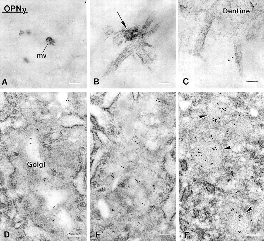
Immunocytochemical preparations with chicken anti-rat osteopontin antibody (OPNy) illustrating the temporal appearance of intracellular labeling for osteopontin (OPNy) in odontoblasts in relation to initiation of mineral deposition in mantle dentine. When the majority of matrix vesicles (mv) exhibit mineral crystals (
Bone sialoprotein and osteopontin are conspicuously present on patches of interfibrillar matrix and over cement lines in bone (reviewed in McKee and Nanci 1995; Ganss et al. 1999; Nanci 1999; Sodek et al. 2000). Although the presence of osteopontin in cement lines is generally accepted, it has been said that they only “sometimes contain bone sialoprotein” (Sodek et al. 2000). The results obtained here with MW processing are consistent with studies that show the presence of both proteins in cement lines (Bianco et al. 1991; Nanci 1999). Although the labeling was generally strong, there were cases in which either of the two proteins appeared to be absent or present in very low amounts in this interfacial structure. This occasional paucity and the presence of cement lines in osteopontin–/– mice (Rittling et al. 1998) suggest that osteopontin and perhaps bone sialoprotein are not the fundamental structural constituents of cement lines, that there are probably other not yet identified components that contribute to their structure, and/or that their absence may be compensated by other matrix molecules.
In summary, MW-processed calcified tissues show good ultrastructural preservation and no difference in distribution of labeling of representative noncollagenous matrix proteins compared to published studies with conventional tissue-processing methods. In addition, immunoreactivity with all the antibodies used was in general stronger and additional sites of labeling were revealed with the anti-osteopontin antibody, supporting the antigen retrieval potential of MW irradiation. We conclude that this approach offers an advantageous alternative for the processing of calcified tissues, in which the mineral phase may complicate the penetration of reagents such as fixatives and decalcification agents.
Footnotes
Acknowledgments
V. E. Arana–Chavez was the recipient of a research fellowship (99/11187–5) from FAPESP, Brazil. This work was supported by an operating grant from Canadian Institutes of Health Research (CIHR).
We thank Sylvia Zalzal for help with immunocytochemical procedures and for preparing the protein A–gold complexes and Micheline Fortin for ultrathin sectioning. We are grateful to Dr L. W. Fisher (National Institutes of Health, Bethesda, MD) for supplying the LF-87 antibody and to Dr P. H. Krebsbach (University of Michigan, Detroit, MI) for the ameloblastin antibody.