
Abstract
Keywords
It is often desirable to obtain muscle samples for direct analyses in order to study the effects of diet, exercise, disuse, or disease on human skeletal muscle. Although invasive, the percutaneous muscle biopsy technique (Bergström 1975) is safe, rapid, and repeatable. It is excellent for allowing the extraction of a small piece of muscle (~80-160 mg) with minimal discomfort and scarring. The vastus lateralis muscle, a portion of the quadriceps femoris muscle group, has been the muscle of choice for biopsies because of its mixed fiber type composition, trainability, and accessibility. Therefore, a large body of data concerning this muscle exists in the literature, potentially facilitating comparisons among studies (Saltin and Gollnick 1983).
However, direct comparisons of morphometric data may be hindered for a number of reasons, including the use of small biopsy samples and/or a small number of subjects (usually less than 12), disregard for possible gender and age differences, different methodologies for fiber type delineation, and different fitness levels (e.g., Edström and Nyström 1969; Edström and Ekblom 1972; Johnson et al. 1973; Gollnick et al. 1974; Edgerton et al. 1975; Tesch and Karlsson 1978; Lexell et al. 1984; Simoneau et al. 1985). Because young men and women are often used as subjects and because the vastus lateralis muscle is most often investigated, it is important to establish mean values for morphometric data from this muscle in this age group. Recently, six fiber types have been delineated in human skeletal muscle and these have been correlated with a specific myosin heavy chain (MHC) profile (Staron 1991).
The purpose of this investigation was to present normative data on muscle fiber types and sizes within the superficial region of vastus lateralis muscle of healthy, untrained young men and women, and to establish specific gender differences using this entire range of histochemically defined fiber types. Biopsy data were gathered from individuals who had participated in various research projects at Ohio University over the past 10 years.
Materials and Methods
Subjects
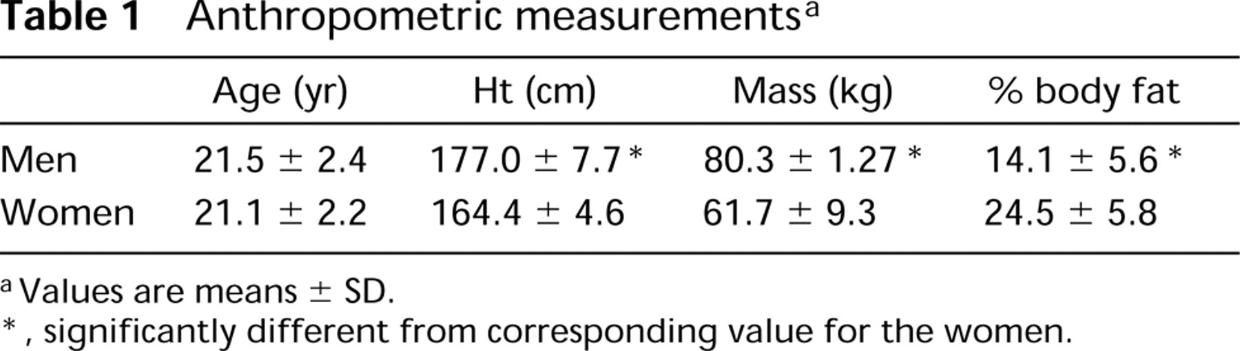
A total of 150 healthy college-aged men and women volunteered to participate in various research projects at Ohio University over the past 10 years (e.g., Staron et al. 1990, 1991, 1994; Allemeier et al. 1994; and recent unpublished data). In all cases, the Ohio University Institutional Review Board gave approval before the beginning of each project. All subjects were informed of the procedures, risks, and benefits, and provided written consent before participation. Subjects consisted of 95 young men and 55 young women (1). Although physically active, all subjects were considered untrained and had not participated in any regular exercise program for at least 6 months before the start of each research project. Anthropometric measurements were taken immediately before the extraction of the muscle biopsies (1). Body composition was estimated using skinfold measurements from three sites for the men (chest, umbilicus, and anterior thigh) (Jackson and Pollock 1978) and women (anterior thigh, posterior brachium, and suprailium) (Jackson et al. 1980).
Muscle Biopsies
Muscle biopsies (80-160 mg) were extracted from the superficial portion of the vastus lateralis muscle by the percutaneous needle biopsy technique (Bergström 1962). The muscle samples were removed from the needle, oriented in tragacanth gum, immediately frozen in isopentane cooled by liquid nitrogen to -159C, and stored at -74C. Because of possible anatomic variations (Willan et al. 1990), as well as potential variations in fiber type distribution and size from superficial to deep and proximal to distal (Blomstrand and Ekblom 1982; Elder et al. 1982; Lexell et al. 1983, 1985; Mahon et al. 1984; Lexell and Taylor 1989), attempts were made to extract samples from each individual at approximately the same location. Biopsies were therefore taken ~16 cm proximal to the superior border of the patella from the superficial portion of the vastus lateralis muscle (25-30 mm deep to the fascia lata). Depth was gauged by interval markings engraved in the biopsy needles. To ensure the procurement of adequate sample sizes, large biopsy specimens were obtained using a double-chop method (Staron et al. 1990) combined with suction (Evans et al. 1982).
Anthropometric measurements a
aValues are means ± SD.
∗, significantly different from corresponding value for the women.
Fiber Type and Cross-sectional Area Determinations
The frozen biopsy specimens were thawed to -24C and serially sectioned (12 ± thick) for histochemical analysis. Myofibrillar adenosine triphosphatase (mATPase) histochemistry was performed using preincubation pH values of 4.3, 4.6, and 10.4 (Brooke and Kaiser 1970) to determine the muscle fiber type composition. Six fiber types (I, IC, IIC, IIA, IIAB, and IIB) were distinguished on the basis of their staining intensities (Figure 1). Fiber Types IIAB and IIB have more recently been referred to as IIAX and IIX, respectively (Smerdu et al. 1994; Ennion et al. 1995). A composite photomontage of each mATPase preparation after preincubation at pH 4.6 was made using Polaroid micrographs (X 56 magnification). These were used in combination with the other mATPase preparations to determine fiber type percentages and total fiber number in each biopsy. Because of the scarcity of hybrid fibers (IC, IIC, and IIAB), only the major fiber types were used for area determinations. Cross-sectional area was determined on at least 50 fibers per major fiber type (I, IIA, and IIB) per biopsy, using either direct tracings (X 200) and a digitizing tablet or an image analysis computer program (NIH Image software program). The six histochemically delineated fiber types were subsequently collapsed into the three major types (I, IIA, and IIB) using the following formulae: I + IC = I, IIC + IIA + 1/2IIAB = IIA, 1/2IIAB + IIB = IIB. These three types were then combined with the respective area measurements to yield fiber type percent area that has been shown to correlate with relative MHC isoform percentages (Fry et al. 1994).
Myosin Heavy Chain Analysis
MHC analysis was performed on all the biopsy samples from the men and on 26 of the 55 samples from the women using sodium dodecyl sulfate (SDS)-polyacrylamide electrophoretic techniques. The protocol for analyzing the specimens was based on the procedures of Perrie and Bumford (1986), with modifications used for single fiber analysis (Staron 1991; Staron and Hikida 1992). Briefly, four to six serial cross-sections (20 μ m thick) from each biopsy were placed in 0.5 ml of a lysing buffer containing 10% (w/v) glycerol, 5% (v/v) 2-mercaptoethanol, and 2.3% (w/v) SDS in 62.5 mM Tris (hydroxymethyl) aminomethane HCl buffer (pH 6.8) and were heated for 10 min at 60C. Small amounts of the extracts (3-5 μl) were loaded on 4-8% gradient SDS-polyacrylamide gels with 4% stacking gels (Bär and Pette 1988), run overnight (19-21 hr) at 120 V, and stained with Coomassie Blue. MHC isoforms were identified according to their apparent molecular masses compared with those of marker proteins and migration patterns from single fiber analyses. Relative MHC isoform content was subsequently determined using a laser densitometer.
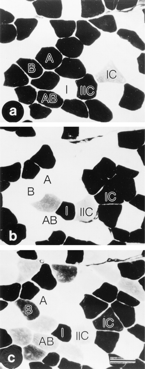
Serial cross-sections of a pretraining muscle sample taken from a subject demonstrating the fiber type delineation by mATPase histochemistry after preincubation at pH 10.4 (
Statistical Analysis
The statistical package SPSS was utilized for all statistical analyses. Descriptive statistics were used to derive mean ± SD for all variables. Anthropometric data were compared between groups using independent t-tests. Muscle fiber characteristics (fiber type distribution, cross-sectional area, percentage fiber type area, and MHC content) were analyzed using repeated-measures one-way and two-way analysis of variance (ANOVA). Significant differences were then determined using Tukey's HSD post hoc test. Correlation analyses were performed to compare percentage fiber type area with MHC content. Differences were considered significant at p 0.05.
Results
Anthropometric Data
The men were taller, heavier, and had a lower percentage of body fat compared to the women (1).
Fiber Type Distribution
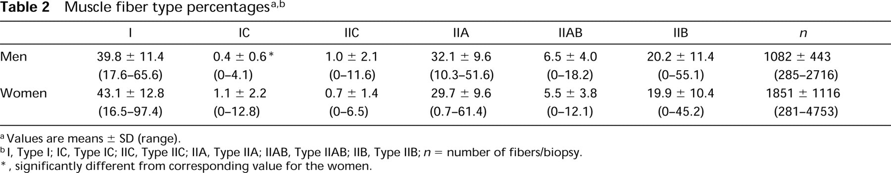
Large sample sizes were obtained for the determination of fiber type composition. Only four of the 150 biopsy specimens contained less than 400 fibers. The distribution of the six fiber types was almost identical for the young men and women (2). The lone exception was a significantly lower percentage of Type IC fibers for the men. The superficial region of the vastus lateralis muscle for both men and women contained a fiber type distribution of approximately 41% I, 1% IC, 1% IIC, 31% IIA, 6% IIAB, and 20% IIB (2). However, there was a wide range for the percentage of Type I fibers for both the men (17.6-65.6%) and the women (16.5-97.4%). Most of the vastus lateralis muscle samples (132 of the 150) had between 25-60% Type I fibers. A total of 6% of the vastus lateralis samples (four women and five men) contained >60% Type I fibers and 6% (three women and six men) contained <25% Type I fibers. Similar to the data of Simoneau and Bouchard (1989), individuals with a percentage of Type I fibers <35% were found almost twice as often for the men (38%) compared to the women (20%).
Cross-sectional Area Measurements
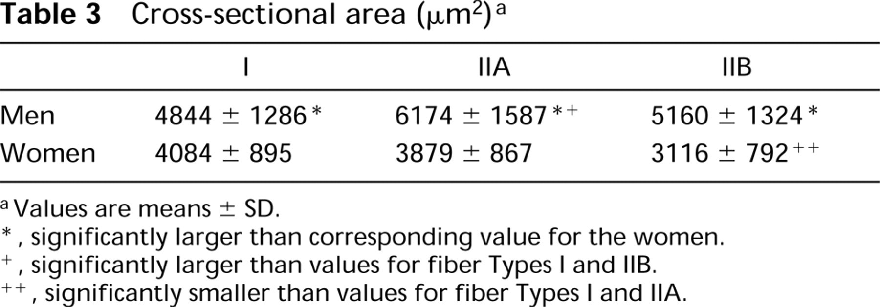
As previously shown for the vastus lateralis muscle (Simoneau and Bouchard 1989), the cross-sectional areas of all three major fiber types (I, IIA, and IIB) were significantly larger for the men compared to the women (Table 3). On average, the Type I, IIA, and IIB fibers were 18.6%, 59.2% and 65.5% larger for the men than the respective fiber types for the women. For the men, the Type IIA fibers were the largest (with no significant difference between Types I and IIB), and for the women Type I and IIA fibers were larger than Type IIB fibers (Table 3).
Muscle fiber type percentages a,b
aValues are means ± SD (range).
bI, Type I; IC, Type IC; IIC, Type IIC; IIA, Type IIA; IIAB, Type IIAB; IIB, Type IIB; n = number of fibers/biopsy.
∗significantly different from corresponding value for the women.
Percentage Fiber Type Area
Percentage fiber type areas were determined following the collapse of the original six fiber types into the three major groups (see Materials and Methods). Although no differences were found between the men and women for fiber type proportions (with the exception of the IC population), the area made up of each of the major fiber types was very different between the men and the women. The percentage area of Type I was significantly smaller and the percentage area of Type IIA significantly larger for the men compared to the women (4). The hierarchy of the percentage fiber type area from largest to smallest was IIA>I>IIB for the men and I>IIA>IIB for the women (4).
Myosin Heavy Chain Content
The relative content of the three MHC isoforms (MHCI, MHCIIa, and MHCIIb) paralleled the fiber type area data. Thus, the relative percentage of MHCI was significantly smaller and the relative percentage of MHCIIa significantly larger for the men compared to the women (5). Likewise, in order from largest to smallest, the relative MHC content for the men was MHCIIa>MHCI>MHCIIb and for the women was MHCI = MHCIIa>MHCIIb (5). As expected, correlations between fiber type area and relative MHC percentages were all significant, including correlations for the men and women separately as well as together (6).
Discussion
Not surprisingly, anthropometric data from the present study revealed gender differences. The young men were significantly taller, heavier, and had a lower percentage of body fat compared to the young women. Similar findings have been previously reported by many others (e.g., Komi and Karlsson 1978; Simoneau et al. 1985; Glenmark et al. 1992; Miller et al. 1993).
Gender differences have also been previously reported for fiber size within the vastus lateralis muscle. Shortly after birth, muscle fibers are small (12-18 diameter), with the Type I fibers slightly larger than the Type II and no difference between male and female (Oertel 1988). However, after puberty, fiber cross-sectional area in males tends to be larger than in females (Brooke and Engel 1969; Edström and Nyström 1969; Saltin et al. 1977; Simoneau et al. 1985; EsséAn-Gustavsson and Borges 1986; Simoneau and Bouchard 1989; Miller et al. 1993). Recently, all three major fiber types (I, IIA, and IIB) have been shown to be larger in the vastus lateralis muscle of men. Simoneau and Bouchard (1989) found that the cross-sectional areas of fiber Types I, IIA, and IIB from the vastus lateralis muscle of young men were 14%, 38%, and 56% larger compared to young women. Similarly, fiber Types I, IIA, and IIB in the present study were approximately 19%, 59% and 66% larger for the men compared to the women. In addition, Type II fibers tend to be larger than Type I in the muscles of men, whereas the reverse is true for women (Brooke and Engel 1969; Saltin et al. 1977; Simoneau et al. 1985; Simoneau and Bouchard 1989). For both men and women, the Type IIA cross-sectional area appears to be larger than the Type IIB, contributing to the hierarchy of fiber sizes (from largest to smallest) found in the present study: IIA>IIB = I for the men and I= IIA>IIB for the women.
Although many studies have found gender differences related to fiber size, conflicting reports have been published regarding the overall proportion of fast and slow fibers in the vastus lateralis muscle of men vs women (see Table 7). Investigations have reported a higher percentage of Type I fibers in women compared to men (Simoneau et al. 1985; Simoneau and Bouchard 1989; Miller et al. 1993), a higher percentage of Type I fibers in men (Komi and Karlsson 1978; EsséAn-Gustavsson and Borges 1986), and no difference between men and women (Saltin et al. 1977; Nygaard 1982). These different findings may be related to differences in sample size, age, methodology, hormone profiles, and/or physical activity level.
Cross-sectional area(±2)a
aValues are means ± SD.
∗, significantly larger than corresponding value for the women.
+, significantly larger than values for fiber Types I and IIB.
++, significantly smaller than values for fiber Types I and IIA.
Percentage fiber type area a
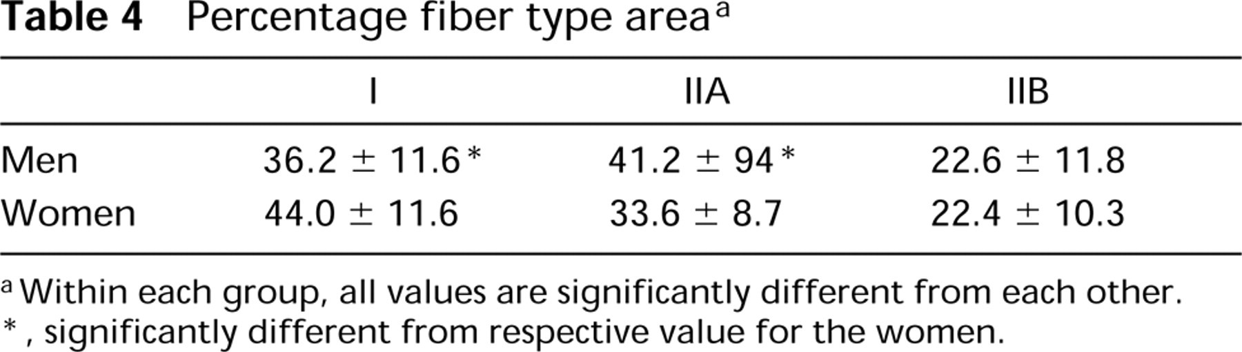
aWithin each group, all values are significantly different from each other.
∗significantly different from respective value for the women.
Relative myosin heavy chain isoform percentages a
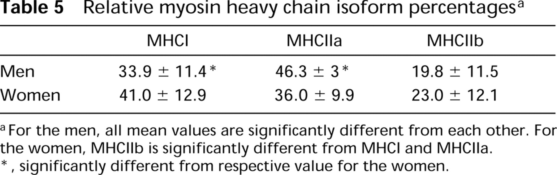
aFor the men, all mean values are significantly different from each other. For the women, MHCIIb is significantly different from MHCI and MHCIIa.
∗significantly different from respective value for the women.
Many studies on human muscle have used small sample sizes and/or have histochemically delineated only two fiber types, I and II (e.g., Brooke and Engel 1969; Edström and Ekblom 1972; Gollnick et al. 1972; Johnson et al. 1973; Komi and Karlsson 1978; Oertel 1988; Willan et al. 1990; Miller et al. 1993). Other studies have combined data from both men and women (Gollnick et al. 1974; Edgerton et al. 1975). Because of the large amount of inter- and intraindividual variations in the percentages of fast and slow fibers in the vastus lateralis muscle, it is important not only to use large biopsy samples but also to use large numbers of subjects. Indeed, even investigations with relatively large numbers of subjects have reported mean values for the percentage of slow fibers ranging from 37 to 55% in women and 36 to 58% men (7).
Although Simoneau and Bouchard (1989) presented cumulative data gathered over a number of years on fiber type composition and area measurements in the vastus lateralis muscle of 270 sedentary young men and women (range 16-33 years old), 148 post-training biopsies were also included in the analysis. In addition, only three muscle fiber types (I, IIA, and IIB) were histochemically delineated using a single-step histochemical technique (Mabuchi and SréAter 1980). Such methods do not allow adequate identification of hybrid fibers (IC, IIC, and IIAB), and will therefore result in fiber misclassification (Staron 1997). It should be made clear, however, that the primary purpose of Simoneau and Bouchard (1989) was to investigate the extent of variation in these morphometric parameters in human skeletal muscle, rather than to establish normative fiber type data. To our knowledge, this is the only study presenting normative data on the entire range of histochemically defined fiber types collected from a large number of untrained young men and women. Data from the current investigation suggest that there may not be a true gender difference between men and women with regard to the fiber type distribution in the vastus lateralis muscle.
Correlation analyses a
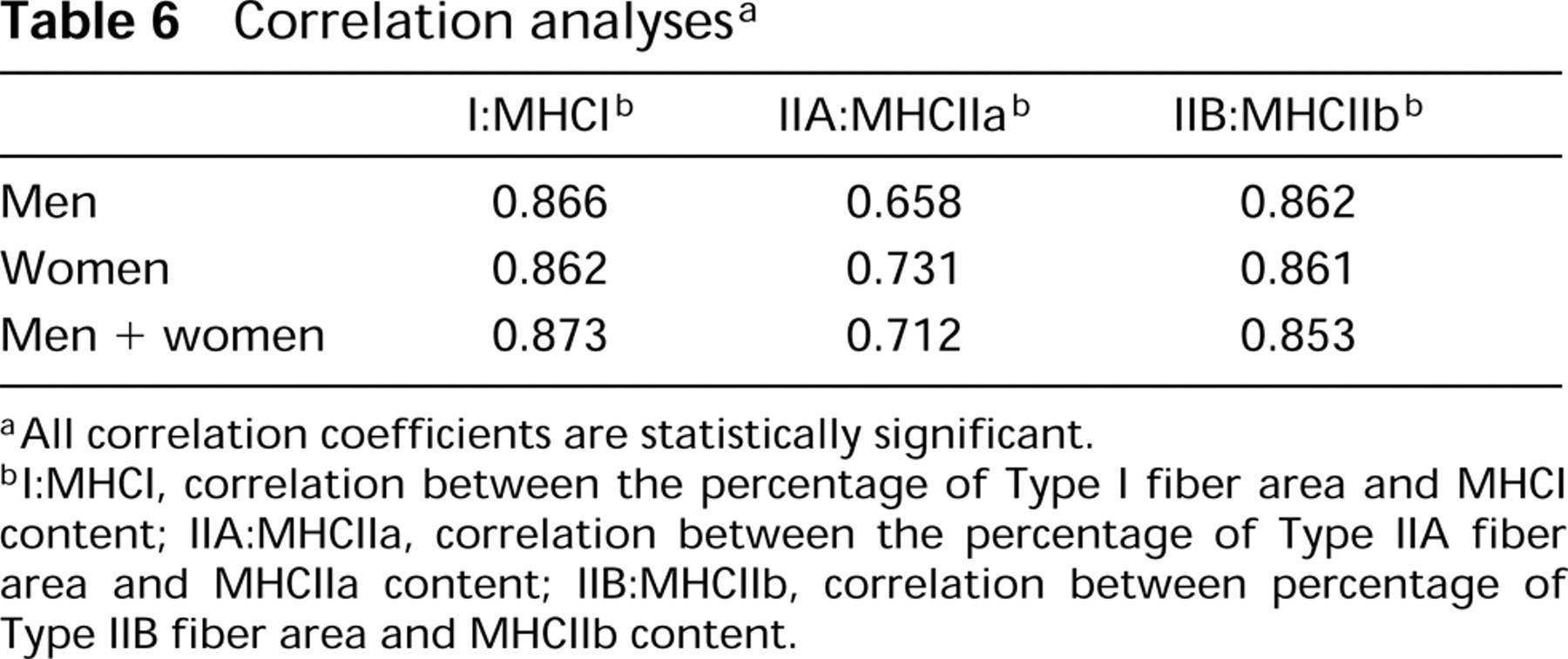
aAll correlation coefficients are statistically significant.
bI:MHCI, correlation between the percentage of Type I fiber area and MHCI content; IIA:MHCIIa, correlation between the percentage of Type IIA fiber area and MHCIIa content; IIB:MHCIIb, correlation between percentage of Type IIB fiber area and MHCIIb content.
Perhaps the most interesting gender difference relates to the percentage area occupied by the major fiber types. Although it appears that men and women have similar overall distributions of fast and slow fiber types in the vastus lateralis muscle, significant gender differences exist with regard to the total area occupied by each fiber type within the muscle. In the present investigation, the slow fibers were found to occupy a greater area in the women, whereas the fast IIA fibers occupied a greater area in the men. Of importance is the fact that these data were verified by the MHC profile. Similar findings with regard to percentage fiber type area have been previously reported for the vastus lateralis (EsséAn-Gustavsson and Borges 1986). On an applied level, these differences in percent fiber type area may result in differences in performance (Gollnick and Matoba 1984).
In conclusion, important aspects of the current investigation are the compilation of data from a large number of healthy untrained individuals, relatively large biopsy specimens, the delineation of the entire range of mATPase-based fiber types, and verification of the histochemical data by comparing percent fiber type area and relative MHC content. These data support previous findings demonstrating that the mean fiber cross-sectional area in the vastus lateralis muscle is smaller in women compared to men and that the Type II fibers of males tend to be larger than the Type I, whereas the reverse is true for the female. The current data suggest that the overall proportion of fast and slow fibers in the vastus lateralis muscle of young men and women is similar. There are, however, gender differences in percent fiber type areas that relate to differences in the hierarchy of cross-sectional areas of the major fiber types. Although the percentages of fast and slow fiber types are similar between young men and women, the slow fibers occupy a greater area in the women compared to the men and the fast fibers occupy a greater area in the men.
Summary of published data on muscle fiber type percentages in the vastus lateralis muscle of untrained young men and women a,b
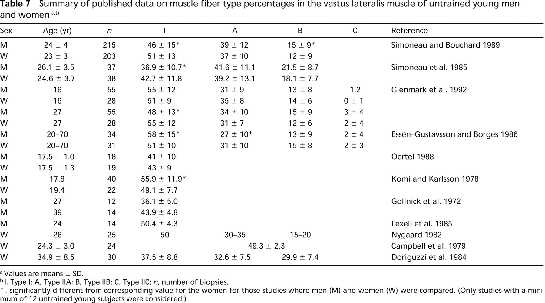
aValues are means ± SD.
bI, Type I; A, Type IIA; B, Type IIB; C, Type IIC; n, number of biopsies.
∗,significantly different from corresponding value for the women for those studies where men (M) and women (W) were compared. (Only studies with a minimum of 12 untrained young subjects were considered.)
Footnotes
Acknowledgements
Acknowledgments
We thank the Ohio University College of Osteopathic Medicine photographic and graphic departments for help with the figures and tables. Special thanks to all those young men and women who participated in various research projects in our laboratory over the past 10 years.