
Abstract
Detection of antigen-antibody interactions in immunocytochemistry relies on a reporter system. The most commonly employed reporter systems used are fluorochromes, enzymes, and particulate probes. This article considers the advantages and disadvantages associated with ultrasmall immunogold particles as the reporter system in immunocytochemical applications.
Demonstration of antigen-antibody binding in situ requires a reporter system. The most commonly used reporters in immunocytochemistry are fluorochromes, enzymes, and particulate probes. Routinely the reporter is conjugated to a secondary antibody or other immunoreagent (e.g., protein A) that is used to detect the primary antibody directed against the antigen of interest. Variations on this theme include labeling the primary antibody directly or labeling a tertiary antibody. The reporter system chosen depends on the requirements of a given procedure or experiment.
Particulate immunoprobes have been especially valuable for localization of cellular antigens at the ultrastructural level. The iron-containing protein ferritin was used as a particulate immunoprobe and was introduced relatively early in the development of immunocytochemistry (Singer 1959). Subsequently, colloidal gold was employed as an electron-dense immunoprobe (Faulk and Taylor 1971). Colloidal gold probes have become the standard for immunolabeling at the electron microscopic level (for review see Roth 1996). The most widely used colloidal gold particles are in the 5–15-nm size range. In addition, ultrasmall colloidal gold particles have been prepared in the 1–3-nm size range (Baschong et al. 1985; Baschong and Wrigley 1990; Chan et al. 1990; De Valck et al. 1991; Van de Plas and Leunissen 1993). Gold cluster compounds of 0.8 or 1.4 nm in size have also been developed for immunolabeling (Hainfeld 1987, 1988; Hainfeld and Furuya 1992). Herein, we refer to immunoprobes in the 0.8–1.5-nm size range as ultrasmall immunogold.
Utility of Ultrasmall Immunogold
In several studies using colloidal gold as the reporter system for immunoelectron microscopy, smaller gold particles have labeled more efficiently than larger ones (e.g., 5-nm>10-nm>15-nm). This labeling has been observed in a variety of situations and appears to be independent of embedding procedures because it occurs in resin-embedded material (e.g., Lackie et al. 1985; Yokota 1988; Ghitescu and Bendayan 1990), in ultrathin cryosections (e.g., Slot and Geuze 1981; Van Bengen en Henegouwen and Leunissen 1986; Takizawa and Robinson 1994), and in un-embedded material (e.g., Horisberger 1981; Kehle and Herzog 1987). Similarly, this observation holds for colloidal gold conjugated with antibodies or other immunological reagents such as protein A (e.g., Slot and Geuze 1981; Gu and D'Andrea 1989; Stierhof et al. 1991).
One reason for the development of ultrasmall immunogold probes was the possibility that these smaller immunogold reagents would yield enhanced labeling efficiency. Experimental evidence supports the contention that ultrasmall immunogold probes label with greater efficiency than do larger 5-, 10-, and 15-nm particles. In a study of the distribution of calcium ATPase in sarcoplasmic reticulum of skeletal muscle, the density of 1-nm immunogold particles was about 20 times greater than with 10-nm particles (Dulhunty et al. 1993). In this case, the tissue was embedded in resin (i.e., LR White). In a recent study, Takizawa (1999) compared the labeling efficiency of ultrasmall gold (i.e., gold cluster compounds) to colloidal gold (5-, 10-, and 15-nm) in immunocytochemical labeling of freeze-fractured neutrophils. HLA class I molecules in neutrophil membranes were more efficiently detected with 1.4-nm immunogold than with colloidal gold probes. These data are summarized in Table 1. The two studies cited illustrate that ultrasmall immunogold probes (≍1-nm) label more efficiently than do he larger colloidal gold particles. This result appears to be independent of the procedures used for sample preparation because disparate methods were employed in these studies (i.e., immunolabeling of resin-embedded sections and freeze-fractured replicas).
Applications Using Ultrasmall Immunogold
We have used gold cluster immunoprobes [Nanogold (NG)] in several applications. In one of these, 1.4-nm NG was used for localization of marker proteins of cytoplasmic granules (e.g., lactoferrin) in ultrathin cryosectioned human neutrophils. We compared the labeling of lactoferrin using 1.4-nm NG with 5-, 10-, and 15-nm colloidal gold immunoprobes. We found an inverse relationship between colloidal gold size and labeling efficiency. In addition, the ultrasmall gold gave heavy labeling in these ultrathin cryosections (Takizawa and Robinson 1994). When thicker cryosections were immunolabeled, we found that the colloidal gold was restricted to lactoferrin-positive granules at the cut surface of the section. On the other hand, the ultrasmall gold labeled these granules throughout the sections. These results, summarized in Figure 1, demonstrate that 1.4-nm immunogold probes penetrate into cryosections to a greater extent than do 5- and 10-nm colloidal gold.
Comparison of the labeling density of neutrophil HLA class I using different sized immunogold probes as the secondary antibodies in freeze-fracture immunocytochemistry.
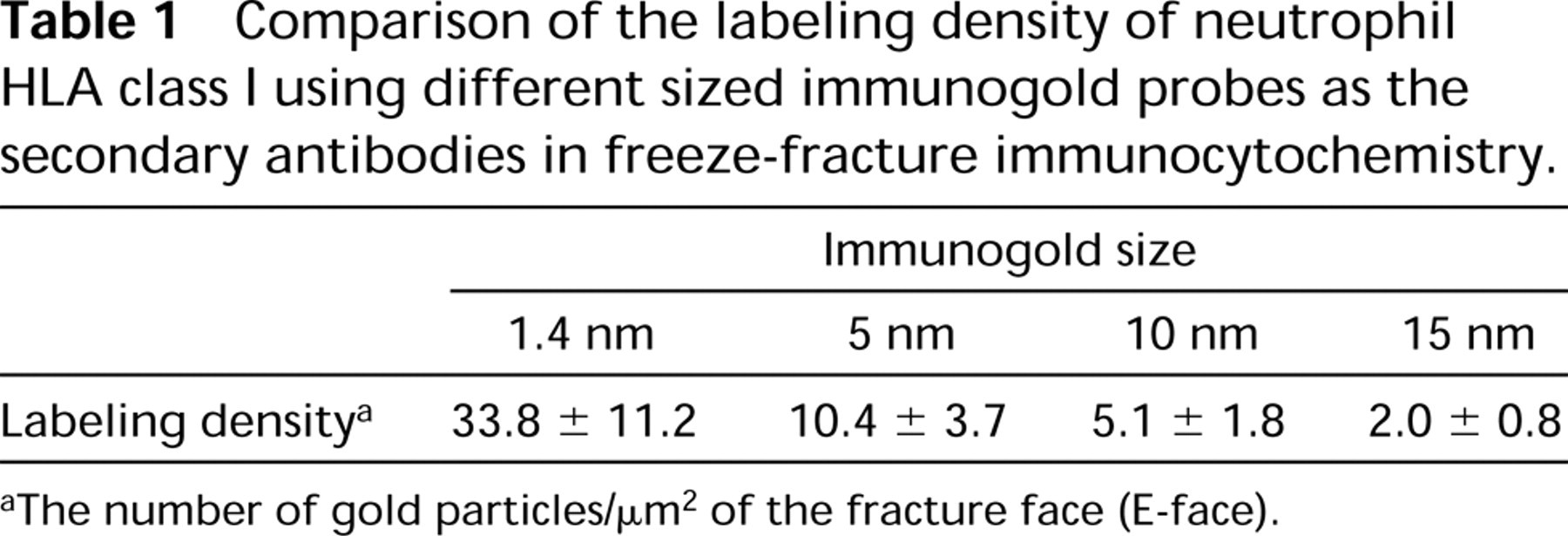
The number of gold particles/μm2 of the fracture face (E-face).
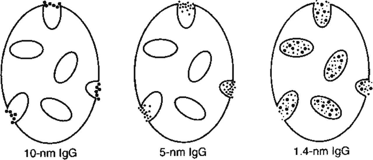
Diagram summarizing the labeling pattern obtained when lactoferrin was detected with immunogold probes of different sizes in the study of Takizawa and Robinson (1994). Cryosections (1–2 μm thick) of paraformaldehyde-fixed neutrophils were used as the substrate for immunolabeling. After labeling the cryosections were embedded in Epon. Thin sections of the resin-embedded material were cut and examined by electron microscopy. The larger 10-nm colloidal gold particles were restricted to the cut surface of the lactoferrin-positive granules, whereas the 5-nm particles penetrated partially into the matrix of the cut granules. The 1.4-nm NG not only detected anti-lactoferrin bound to the cut granules but also granules away from the cut surface. Therefore, the ultrasmall immunogold probe penetrates into fixed and cryosectioned cells to a greater extent than the larger colloidal gold particles.
We have also compared the ability of 1.4-nm immunogold and 5-nm colloidal gold to penetrate into other types of specimens. In this case, we employed tissue culture cells and isolated leukocytes for localization of tubulin in microtubules. When cells were fixed with glutaraldehyde and subsequently permeabilized with detergent, microtubules could be demonstrated with 1.4-nm immunogold but not with 5-nm colloidal gold. Alternatively, if cells were first permeabilized with detergent and then fixed with glutaraldehyde, then microtubules were localized with both 1.4-nm immunogold and 5-nm colloidal gold (Robinson and Vandré 1997). These results demonstrate that 1.4-nm immunogold penetrates into cells under conditions that preclude penetration of 5-nm colloidal gold (Figure 2).
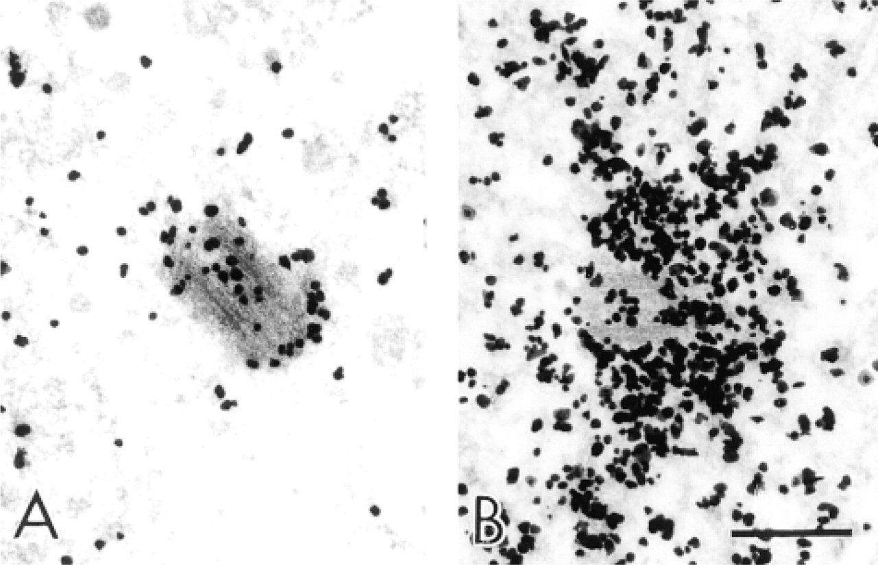
We have also investigated the efficacy of 1.4-nm immunogold for labeling centrosomes and centrioles. The structure of centrioles is well known from morphological studies and several centrosome-associated proteins have been identified (e.g., Gowen et al. 1995). The central part of the centrosome is the centriole, consisting of a characteristic array of microtubules. However, it has been virtually impossible to document this by immunolabeling of tubulin at the ultrastructural level (e.g., Gowen et al. 1995). It has been believed that this lack of successful immunolabeling was due, at least in part, to the poor penetration of colloidal gold probes into this structure. We have tested the ability of 1.4-nm immunogold to penetrate into these structures and to detect antibodies to tubulin associated with the centrioles of phagocytic leukocytes. In these studies, tubulin was localized according to the protocol of Ding et al. (1995). These experiments were successful, and 1.4-nm immunogold was able to gain entry into the centriole and to detect the anti-tubulin associated with the centriole (Figure 3).
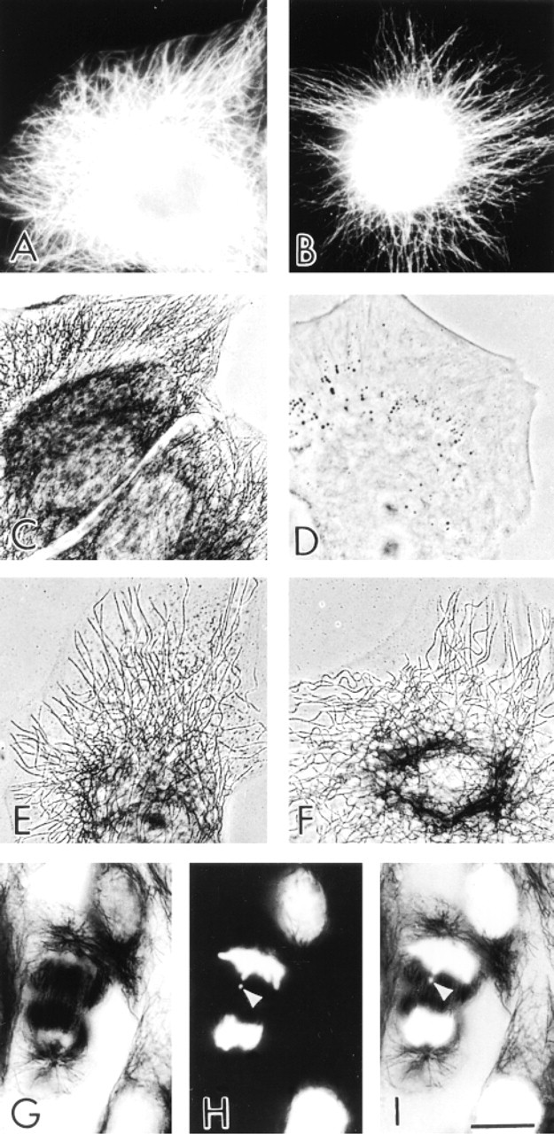
Comparison of 1.4-nm FNG and 5-nm immunogold probes for labeling LLC-PK cell microtubules. Detection of microtubules by fluorescence microscopy using FNG (
Immunocytochemical detection of tubulin in centrioles of human neutrophils with 1.4-nm immunogold and silver enhancement. (
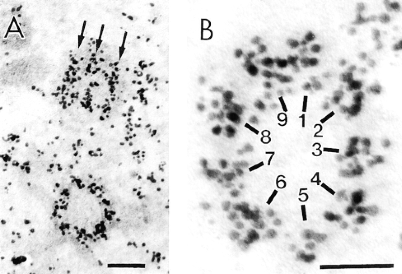
In other experiments, centrosomal proteins were localized with the MPM-2 antibody. This monoclonal antibody recognizes a subset of mitotic phosphoproteins (Davis et al. 1983). Some of these proteins are components of the centrosome and increase in amount during mitosis (reaching a maximum at metaphase) (Vandré and Borisy 1989). The binding of MPM-2 to its cellular targets was demonstrated at the ultrastructural level with 1.4-nm immunogold. Gold particle intensity surrounding the MPM-2-labeled centrosomes correlated with the stage of the cell cycle (Vandré and Burry 1992). Whereas only a few gold particles were associated with the interphase centriole, the mitotic centriole and surrounding pericentriolar material were heavily labeled (Figure 4). These results demonstrate that the 1.4-nm immunogold particles can readily penetrate into the centrosome. When the MPM-2 labeling is coupled with the detection of tubulin within the centriole, we show that 1.4-nm immunogold probes are useful for ultrastructural immunolabeling of centrosomes (structures difficult to label by other means).
Applications Using FluoroNanogold
Additional tools for immunolabeling are fluorescent derivatives of NG known as FluoroNanogold (FNG) (Powell et al. 1997). These bifunctional probes consist of a 1.4-nm gold cluster compound to which an antibody [IgG, F(ab′)2, or Fab] is conjugated; fluorochromes are conjugated to the antibody. These are unique immunoprobes because they facilitate combined fluorescence and electron microscopy.
We have been interested in microtubules of phagocytic leukocytes. However, microtubules in these cells have been difficult to visualize by immunocytochemistry; the reasons for this are incompletely understood. We have developed preparative procedures that facilitate the reliable immunocytochemical detection of these structures in phagocytes (Ding et al. 1995). Recently, we have applied FNG as the secondary antibody for detection of anti-tubulin bound to microtubules in phagocytic leukocytes (Robinson and Vandré 1997). In this case, the fluorescence signal from FNG was comparable to that from conventional fluorescently labeled secondary antibodies. After silver enhancement, the microtubules could be detected by transmitted light microscopy (i.e., bright field, phase-contrast, and differential interference contrast) and by epipolarization optics (Figure 5).
Immunoelectron microscopy detection of centrosome-associated MPM-2 immunoreactivity during the cell cycle with 1.4-nm immunogold (for methods see Vandré and Burry 1992). Interphase centrosomes had relatively little immunoreactivity. However, note that silver-enhanced gold particles are closely associated with the centriole and are even present in the central portion of the cylinder (
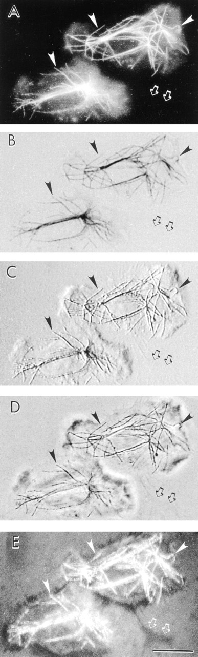
Immunocytochemical detection of microtubules in human neutrophils using FNG (for methods see Robinson and Vandré 1997). After incubation with FNG, the samples were subjected to a short silver enhancement reaction (2 min). The same neutrophils are shown in each panel. (
The microtubule labeling illustrates the versatility of FNG. Furthermore, those results suggest the potential for FNG in correlative microscopy (i.e., examination of the same sample with two or more imaging techniques). We have tested the usefulness of FNG for correlative microscopy with ultrathin cryosections of neutrophils as the model system. Immunolabeling of marker proteins for intracellular granules revealed a precise one-to-one relationship between the fluorescence signal and the silver-enhanced gold signal (Takizawa et al. 1998). Therefore, we find FNG to be very useful in this case and to be an important tool for correlative microscopy. A fuller discussion of FNG and its use in correlative microscopy is not given here because that topic is dealt with in another article from this symposium (Takizawa and Robinson 2000).
Disadvantages Associated with Ultrasmall Immunogold
The major drawback to the use of ultrasmall immunogold relates to the difficulty of detection in sections by conventional electron microscopy. This problem can be overcome to a large extent by using procedures to increase the size of the particles (Humbel et al. 1995). Usually this is accomplished with a silver enhancement reaction in which metallic silver is deposited on the gold (for reviews see Burry 1995; Danscher et al. 1995; Hacker et al. 1995). It should be noted that prolonged silver enhancement leads to increased background levels. Alternatively the ultrasmall gold particles can be rendered larger with gold chloride in a process known as “gold toning” (Marshak 1992; Pohl and Stierhof 1998). Whereas 5-, 10-, and 15-nm colloidal gold particles are uniform in diameter, enhancement of ultrasmall immunogold typically produces particles of various sizes. Therefore, improvements in methods for increasing the size of ultrasmall gold in a predictable and linear manner are highly desirable.
Summary
We discuss the utility of ultrasmall immunogold probes in immunocytochemistry while focusing on the gold cluster compounds (i.e., NG and FNG). These immunoprobes label more efficiently in immunocytochemical applications than do the larger colloidal gold particles (≥5-nm). We demonstrate that at least one of the reasons for the high labeling efficiency of the ultrasmall immunogold relates to the increased penetration of this probe into samples compared to larger gold particles. In addition, FNG with its dual signaling capability increases our ability to carry out combined light and electron microscopy.