
Abstract
Background
To date, the difference in microRNA expression profiles in tears of dry eye patients and healthy people has not been reported. In current study, we evaluated the significance of microRNAs and transforming growth factor beta2 (TGF-β2) in distinguishing dry eye.
Methods
A total of 138 patients with dry eye from October 2017 to October 2018 were selected. During the same period, 138 healthy persons were collected. All patients were followed up for 12 months through outpatient, telephone or medical records and the time of corneal injury was recorded.
Results
Compared with healthy people, TGF-β2 concentrations in dry eye patients were significantly decreased (P < 0.05). Array analysis, predictive software and dual-luciferase reporter assays showed that miR-450b-5p, miR-1283 and miR-3671 can target TGF-β2 expression. Tear miR-450b-5p, miR-1283 and miR-3671 concentrations were significantly higher in dry eye patients than healthy people. A logistic regression model combining miR-450b-5p, miR-1283, miR-3671 and TGF-β2 was performed. This model presented a high discriminating value (AUC: 0.907, 0.876–0.939, P < 0.001) than any single indicator, and the sensitivity and specificity were 77.7% and 92.7%, respectively. Compared with the low miR-450b-5p, low miR-1283, low miR-3671 and high TGF-β2 groups, the high miR-450b-5p, high miR-1283, high miR-3671 and low TGF-β2 groups had a significantly higher probability of corneal injury (TGF-β2: χ2 = 5.762, P = 0.016; miR-450b-5p: χ2 = 13.267, P < 0.001; miR-1283: χ2 = 19.431, P < 0.001; miR-3671: χ2 = 8.131, P = 0.004).
Conclusion
Current model combining tear miR-450b-5p, miR-1283, miR-3671 and TGF-β2 had important values in the identification of dry eye and was of great value in evaluating the risk of corneal injury.
Introduction
Dry eye syndrome is a common eye disease. A survey in the United States showed that 14.6% (approximately 4.3 million) of the population had dry eyes, compared with 17.0% in the Japanese population and 10.3% in Australia. 1 At present, it is believed that autoimmune diseases, hormone deficiency, dry air, environment, age and other factors form a comprehensive cause of dry eye syndrome.2,3 An important manifestation of dry eye is that the amount of water, lipid and mucin secreted on the surface of the eyeball is reduced, breaking the balance between the quality and quantity of tear fluid, which causes eye discomfort. 4 The concentration of transforming growth factor beta2 (TGF-β2) in tears can be used as an index to evaluate the amount of tears and can judge the tear secretion function to a certain extent. 5 In recent years, there are various diagnostic methods for dry eye syndrome, but it is currently the focus of attention to seek a diagnostic method that has both high practical value and economical. 6
MicroRNA (miR) is a type of endogenous non-protein-encoded RNA molecule, about 22 nucleotides in length. 7 It is an important molecule involved in the regulation of gene expression that has received much attention in recent years. A large amount of evidence shows that miR plays an important regulatory role in the signal transduction of innate immunity, adaptive immunity and inflammatory factors.8,9 Studies on the correlation between miR and dry eye syndrome have been reported. Wang et al. 10 pointed out that the miR-103/107 family plays an important role in regulating the biological processes of stem cells in the corneal limbal epithelial cells. They also found that miR-103/107 can simultaneously regulate the following two important processes, which can not only ensure that the autophagy process persists to the final stage of cell division but also prevent excessive fluid intake from outside the cell during the giant cell drinking process. 10 However, the difference in miR expression profiles in tears of dry eye patients and healthy people has not been reported.
In current study, firstly, the miR microarray chip was used to initially investigate the miR spectrum of dry eye patients, and the differential miR expression spectrum between healthy people and dry eye patients was found. Moreover, we identified the miRs that are targeted for downregulating TGF-β2 expression through bioinformatics software and luciferase reporter gene assay. Then, we evaluated the significance of miRs and TGF-β2 in distinguishing dry eye. In addition, logistic regression analysis was used for corneal injury in dry eye.
Materials and methods
Inclusion of subjects
A total of 138 patients with dry eye who received treatment in The Heji Hospital Affiliated to Changzhi Medical College from October 2017 to October 2018 were selected. The enrolment criteria were: (1) Patients were 24 to 70 years old. (2) All enrolled patients signed informed consent. (3) Eyes have obvious dryness, foreign body sensation, burning sensation and they are easily fatigued. (4) The eyes are photophobic, reddish and blurred vision. (5) Tear break-up time (BUT) ≤5 s. (6) Schirmer test ≤5 mm. (7) Corneal fluorescence staining ≥1. (8) All patients received a standardized treatment plan, namely the use of artificial tears combined with oral vitamin A. The exclusion criteria were: (1) Subject with a history of surgery on the eye. (2) Subject with a history of allergies in the eyes. (3) Patients with severe lung, kidney and liver abnormalities. (4) Those with mental illness. (5) Women who are breastfeeding or pregnant. (6) Those have not completed the 12-month follow-up. During the same period, 138 healthy persons were collected as a control group. This study was approved by the ethics committee of The Heji Hospital Affiliated to Changzhi Medical College (CZ20171006).
Prognosis assessment of patients with corneal injury
All patients were followed up for 12 months through outpatient, telephone or medical records, and the time of corneal injury was recorded.
Tear specimen collection
The tears of the enrolled subjects were collected, and 60 µL of 0.9% saline was dripped into the conjunctival sac to mix the tears with the saline. Then, a capillary glass tube was used to collect the left and right tears into a centrifuge tube and stored at –20°C to be tested.
Instruments and kits
Platinum SYBR Green qPCR SuperMix-UDG (Invitrogen, 11733–038), dNTP (Takara, D4030RA), Nase Inhibitor (Takara, 2313 A), 50 bp DNA Ladder Marker (LC-Bio, DL-1004B) and total RNA extraction kit were purchased from Norgen Biotek. The miRNA chips were purchased from LC Sciences. TurboFect TM siRNA transfection reagents were purchased from Fer-mentas. The reverse transcription kit ReverTra Ace Qpcr RT Kit was purchased from Takara. SYBRGreen Mix Plus fluorescent dye was purchased from TOYOBO Company. Human TGF-β2 kit was purchased from Nanjing Jinyibai Biotechnology Co., Ltd.
Detection of TGF-β2 concentration
First, 80 µL of the sample diluent was added to the 1.5 mL polypropylene tube, and then 10 µL of the specimen was added. Then, 5 µL of 1 mol/L HCL was added and stored at 2–8°C for 1 h; later, 5 µL of 1 mol/L NaOH was added and shaken. The kit was removed 20 min before the test and equilibrated to room temperature. Then, eight standard wells were set up and 100 µL of sample diluent was added to each well. Starting from the first well, 100 µL of the standard solution was added, mixed thoroughly and then aspirated 100 µL to the second well, then the procedure was repeated till the seventh well. The eighth well is a blank control. One hundred microliters of activated specimens were added to the wells of the product to be tested, and then the reaction plate was stored at 37°C for 2 h. Then, the absorbance of each sample was measured.
Microarray hybridization and data analysis
The chip is made by LC Sciences of the United States. The hybrid image was collected by laser scanner, and the hybrid image was digitally converted by Array-Pro software. Data processing and analysis first deducts the background, calculates the repeat point value and standard deviation, and then uses the LOWESS algorithm to standardize the original chip data. After standardization analysis, the chip data were subjected to Chi-square test. In order to reduce the error of Chi-square test, Bonferroni was used to correct the error rate of the Chi-square test results. Finally, FDR < 0.05 was used as the differential standard for differential expression screening.
Cell culture and transfection
Human bulbar conjunctiva fibroblasts (hBCFs) cell line was cultured in a DMEM medium containing 10% fetal bovine serum at 37°C, 5% CO2 incubator. Lipofectamine 2000 was used to transfect miR-450b-5p inhibitor, miR-1283 inhibitor, miR-3671 inhibitor and negative control (NC) into hBCFs cells.
Quantitative real-time polymerase chain reaction
This study complies with the Minimum Information for Publication of Quantitative Real-Time PCR Experiments (MIQE) guidelines 2009. 11 Total RNA of cell line or tears was extracted according to the instructions of the Trizol kit. U6 was used as housekeeping gene. The primers sequences were as follows: miR-450b-5p (forward: 5′-TACGCGTCCTGAACCGTTTAG-3′ and reverse: 5′-CGCCTACTAACGCCGTT-3′); miR-1283 (forward: 5′-CGGTCAATCGCGATCATCGCAG-3′ and reverse: 5′-GGGACCTGAAGTTAGTCA-3′); miR-3671 (forward: 5′-GCGTCGTGGCGCGGATATACGGG-3′ and reverse: 5′-GCCTACCTGAGTACTGGGGCA-3′); U6 (forward: 5′-CGCTCGCGGCATTAGGCATCC-3′ and reverse: 5′-AAGGCCTCCATCGCATGGCCTT-3′). The amplification efficiencies of miR-450b-5p, miR-1283, miR-3671 and U6 were 96.8%, 95.4%, 96.3% and 97.2%, respectively.
Western blot
The proteins of TGF-β2 and glyceraldehyde-3-phosphate dehydrogenase (GAPDH) were tested by Western blot. The rabbit anti-TGF-β2 and GAPDH antibody were added overnight at 4°C. The secondary antibody was then added and incubated at room temperature for 0.5 h the next day.
Luciferase reporter gene assay
MiR-450b-5p, miR-1283 and miR-3671 mimics/inhibitors were transferred to the pmiR-RB-ReportTM reporter gene. Luciferase reporter plasmids (TGF-β2 wild type and mutant type) were co-transfected into hBCFs cells with miR-450b-5p, miR-1283, miR-3671 mimics/inhibitors and NC, respectively. Then, luciferase activity was detected.
Statistical analysis
Statistical analysis was performed using GraphPad Prism 8. Measurement data are expressed as mean ± standard deviation. A comparison between two groups was performed by t test. Spearman test was used to calculate the correlation between tears miR-450b-5p, miR-1283 and miR-3671 concentrations and clinical features. Binary logistic regression analysis was used to calculate the prognostic factors of dry eye patients with corneal injury. Receiver operator characteristic curve (ROC) and Kaplan–Meier method were used to calculate the diagnosis and prognostic values of miR-450b-5p, miR-1283 and miR-3671 for dry eye, respectively. P < 0.05 was considered statistically significant.
Results
The basic information and TGF-β2 concentrations in tears of the two groups
There was no significant difference in age, gender, hypertension, diabetes, hyperlipidaemia, smoking history and drinking history between patients with dry eye and healthy people (P > 0.05, Supplemental Table 1). Compared with healthy people, TGF-β2 concentrations in tears of patients with dry eye were significantly reduced (P < 0.05, Supplemental Table 1).
Array analysis and the prediction and verification of miRs targeting TGF-β2
According to array analysis (Figure 1(a)), miRs with a statistically significant (P < 0.05) increase or decrease of two-fold or higher expression between tears of dry eye patients and healthy people were analysed. It was found that 32 of 2038 miRs were up- or downregulated in patients with dry eye tear compared with healthy people tear. The expression levels of four miRs were upregulated. Among these four increased miRs, the fold-change value of miR-450b-5p, miR-1283, miR-5700 and miR-3671 was 3.11, 2.69, 2.31 and 2.32, respectively. Using miRanda, miRDB and Targetscan software to predict the miRs targeting TGF-β2, a total of three miRs (miR-450b-5p, miR-1283 and miR-3671) were predicted, Figure 1(b). Therefore, we selected miR-450b-5p, miR-1283 and miR-3671 for further verification.
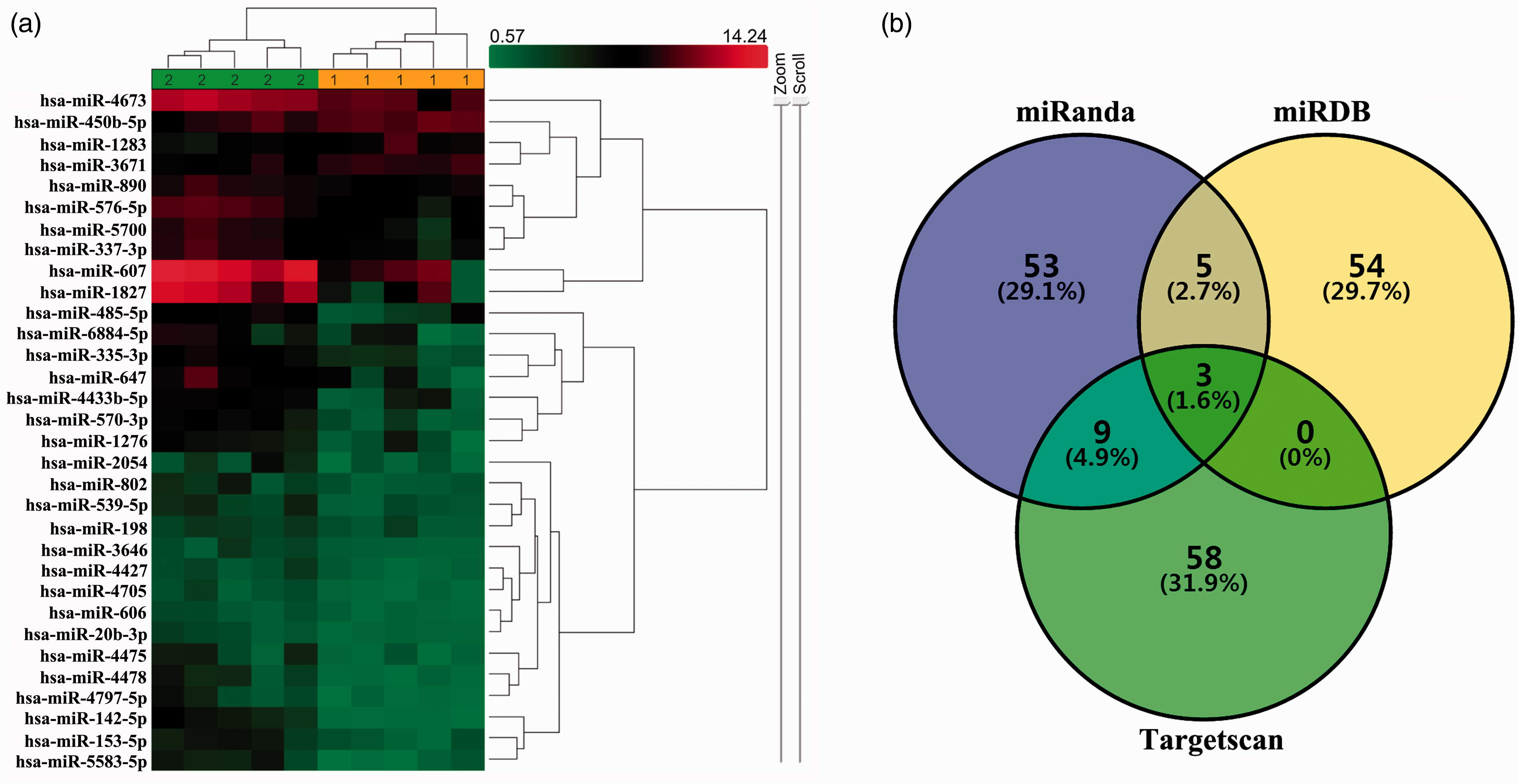
Array analysis and the prediction of miRs targeting TGF-β2. (a) Array analysis found that 32 miRs were differentially expressed between tears of dry eye patients and healthy people. Dry eye: 1 (red labelled) and Healthy people: 2 (green labelled) sample fall in separate clusters. (b) miRanda, miRDB and Targetscan software were used to predict the miRs targeting TGF-β2.
Luciferase reporter gene assay and Western blot were used. We found that the luciferase activity of the miR-450b-5p mimic group (0.41 ± 0.05), miR-1283 mimic group (0.49 ± 0.07) and miR-3671 mimic group (0.23 ± 0.04) was significantly lower than that of the NC group (1.01 ± 0.11, 0.98 ± 0.08 and 1.03 ± 0.10) in the wild-type of TGF-β2 (P < 0.05), but there was no significant difference in the mutant-type of TGF-β2 (P > 0.05), Figure 2(a), (d) and (g). Western blot results showed that after transfecting si-miR-450b-5p, si-miR-1283, si-miR-3671 and si-NC into hBCFs cells, compared with the NC group, the concentrations of TGF-β2 in the si-miR-450b-5p, si-miR-1283 and si-miR-3671 groups were significantly increased (P < 0.05, Figure 2(b), (c), (e), (f), (h) and (i)).
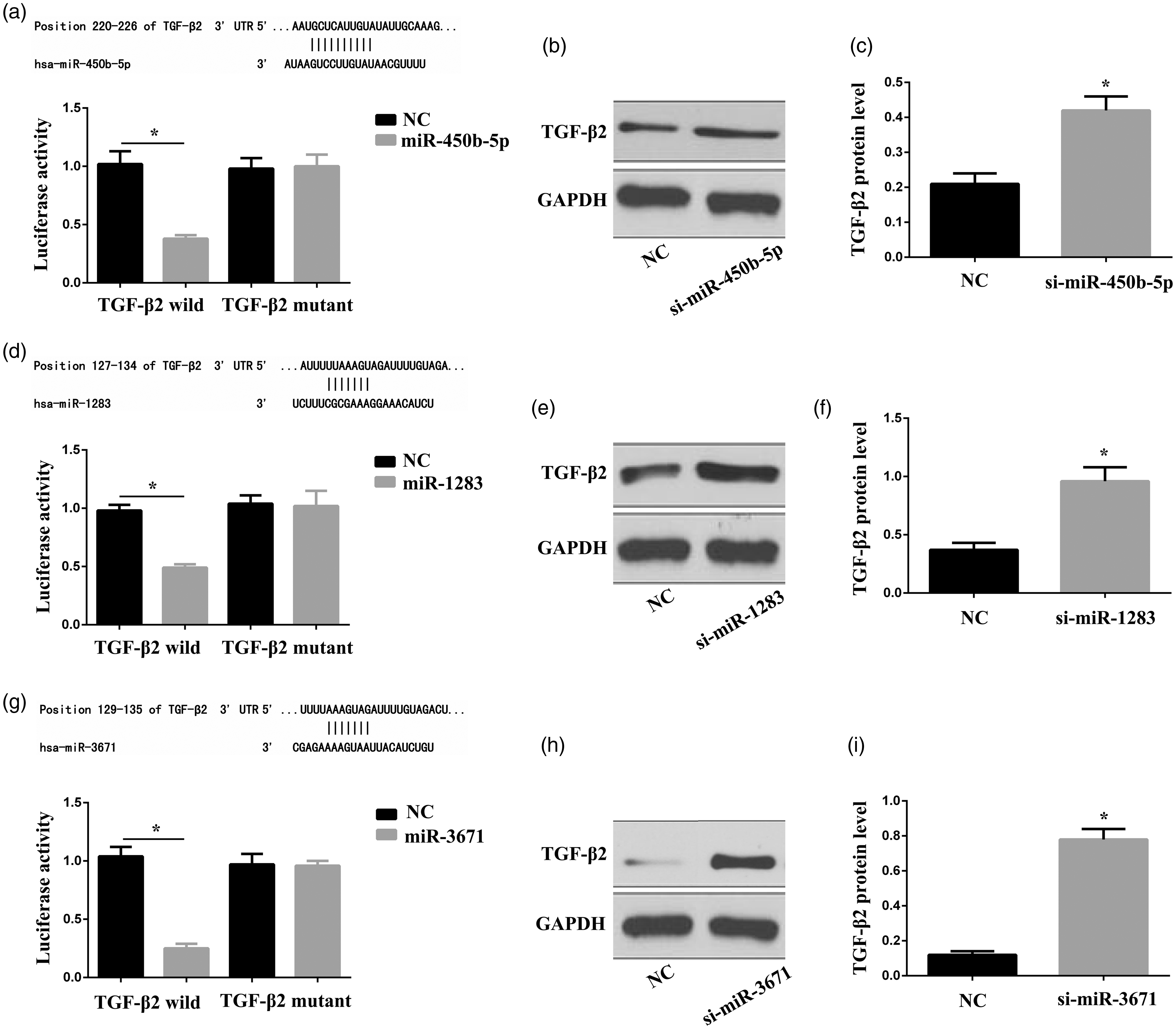
Verification of miRs targeting TGF-β2. (a) Luciferase reporter gene assay of miR-450b-5p. (b and c) Compared with the NC group, the concentrations of TGF-β2 in the si-miR-450b-5p group are significantly increased (P < 0.05). (d) Luciferase reporter gene assay of miR-1283. (e and f) Compared with the NC group, the concentrations of TGF-β2 in the si-miR-1283 group are significantly increased (P < 0.05). (g) Luciferase reporter gene assay of miR-3671. (h and i) Compared with the NC group, the concentrations of TGF-β2 in the si-miR-3671 group are significantly increased (P < 0.05).
The concentrations of miR-450b-5p, miR-1283 and miR-3671 in the tears of the two groups
We applied quantitative real-time polymerase chain reaction (qRT-PCR) to detect miR-450b-5p, miR-1283 and miR-3671 concentrations in the tears of healthy people and patients with dry eye. Compared with healthy people (miR-450b-5p: 90.4 ± 8.2, miR-1283: 6.8 ± 1.7, miR-3671: 7.6 ± 1.5), miR-450b-5p (137.8 ± 19.3), miR-1283 (12.5 ± 2.8) and miR-3671 (12.0 ± 3.3) of dry eye patients were significantly increased (P < 0.05, Figure 3(a) to (c)). As shown in Figure 3(d) to (l), the concentrations of miR-450b-5p, miR-1283 and miR-3671 were significantly correlated with BUT, Schirmer test and Osmotic pressure (P < 0.05).
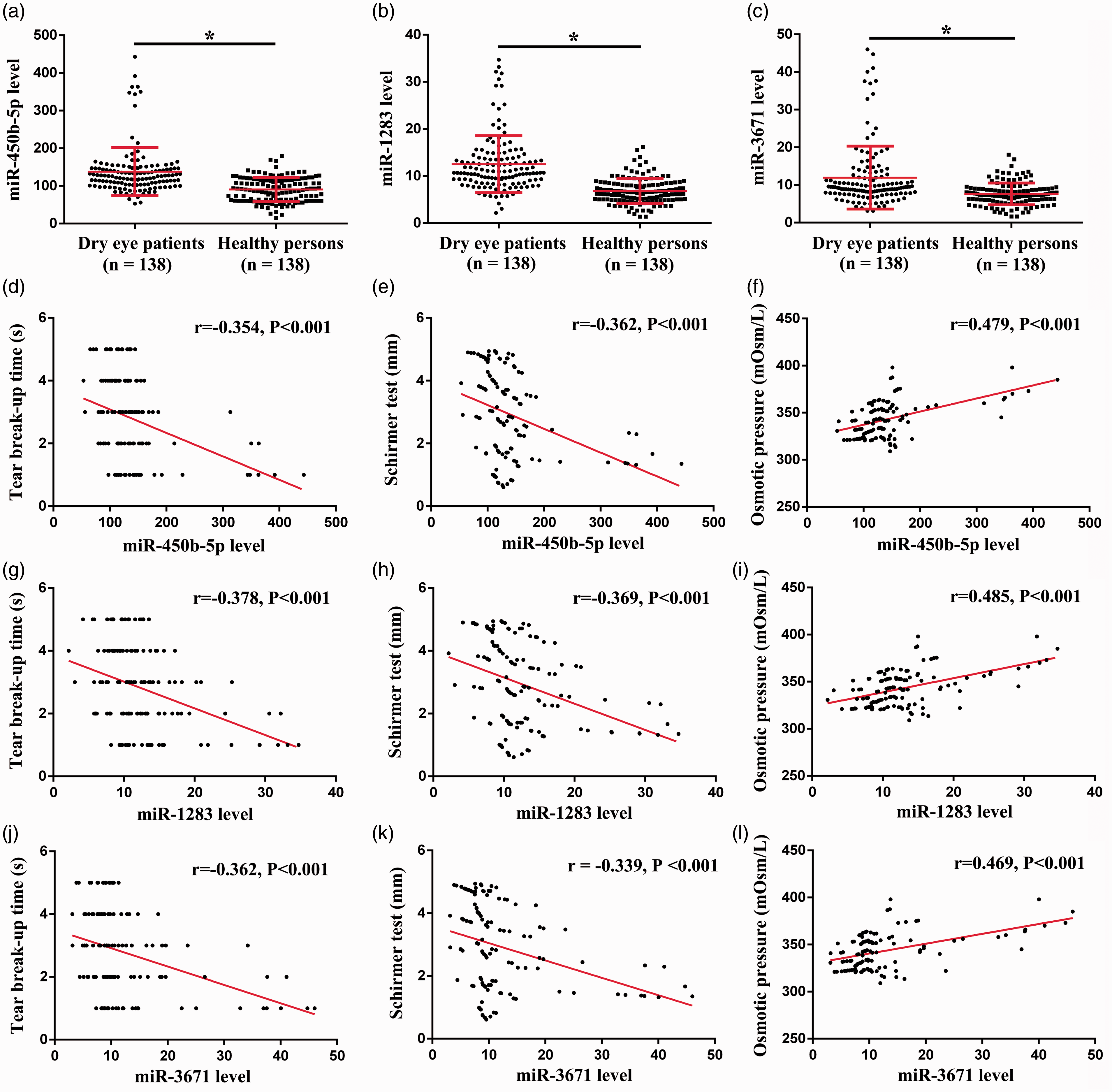
The concentrations of miR-450b-5p, miR-1283 and miR-3671 in the tears and their correlations with BUT, Schirmer test and Osmotic pressure. (a) The concentrations of miR-450b-5p in the two groups. (b) The concentrations of miR-1283 in the two groups. (c) The concentrations of miR-3671 in the two groups. (d) Correlation between miR-450b-5p concentration and BUT. (e) Correlation between miR-450b-5p concentration and Schirmer test. (f) Correlation between miR-450b-5p concentration and osmotic pressure. (g) Correlation between miR-1283 concentration and BUT. (h) Correlation between miR-1283 concentration and Schirmer test. (i) Correlation between miR-1283 concentration and osmotic pressure. (j) Correlation between miR-3671 concentration and BUT. (k) Correlation between miR-3671 concentration and Schirmer test. (l) Correlation between miR-3671 concentration and osmotic pressure.
ROC analysis of the identification of dry eyes by miR-450b-5p, miR-1283, miR-3671 and TGF-β2 in tears
The ROC results are shown in Figure 4. The AUC of miR-1283, miR-450b-5p, miR-3671 and TGF-β2 in tears in predicting dry eye were 0.808 (0.761–0.852, P < 0.001), 0.805 (0.759–0.849, P < 0.001), 0.781 (0.731–0.828, P < 0.001) and 0.706 (0.651–0.760, P < 0.001). The sensitivity and specificity of each index were 80.4%/66.9%, 87.3%/58.2%, 65.6%/79.7% and 65.1%/69.9%. Using the logistics regression model combined with miR-1283, miR-450b-5p, miR-3671 and TGF-β2, the AUC of the four indicators in predicting dry eye was 0.907 (0.876–0.939, P < 0.001). The sensitivity and specificity were 77.7% and 92.7%, respectively. The ‘critical value’ for the combined detection of miR-1283, miR-450b-5p, miR-3671 and TGF-β2 were 114.2, 10.3, 9.8 and 12.7 pg/mL, respectively.
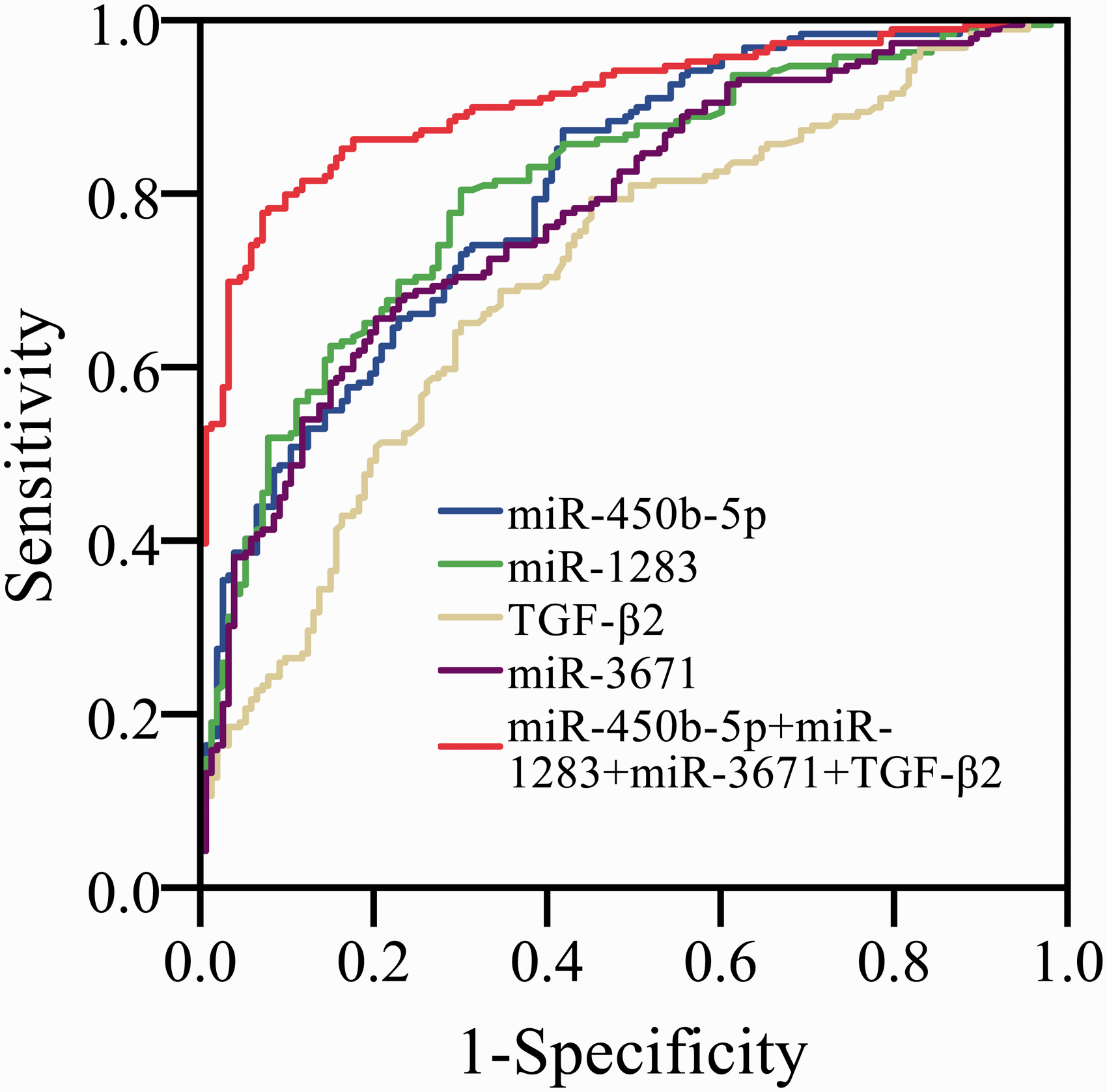
ROC analysis of the identification of dry eyes by miR-450b-5p, miR-1283, miR-3671 and TGF-β2 in tears.
The prognostic value of miR-450b-5p, miR-1283, miR-3671 and TGF-β2 concentrations in tears for corneal injury in dry eye
During the follow-up process, 32 patients had corneal injury. Dry eye patients were divided into the high/low expression groups according to the median tear miR-450b-5p (141.2), miR-1283 (12.8), miR-3671 (11.9) and TGF-β2 (14.7 pg/mL) concentration. Compared with the low miR-450b-5p, low miR-1283, low miR-3671 and high TGF-β2 groups, the high miR-450b-5p, high miR-1283, high miR-3671 and low TGF-β2 groups had a significantly higher probability of corneal injury (TGF-β2: χ2 = 5.762, P = 0.016, Figure 5(a); miR-450b-5p: χ2 = 13.267, P < 0.001, Figure 5(b); miR-1283: χ2 = 19.431, P < 0.001, Figure 5(c); miR-3671: χ2 = 8.131, P = 0.004, Figure 5(d)). Corneal injury or not was treated as a dependent variable, age, sex, hypertension, hypercholesterolemia, diabetes mellitus, smoking, alcoholism, TGF-β2, miR-450b-5p, miR-1283 and miR-3671 were used as independent variables for multiple linear regression analysis. The assignment of counting data is as follows: sex (male: 1, female: 0), hypertension (yes: 1, no: 0), hypercholesterolemia (yes: 1, no: 0), diabetes mellitus (yes: 1, no: 0), smoking (yes: 1, no: 0), alcoholism (yes: 1, no: 0). Logistic regression analysis for corneal injury was shown in Table 1. Tear miR-450b-5p, miR-1283, miR-3671 and TGF-β2 concentrations were the risk markers.
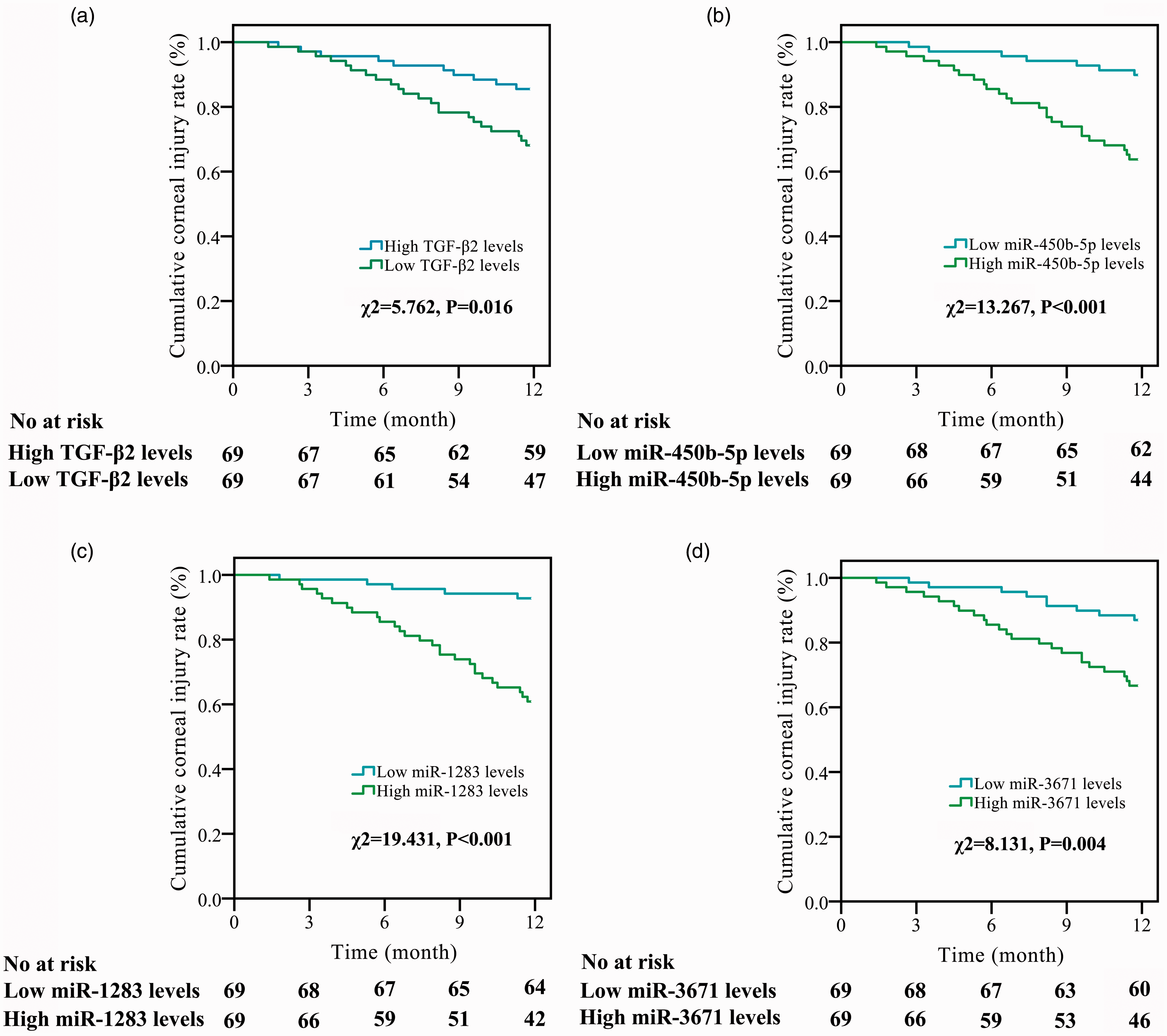
The prognostic value of miR-450b-5p, miR-1283, miR-3671 and TGF-β2 concentrations for corneal injury in dry eye. (a) Compared with high TGF-β2 group, the low TGF-β2 group had a significantly higher probability of corneal injury (χ2=5.762, P = 0.016). (b) Compared with low miR-450b-5p group, the high miR-450b-5p group had a significantly higher probability of corneal injury (χ2=13.267, P < 0.001). (c) Compared with low miR-1283 group, the high miR-1283 group had a significantly higher probability of corneal injury (χ2=19.431, P < 0.001). (d) Compared with low miR-3671 group, the high miR-3671 group had a significantly higher probability of corneal injury (χ2=8.131, P = 0.004).
Univariate and multivariate logistic regression of clinicopathological factors for corneal injury in dry eye.
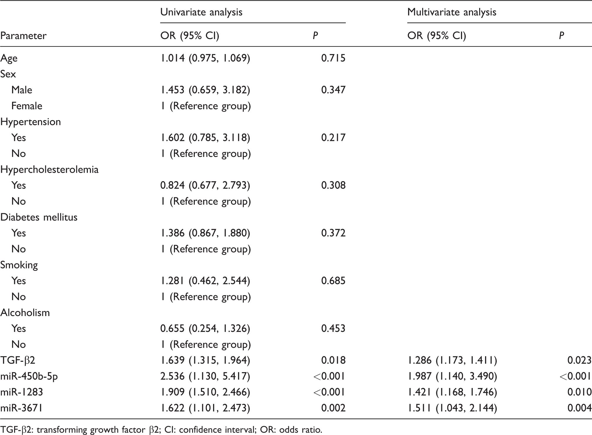
TGF-β2: transforming growth factor β2; CI: confidence interval; OR: odds ratio.
Discussion
The pathogenic factors of dry eye syndrome are more complicated, and the environment and personal habits may cause pathological and physiological changes on the surface of the eye.12,13 Studies have shown that all factors that damage the function of the lacrimal gland may cause changes in the composition of the tear film and cause tear film instability. 14 If the tear film is abnormal for a long time without intervention, it may be converted into inflammation. 14 The diagnosis of dry eye disease is mainly based on the patient's self-reported symptoms, and the patient's condition is comprehensively judged according to the traditional examination methods such as Schirmer I test, fluorescein staining test and BUT test. However, it is susceptible to factors such as environment, filter paper and examinee, which can easily lead to false-positive or false-negative results of misdiagnosis and misjudgment. 6 At present, there are still some shortcomings in the diagnosis and treatment of dry eye syndrome in China, mainly manifested in the following aspects: (1) Diagnostic standards are not uniform. 15 (2) The treatment of dry eye is more confusing. 15 (3) Lack of inspection methods with high specificity and sensitivity. 15 (4) Lack of national authoritative epidemiological data. 16
The relative molecular weight of TGF-β2 is about 25,000. Generally, TGF-β2 has multiple biological effects, including the following aspects: (1) It can stimulate extracellular matrix protein synthesis. 17 (2) Promoting the proliferation of fibroblasts and interstitial cells. 18 (3) It has the ability to inhibit the proliferation of many types of cells derived from lymph, endothelium and epithelium. 19 (4) It has a certain regulatory role in cell adhesion and chemotaxis and can activate some functions of macrophages and monocytes. 20 Studies have pointed out that TGF-β2 in the eye plays an important role, and the secretion of TGF-β2 by the lacrimal gland has high expression in tears. 21 miR is a type of non-coding small RNA molecule. Studies have shown that it plays an important role in immune homeostasis. 7 It mainly acts on the post-transcriptional level of genes. 9 The development and function of immune cells regulated by miR are related to autoimmune diseases.8,9 The role of miR and immunoregulatory mechanisms has received increasing attention and has also played an important role in the development of autoimmune diseases. In this study, we identified the miRs that are targeted for downregulating TGF-β2 expression through bioinformatics software and luciferase reporter gene assay. Then, we evaluated the significance of miRs and TGF-β2 in distinguishing dry eye.
First, the microarray chip technology was used to preliminarily study the miRNA spectrum in tears of patients with dry eye. We found that there were 32 differentially expressed miRNAs between healthy people and patients with dry eye, 4 miRNAs were upregulated and 28 miRNAs were downregulated in dry eye patients. This result suggested that miR in tears may be used as a molecular biomarker for the diagnosis of dry eye. Finally, three miRs (miR-450b-5p, miR-1283 and miR-3671) were finally determined by the prediction software. Correlation analysis results showed that the concentrations of miR-450b-5p, miR-1283 and miR-3671 were significantly correlated with BUT, Schirmer test and Osmotic pressure. This results suggested that the concentrations of the above three miRs in the tears of dry eye patients were associated with the severity of the disease. Deregulated expression of several miRs in tears of patients with Sjögren syndrome has been reported. Kim et al. 4 found that 10 differentially expressed miRs (miR-30b-5p, miR-30c-5p, miR-30d-5p, miR-92a-3p, miR-134-5p, miR-137, miR-302d-5p, miR-365b-3p, miR-374c-5p and miR-487b-3p) may be involved in the pathogenesis of Sjögren syndrome, in particular, related to autoimmunity and neuropathy. It is suggested that the differential expression of miRs in tears may be related to dry eye. In the current study, a logistic regression model was established, which includes miR-450b-5p, miR-1283, miR-3671 and TGF-β2. It presented a high discriminating value (AUC: 0.907, 0.876–0.939, P < 0.001) than any single indicator. In conclusion, current logistic regression model combining tear miR-450b-5p, miR-1283, miR-3671 and TGF-β2 has potential significance for the non-invasive differential diagnosis for dry eye. Finally, compared with the low miR-450b-5p, low miR-1283, low miR-3671 and high TGF-β2 groups, the high miR-450b-5p, high miR-1283, high miR-3671 and low TGF-β2 groups had a significantly higher probability of corneal injury. The above results further suggested that the combined detection of miR-450b-5p, miR-1283, miR-3671 and TGF-β2 concentrations in tears of patients with dry eye had certain significance for predicting long-term corneal injury in patients.
In summary, we found that combining the miR-450b-5p, miR-1283, miR-3671 and TGF-β2 had important values in the identification of dry eye and was of great value in evaluating the risk of corneal injury.
Supplemental Material
sj-pdf-1-acb-10.1177_0004563220961746 - Supplemental material for Discovery of microRNA expression profiles involved in regulating TGF-β 2 expression in the tears of dry eye patients
Supplemental material, sj-pdf-1-acb-10.1177_0004563220961746 for Discovery of microRNA expression profiles involved in regulating TGF-
Footnotes
Declaration of conflicting interests
The author(s) declared no potential conflicts of interest with respect to the research, authorship, and/or publication of this article.
Funding
The author(s) received no financial support for the research, authorship, and/or publication of this article.
Ethical approval
This study was approved by the ethics committee of The Heji Hospital Affiliated to Changzhi Medical College (CZ20171006). Written informed consent was provided in accordance with the Declaration of Helsinki.
Guarantor
SH.
Contributorship
SH conceived this study. QW, XX and HL were involved in gaining ethical approval, patient recruitment and data analysis. QW and SH wrote the first draft of the article. All authors approved the final version of the article.
Supplemental material
Supplemental material for this article is available online.
References
Supplementary Material
Please find the following supplemental material available below.
For Open Access articles published under a Creative Commons License, all supplemental material carries the same license as the article it is associated with.
For non-Open Access articles published, all supplemental material carries a non-exclusive license, and permission requests for re-use of supplemental material or any part of supplemental material shall be sent directly to the copyright owner as specified in the copyright notice associated with the article.