
Abstract
A significant number of cancer-related deaths are recorded globally each year, despite attempts to cure this illness. Medical science is working to develop new medication therapies as well as to find ways to identify this illness as early as possible, even using noninvasive techniques. Early detection of cancer can greatly aid its treatment. Studies into cancer diagnosis and therapy have recently shifted their focus to exosome (EXO) biomarkers, which comprise numerous RNA and proteins. EXOs are minuscule goblet vesicles that have a width of 30 to 140 nm and are released by a variety of cells, including immune, stem, and tumor cells, as well as bodily fluids. According to a growing body of research, EXOs, and cancer appear to be related. EXOs from tumors play a role in the genetic information transfer between tumor and basal cells, which controls angiogenesis and fosters tumor development and spread. To identify malignant activities early on, microRNAs (miRNAs) from cancers can be extracted from circulatory system EXOs. Specific markers can be used to identify cancer-derived EXOs containing miRNAs, which may be more reliable and precise for early detection. Conventional solid biopsy has become increasingly limited as precision and personalized medicine has advanced, while liquid biopsy offers a viable platform for noninvasive diagnosis and prognosis. Therefore, the use of body fluids such as serum, plasma, urine, and salivary secretions can help find cancer biomarkers using technologies related to EXOs.
Introduction
Recent years have seen an increase in reports on the relevance of exosomes (EXOs) to cancer biology. 1 EXOs are membrane sacs and nanoscale vehicles of endosomal origin produced by almost all normal and pathological cells and are found in all body fluids. They contain a wide range of biological molecules such as proteins, lipids, and nucleic acids (DNA and miRNA) and reflect the physiological conditions and secretory functions of the cell.2,3
The microRNA (miRNA) is a single-stranded molecule approximately 18 to 25 nucleotides in length 4 that is responsible for regulating gene expression at the posttranscriptional level. It also plays an important role in cell proliferation. 5 In cancer, a series of genetic deviations occur, and the control mechanism of cell proliferation is out of regulation. 6 Furthermore, metastasis is a cause of death in cancer patients, and it is not unthinkable that exosomal miRNAs (ExomiRs) play a role in metastasis and are the link between cancer and the host. 7 Evidence has shown that miRNAs enclosed in EXOs can cross the blood–brain barrier and are safe from immune attacks. 8 Because of their double-layered membrane and small size, high stability, and, correspondingly, a higher half-life are among their characteristics. 9
Currently, imaging techniques and morphological analysis of tissues (histology) or cells (cytology) can detect cancer only when there is a visible change in the tissue; for example, when thousands of cancer cells have multiplied and even metastasized. Moreover, cancer biomarkers currently in use suffer from issues such as false negatives or false positives because of a lack of specificity. Therefore, ExomiRs related to the production and progression of tumors can be proposed as potential diagnostic, prognostic, and predictive biomarkers of cancer. 10 The main advantage of using EXOs as biomarkers is their presence in various body fluids, for example, blood, urine, saliva, breast milk, etc, which makes them easily and noninvasively collectible and utilizable in clinical trials 11 (Figure 1). Due to their biocompatibility, EXOs can be measured in such a way that they can be used to diagnose cancer in the future.
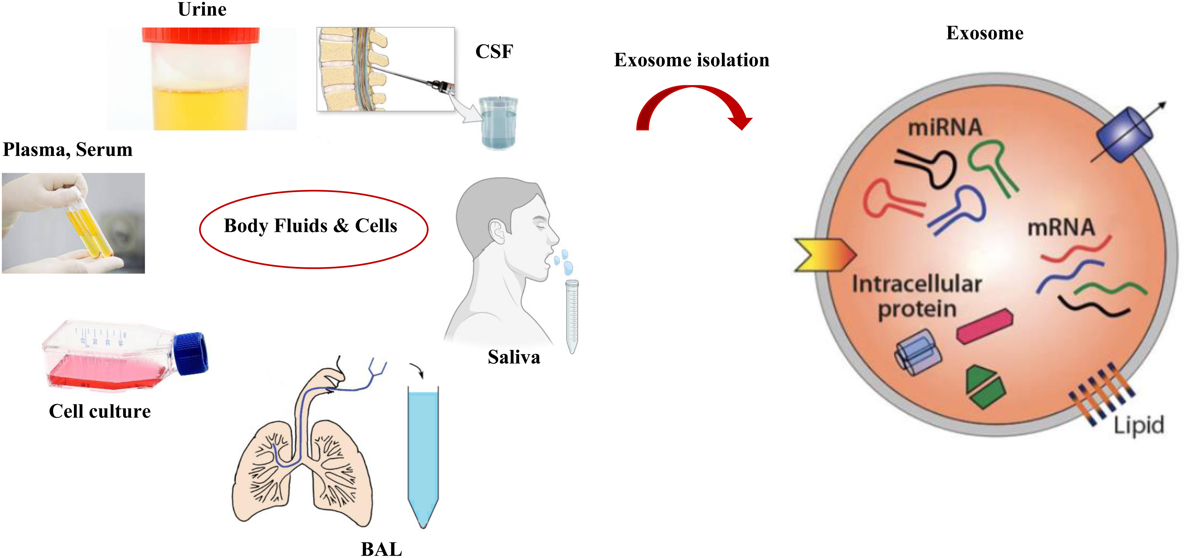
Exosomes isolation from body fluids, EXOs can be extracted from different body fluids such as urine, saliva, plasma, serum, CSF, and BAL. These nanoparticles contain various compounds, including miRNA, which can be used as biomarkers in medical diagnostics.
Definition and Biogenesis of EXOs
An EXO is a membranous sac released into the extracellular matrix (ECM) after fusing with a cell membrane. Its surface is made up of protein receptors, polysaccharides, and other lipids and has a double structure containing many biologically active substances. 12 miRNAs, proteins, and coding-protein mRNAs are all present in EXOs and represent the physiological and functional conditions of secretory cells. According to recent research, blood, saliva, cerebrospinal fluid, tumor cells, and other body fluids contain EXOs.3,13
The biogenesis of EXOs occurs in 3 distinct phases:
Formation of endocytic vesicles (early endosome) through the invagination of the plasma membrane. Formation of multivesicular bodies (MVBs) with intraluminal vesicles (ILVs) generated by cytoplasmic components budding inside the endosomal membrane. MVB fusion with the plasma membrane and extracellular release of ILVs as EXOs.14–16
MVBs may potentially decay through fusion with lysosomes or autophagosomes.
17
Many different proteins, including the endosomal sorting complexes required for transport (ESCRT) proteins, are involved in the development of MVBs and ILVs, which contain 4 protein complexes, ESCRT-0, -I, -II, and -III.14,18,19 In addition to ESCRT proteins, the X protein that interacts with apoptosis-linked gene 2 (ALIX),
20
the soluble N-ethylmaleimide-sensitive factor attachment protein receptor (SNARE) proteins,
21
the vesicle trafficking 1 (VTA1) protein,
22
and guanosine triphosphatases (GTPases) are also thought to be important players in the biogenesis and secretion of EXOs.
23
Current Challenges in EXO Isolation
EXO application in medicine has technological difficulties, much like any other novel nanobiotechnology that is under development. One issue is the uniformity of EXO separation methods, which needs special consideration. EXO isolation has no industry-accepted gold standard, and new techniques are developed practically daily. EXO extraction using ultracentrifugation is by far the commonly most used approach, although it has several limitations, including the simultaneous isolation of nonexosomal contaminants, poor repeatability, low RNA yield, and probable EXO destruction. Additionally, the majority of separation techniques make it impossible to achieve EXO purity at all levels.24–26 EXOs content, purity, and size vary depending on the technique employed to separate them from body fluids. Information suggests that the isolation technique may alter the miRNA profile in EXOs. 27
According to the findings, EXO separation techniques from plasma or body fluids are the most important factor for defining the molecular or genetic payload of EXOs. Therefore, it is important to be cautious when selecting the best procedure for isolating EXOs and to distinguish between the EXO's true composition and its contamination with nucleic acids or plasma proteins. 28 Additionally, a study of various separation methods revealed that there were differences in the amounts of numerous proteins, including tetraspanins, that are frequently employed as “markers” of EXOs. It comes from several tumor cells. EXO purity is greatly influenced by the separation technique and has an impact on how the results should be interpreted. The highest purity, however, typically comes at the expense of poor yield and may not be significant for studies in which biomarkers can be reliably detected, but it is significant for other applications, such as proteomic analysis.29,30
Definition and Biogenesis of miRNAs
Much evidence has shown that miRNAs, a small part of ncRNAs, regulate posttranscriptional gene expression, intracellular transcription, and mRNA degradation to manage cancer cell proliferation, apoptosis, differentiation, metastasis, and stem cell features. It is estimated that miRNAs target more than 50% of all mRNAs, and each miRNA is expected to influence hundreds of target mRNAs.31,32
Various studies have shown that miRNAs in body fluids maintain their stability under a variety of severe situations, including boiling, extremely low or high pH, multiple freeze-thaw cycles, and long-term room temperature storage. 33
A well-known process is responsible for the synthesis of these small single-strand RNAs; RNA polymerase II transcribes the miRNA genes in the nucleus to create a primary transcript, also known as a pri-miRNA. Exportin 5 is a protein that then transports the pre-miRNA to the cytoplasm, where the Dicer complex turns it into a mature miRNA duplex around 20 to 22 nucleotides in length. Once the strands have been separated, the passenger strand is destroyed, and the second strand is incorporated in the RNA-induced silencing complex (RISC), which targets the specific mRNA and inhibits gene expression through mRNA degradation or translational suppression based on the 3ʹ-untranslated region (3ʹUTR) and complementary sequence of the target.34,35
Role of ExomiRs in Cancer
ExomiRs play an important role in controlling the development of cancer. 36 Cancer cells secrete at least 10-fold more EXOs than normal cells and tumor-derived EXOs (TDEs) can transport chemokines, ExomiRs, growth factors, and various small molecules, thereby enhancing intercellular communication.37,38 EXOs are released and then ingested by both nearby and distant cells, where the miRNAs they contain regulate processes like sabotaging tumor immunity and the microenvironment, potentially promoting tumor proliferation, invasion, metastasis, angiogenesis, and drug resistance. 36 Furthermore, evidence suggests that the tumor microenvironment significantly participates in the metabolic rewiring of cancerous cells through extracellular vesicles, promoting complete nutrient exploitation and changing the microenvironment from a normal to a tumor-favorable state. This alteration allows for invasion, drug resistance, and tumor growth. 39
Additionally, ExomiRs produced by cancer help recruit and modify elements of the tumor environment. 40 These molecules unquestionably play a part in cancer as tumor suppressors and promoters, influencing angiogenesis, tumor growth, proliferation process, metastasis, and cell migration as well as the epithelial–mesenchymal transition (EMT). 38 ExomiRs can also impact the ECM, immune system activation, and recruitment in the area around the tumor. 41
General Roles of ExomiRs in Cancer
ExomiRs are exported by cancer cells to neighboring cancer cells around them. In the tumor microenvironment, they facilitate communication between primary tumor cells and other cells. EXOs produced by healthy cells can influence the behavior of malignant cells, and those produced by virus-infected cells can impact not only their oncology but also that of normal cells. 36
ExomiRs and Tumor Immunity
Tumor-derived EXOs aid tumor immune escape by transporting immunosuppressive factors and molecules. 42 ExomiRs act as information carriers and can modulate the behavior of certain immunological activity factors and immune target cells, such as T lymphocytes, dendritic cells (DCs), and natural killer (NK) cells. 43 They are also involved in the biology of T lymphocytes and NK. 44 Additionally, the process by which ExomiRs affect NK cells’ immunological activity and, consequently, cause tumor immunology resistance involves a multifaceted, multitargeted, and multifactored effect. 36
ExomiRs and Tumor Proliferation
Malignant cells can transport genetic information to different cells within the tumor microenvironment through EXOs. 36 ExomiRs play a role in the metastasis, angiogenesis process, proliferation, drug resistance, and tumor suppression of cancer cells, as some of them are transferred between donors and recipients. 36 Proliferation is a critical factor in the development and spread of cancer, characterized by changes in the expression and function of proteins involved in the cell cycle. 36 Additionally, cell proliferation is stimulated by the constitutive activation of several signal transduction pathways. 45
ExomiRs and Tumor Angiogenesis
The tumor angiogenesis process consists of numerous phases, including the enzymatic breakdown of the vessel's basement membrane and the sprouting, proliferation, branching, migration, and tube creation of endothelial cells. EXOs produced by several cell types, such as stromal, endothelial, and mesenchymal stem cells, have been demonstrated to serve as beneficial mediators in the tumor microenvironment.46,47 One key variable affecting tumor angiogenesis is hypoxia, which can influence the activities of many different chemicals and encourage the development of ExomiRs. 36
ExomiRs and Tumor Metastasis
Numerous signaling molecules participate in intercellular communication. The several stages of the metastatic process require EXOs produced by the tumor. 48 Research has identified 4 common ways in which ExomiRs distribution occurs when a tumor is developing in the microenvironment. 49
First, miRNAs released by more invasive tumor cells may be absorbed by less invasive tumor cells through TDEs, which could lead to the worsening of a primary tumor. 36
Second, ExomiRs in the tumor microenvironment allow primary tumor cells to communicate with other cells. 50
Third, EXOs produced by healthy cells or common biological processes serve as a means of intercellular communication and can influence the behavior of tumor cells. 46
The fourth and final mode focuses on tumors induced by viral infections. Abnormally released ExomiRs from virus-infected cells create precancerous conditions in both healthy cells and the infected cells themselves. 51
ExomiRs as a Cancer Biomarker
Typically used cancer biomarkers suffer from false negatives or false positives and are not specific to tumors. 52 Therefore, the only accurate diagnostic option currently available is tumor biopsy, which is an invasive and potentially harmful procedure. 10 In contrast, miRNAs are commonly found in circulation and can be released into the bloodstream by extracellular vesicles like EXOs. 53 According to mounting evidence, they offer significant advantages over blood-free miRNAs as diagnostic markers for cancer. 54 ExomiRs show more resistance to degradation than free miRNAs, primarily due to their lipid bilayer structure, which protects the miRNAs from degradation. 55 They are resistant to freeze-thaw cycles and can remain stable at −20 °C for up to 5 years. Additionally, ExomiRs remain almost unchanged even after 2 weeks at 4 °C. 56 Consequently, EXOs provide a source of miRNAs that allows for effective preservation and recovery, even under circumstances that would typically cause free miRNAs to degrade. 10 As free miRNAs originate from various cell types, their intrinsic heterogeneity may reduce their sensitivity and specificity as cancer biomarkers. ExomiRs, on the other hand, are more reliable than circulating serum miRNAs, according to many studies.57,58
Naturally, however, ExomiRs may face some of the same challenges as more conventional tumor biomarkers. For example, they may be released by other cell types, potentially concealing signals that are exclusive to cancer. However, it is anticipated that by profiling a diverse set of ExomiR markers and distinguishing those associated with tumor-specific protein markers, it will be feasible to enhance sensitivity and specificity, addressing the issues associated with existing cancer biomarkers. Through such efforts, ExomiRs hold promising potential for improving cancer diagnostics and contributing to a more accurate and effective approach to cancer detection and management. 10
In the diagnosis of different cancers, false positive cases should also be considered. Some diseases with inflammatory causes report cancer in test results, which should be noted. People with benign bladder diseases such as infection, stones, inflammation, and hematuria can have false-positive findings for bladder cancer. 59 Glioblastoma has significant levels of exosomal miR-21 expression. However, patients with the Japanese encephalitis virus (JEV) have been found to have a positive expression of the miR-21 gene. Therefore, while diagnosing glioblastoma in virus-infected regions, it is important to pay more attention to the false positive results caused by exosomal miR-21. 60 The presence of “false” positive serum EXOs in chronic pancreatitis patients is one of the field's limitations. To significantly reduce medical mistakes, it could be required to include another panel that can identify inflammatory markers. 61
Lung Cancer
In a study conducted by Cazzoli et al. 62 plasma ExomiR expression levels were examined in patients with lung adenocarcinoma (LAC), pulmonary granuloma, and healthy smokers. The researchers discovered that by utilizing ExomiRs miR-200b-5p, miR-379, miR-378a, and miR-139-5p, they could distinguish lung cancer (LC) patients from healthy individuals. Moreover, by using ExomiRs miR-154-3p, miR-629, miR-151a-5p, miR-100, miR-200b-5p, and miR-30a-3p, they could differentiate between patients with LAC and those with lung granulomas. These findings suggest that ExomiRs hold promise as potential biomarkers for diagnosing and differentiating LC from other lung-related conditions, providing a noninvasive and potentially more accurate approach to LC detection and classification.
In the study conducted by Zhou et al. 63 6 ExomiR groups (miR-425-5p, miR-19b-3p, miR-409-3p, miR-221-3p, miR-21-5p, and miR-584-5p) were identified, which could be used to differentiate patients with LAC from healthy individuals. Furthermore, the researchers found that all of the identified miRNAs, except miR-584-5p, were significantly up-regulated in LAC tissues. Indeed, joint diagnoses using several ExomiRs can potentially enhance diagnostic effectiveness. For example, the combination of exosomal markers let-7e-5p, let-7b-5p, miR-21-5p, and miR-24-5p collected from plasma could be valuable in differentiating patients with nonsmall-cell LC (NSCLC) from controls, even in the early stages. Additionally, these ExomiRs can also distinguish between squamous cell carcinoma (SCC) and LAC. 64
According to Shan et al. 65 the combination of ExomiRs miR-93-5p, miR-21-5p, miR-181a-5p, and miR-106a-5p could be valuable in detecting SCC. Similarly, Zhang et al. 66 demonstrated that a combination of 3 ExomiRs, miR-20a-5p, miR-106a-5p, and miR-93-5p, is useful for diagnosing SCC in male patients. They also observed that combinations of these 3 miRNAs were highly effective in distinguishing lung SCC from lung hematoma.
Similarly, Feng et al. 67 reported that serum EXOs from LAC patients had higher expression levels of miR-140-5p, miR-21-5p, and miR-126-3p compared to healthy controls. In the study by Zhang et al. 68 ExomiR-17-5p expression was found to be significantly elevated in patients with NSCLC compared to controls. Furthermore, Grimolizzi et al. 69 found in their study that ExomiRs-126 can be used to differentiate healthy people from patients with early-stage NSCLC.
Wu et al. 70 recently found that serum levels of miRNAs (miR-486-5p, miR-21-5p, miR-222-3p, and miR-141-3p) and ExomiRs (miR-486-5p and miR-146a-5p) were significantly elevated in the early-stage NSCLC patients. These miRNAs can be combined to aid the early diagnosis of NSCLC. Additionally, Sun et al. 71 demonstrated that serum ExomiR-106b levels were correlated with lymph node metastases and TNM (tumor, nodes, metastases) staging and that they were higher in LC patients than healthy individuals.
Using qRT-PCR, Chen et al. 72 confirmed that patients with LAC demonstrated elevated levels of ExomiR-7797 in serum and decreased miR-98-3p levels. Additionally, the combination of 2 miRNAs provided more accurate diagnoses.
According to the study by Kim et al. 73 patients with LAC showed increased levels of let-7a and ExomiRs-126 in both their tumor tissues and bronchoalveolar lavage (BAL) samples. The work of Roman-Canal et al. 74 provides additional pertinent evidence. They made it possible to employ ExomiRs from pleural and lavage fluids. Moreover, miR-150-5p, miR-144-5p, and ExomiRs-1-3p were used to diagnose LC specifically (Table 1).
ExomiRs Are Used as LC and CRC Diagnostic Biomarkers.

Abbreviations: ExomirRs, exosomal miRNAs; LC, lung cancer; CRC: colorectal cancer; BAL, bronchoalveolar lavage; miRNA, microRNA; NSCLC: nonsmall cell lung cancer.
Colorectal Cancer
The plasma expression levels of ExomiR-130a and ExomiR-27a are significantly higher in early-stage colorectal cancer (CRC) patients compared to healthy individuals, with area under the curve (AUC) values of 0.742 and 0.773, respectively. Importantly, high expression levels of ExomiR-130a and ExomiR-27a are correlated with poor patient prognoses. 75 Furthermore, CRC patients exhibit significantly lower levels of plasma ExomiR-92b compared to noncancerous controls (NC), indicating the potential of this EXO as a promising biomarker for early CRC diagnosis, particularly in patients with the TNM stage II (AUC = 0.793). The accuracy of miR-92b reaches 0.867, even in patients of different ages. 76 Plasma EXOs were found to contain 4 miRNAs (miR-139-3p, miR-145-3p, miR-150-3p, and let-7b-3p), all of which showed enrichment in the early-stage CRC. These miRNAs exhibited good diagnostic efficacy with AUC values of 0.692, 0.679, 0.686, and 0.792, respectively. 77 miR145-3p combined with miR-139-3p and miR let-7b-3p can diagnose early-stage CRC with an AUC value of 0.927. 77 In addition, patients with early-stage CRC have significantly higher levels of ExomiR-320c and miR-125a-3p in their plasma than healthy volunteers. 78 Even though miR-125a-3p on its own has an AUC of 0.685, combined with carcinoembryonic antibody (CEA), its AUC value reaches 0.855 78 (Table 1).
Hepatocellular Cancer
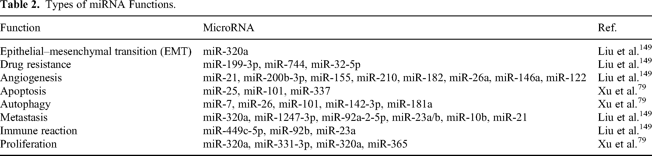
miRNAs play a critical role in the progression and development of hepatocellular carcinoma (HCC) by functioning as regulators. 79 They are involved in various regulatory mechanisms such as apoptosis, metastasis, angiogenesis, autophagy, invasion, EMT, drug resistance, and proliferation in HCC. By controlling gene expression in target cells, ExomiRs are also crucial in HCC invasion, metastasis, proliferation, and drug resistance. Additionally, some miRNAs, such as ExomiRs, can be used as possible diagnostic and prognostic markers for HCC 79 (Tables 2 and 3).
Types of miRNA Functions.
ExomiRs Used as HCC Diagnostic Biomarkers.

Abbreviations: ExomirRs, exosomal miRNAs; HCC, hepatocellular carcinoma; CCS, cell cultural supernatants; miRNA, microRNA.
Gastric Cancer
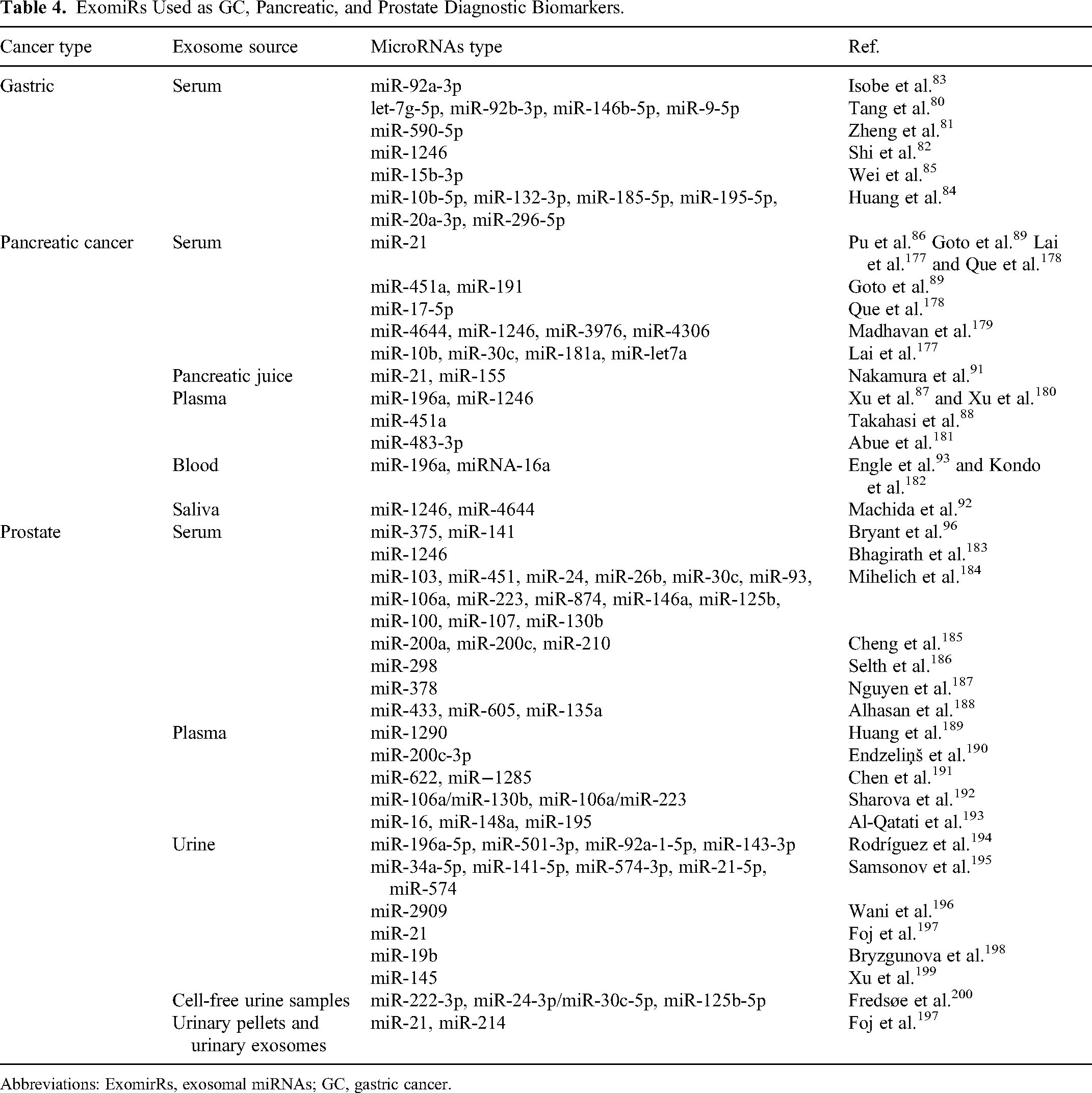
According to Tang et al. 80 serum levels of ExomiR-9-5p, let-7g-5p, miR-146b-5p, and miR-92b-3p can be employed as possible markers in patients with early-stage gastric cancer (GC). Similarly, serum levels of ExomiR-590-5p have been observed to be significantly higher in some patients diagnosed with early-stage GC (stage I/II) compared to healthy individuals. 81
Shi et al. 82 discovered that serum levels of ExomiR-1246 were significantly higher in patients with GC, allowing for the differentiation of healthy individuals from those with early-stage GC. Additionally, GC patients were found to have considerably lower serum levels of ExomiR-92a-3p compared to healthy individuals. 83 In a study by Huang et al. 84 the diagnostic efficacy of 58 circulating miRNAs in the sera of GC patients was investigated in a three-step investigation. They found that the expression of miR-296-5p, miR-20a-3p, miR-10b-5p, and miR-195-5p was noticeably increased in EXOs from serum samples of GC patients. According to Wei et al. 85 ExomiR-15b-3p expression was seen to be elevated, and it may be used as a prognostic and diagnostic marker in GC (Table 4).
ExomiRs Used as GC, Pancreatic, and Prostate Diagnostic Biomarkers.
Abbreviations: ExomirRs, exosomal miRNAs; GC, gastric cancer.
Pancreatic Cancer
Early-stage pancreatic cancer patients exhibit significantly elevated serum levels of ExomiR-21 when compared to healthy individuals. 86 Importantly, clinical risk factors like blood type, gender, smoking history, drinking history, age, body mass index, and diabetes mellitus have no significant impact on ExomiR-21 levels. Researchers have discovered that in stages I and IIa PDAC patients, intraductal papillary mucinous neoplasms (IPMN) have significantly elevated plasma ExomiR-196a and ExomiR-1246 levels compared to healthy people. 87 In addition, the plasma EXOs of stages I and II patients show a significant increase in miR-451a compared to healthy individuals, implying that ExomiR-451a could be used for the early detection of PDAC. 88
Likewise, Goto et al. 89 discovered that early-stage pancreatic cancers can be diagnosed using serum ExomiR-191, ExomiR-451a, and ExomiR-21. The receiver operating characteristic (ROC) analysis of these EXOs is more accurate than that of carcinoembryonic antigen (CEA) (AUC of 0.754, 0.935, 0.741, and 0.601, respectively). Additionally, a high ExomiR-21 expression level is an independent predictor of overall survival. With a 0.9% positive prediction rate, ExomiRs are more effective than CA19-9 in making an early diagnosis of pancreatic cancer. 90
ExomiRs have been also discovered in other body fluids, such as pancreatic juice (miR-21 and miR155), 91 saliva (miR-1246 and miR-4644), 92 and blood (miR-196a and miR-16a)93,94 (Table 4).
Prostate Cancer
It has been demonstrated that EXOs, which include proteins, DNAs, and RNAs (miRNAs), play a crucial role in tumor development and represent a rich source of potential biomarkers, especially for miRNA content profiling 95 (Table 4). ExomiRs such as miR-375 and miR-141 have been studied in plasma for prostate cancer. 96
Brain Cancer
Santangelo et al. 97 reported an AUC of 0.87 for a glioblastoma multiforme (GBM) serum panel made up of ExomiR-21, miR-222, and miR-124-3p.
In patients with advanced gliomas, the expression of these miRNAs dropped dramatically after tumor excision. In another study, miR-320 and miR-574-3p levels were shown to be significantly higher in EXOs extracted from the sera of 75 individuals with GBM, and they were associated with the diagnosis of this disease. 98
Serum ExomiR-301a levels are considerably higher in glioma patients than in controls, corresponding with advancing pathological stages. It was also discovered that serum ExomiR-301a levels are dramatically decreased following surgical removal of tumors but then rise again after disease recurrence. 99 It has been shown that miR-1246 collected from cerebrospinal fluid (CSF) can be used as a possible diagnostic biomarker in gliomas and that miR-1246 targeting therapy may damage the immunosuppressive tumor microenvironment and provide insight into anticancer immunotherapy. 95 According to Shao et al. 100 ExomiR-454-3p has a sensitivity and specificity of 79.17% and 91.67%, respectively, showing it to potentially be a glioma diagnostic biomarker (Table 5).
ExomiRs Are Used as Brain Cancer, OC, Breast Cancer, OSCC, and Bladder Cancer Diagnostic Biomarkers.

Abbreviations: ExomirRs, exosomal miRNAs; OSCC, oral squamous cell carcinoma; OC: ovarian cancer; CSF, cerebrospinal fluid.
Ovarian Cancer
According to Yokoi et al. 101 ExomiRs miR-766-3p, miR-200a-3p, miR-26a-5p, miR-374a-5p, miR-142-3p, miR-328-3p, and let-7d-5p show higher expression levels in the serum of early-stage ovarian cancer (OC) patients (n = 15) compared to healthy individuals. These findings suggest that these miRNAs have the potential to distinguish between early-stage OC patients and healthy individuals. Other researchers Kobayashi et al. 102 have analyzed the combination of miR-200a, miR-200b, and miR-200c and found that this EXO compound could differentiate benign ovarian disorders from EOC patients with a specificity of 90% and a sensitivity of 88%.
According to Cappellesso et al. 103 miR-21 contributes to the oncogenesis of ovarian serous carcinoma (OSC). ExomiR-21 has the potential to stimulate neoplastic transformation in target cells and might be employed as a diagnostic tool. Urine ExomiRs are easily accessed and have lately been more extensively studied, especially in gynecological and urological illnesses. Zavesky et al. 104 demonstrated a significant increase in miR-92a levels in the urine of OC patients, suggesting that it could serve as a diagnostic tool.
According to miRNA microarray data, miR-30a-5p shows higher levels in urine samples from ovarian serous adenocarcinoma (OSA) patients compared to healthy individuals. Conversely, decreased concentrations of miR-30a-5p were observed in the urine of individuals with colon cancer and GC, indicating that urinary levels of miR-30a-5p might be specific to OC. These findings suggest that exosomal urinary miR-30a-5p could potentially serve as a specific diagnostic biomarker for OC. 105 The expression of miR-205 was considerably greater in the plasma EXOs of OC patients than in the benign tumor group and healthy individuals, and throughout stages III and IV of OC and lymph node metastases, miR-205 levels were raised. Thus, miR-205 concentration in plasma EXOs is a promising tumor biomarker to help diagnose OC. 106
ExomiR-4732-5p extracted from plasma has a specificity of 82.4% and a sensitivity of 85.7% for distinguishing EOC patients from healthy individuals. Thus, it could act as a possible marker for tracking the development of EOC from early to late stages. It may also be a potential new biomarker for detecting EOC 107 (Table 5).
Breast Cancer
miR-372, 108 miR-18a-3p, 109 miR-101, miR-423-5p, 110 and 8 miRNAs of the miR-106a-363 cluster 111 can differentiate breast cancer patients from healthy individuals and are associated with cancer proliferation, cell properties, and migration. Triple-negative patients have higher levels of other miRNAs, like miR-373, than healthy controls or even luminal cancer patients; miR-223-3p 112 levels are higher in invasive ductal carcinoma patients than in those preoperatively diagnosed with ductal carcinoma in situ (DCIS); miR-93 113 is also upregulated in DCIS.
Breast cancer patients have higher levels of plasma ExomiR-223-3p, which can differentiate them from the general population with early breast cancer. Moreover, ExomiR-223-3p expression is significantly increased in biopsy-proven invasive DCIS (n = 13) compared to early ductal carcinoma, suggesting that ExomiR-223-3p can be used to predict the risk of invasive lesions in DCIS patients. 112 Additionally, DCIS patients have significantly higher levels of plasma ExomiR-93 expression than healthy individuals (n = 80) 113 (Table 5).
Oral and Oropharyngeal Cancer
Several ExomiRs related to oral SCC (OSCC) have been identified, such as miR-342-3p 114 and miR-138 115 (Table 5).
Bladder Cancer
miR-1285-3p, miR-142-3p, miR-16-1-3p, miR-195-3p, miR-196b-5p, miR-23b-3p, miR-28-5p, and miR-299-3p 116 are identified as ExomiRs that can distinguish bladder cancer patients from healthy individuals. These miRNAs are associated with cancer proliferation, cell properties, and migration (Table 5).
Conclusion
EXOs serve as efficient and dependable carriers of miRNAs found in various body secretions. Moreover, the expression patterns of ExomiRs in tumor cells differ significantly from those in normal cells. As a result, ExomiRs offer a promising noninvasive approach to cancer detection. This innovative approach may lead to improved cancer therapy. Various industries are currently exploring the applications of EXOs in biotechnology. EXOs Diagnostics, now a part of Bio-Techne, has been at the forefront of developing molecular diagnostics using biological fluid samples. Their precision EXO technology has enabled liquid biopsy for the detection of lung and prostate malignancies. However, there are still challenges to overcome before this technology can be widely applied in clinical settings. One of the major hurdles is the standardization of EXO extraction methods from different body fluids. Future applications should focus on more efficient techniques that require minimal biofluid volume while ensuring high purity and yield of EXOs.
Footnotes
Abbreviations
Declaration of Conflicting Interests
The author(s) declared no potential conflicts of interest with respect to the research, authorship, and/or publication of this article.
Funding
The author(s) disclosed receipt of the following financial support for the research, authorship, and/or publication of this article: Funding for this work was provided by Kermanshah University of Medical Sciences, Kermanshah, Iran. This study was carried out under the approval code IR.KUMS.REC.1402.039 at Kermanshah University of Medical Sciences, Kermanshah, Iran.