
Abstract
Introduction:
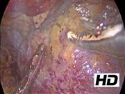
Internal suspension technique in retroperitoneal laparoscopic partial nephrectomy for the management of renal tumors was described with good perioperative results. The objective of this video is to show the steps to perform this technique and the results obtained in the patients treated.
Methods:
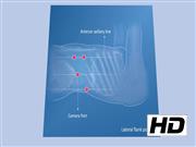
Between January 2013 and January 2021, a total of 301 patients with a renal tumor underwent retroperitoneal laparoscopic partial nephrectomy. In patients who were treated with the internal suspension technique, the tumors were either ventral or in the lateral border. The surgeon preserved the perinephric fat with the renal tumor as a suspension traction measure when separating the kidney, so it exerted traction on the tumor during its resection. Patients' characteristics as well as intraoperative and postoperative outcomes were described.
Results:
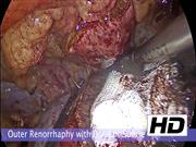
Twelve patients were treated with the internal suspension technique. The average age was 59.7 (SD 10.22) years. Of the patients, 65% were women and 58% presented a tumor in the right kidney. A total of 58.3% patients had low-complexity tumors (R.E.N.A.L score <6) and none had high-complexity tumors; 65% of the tumors were located between the polar lines. The estimated glomerular filtration rate (eGFR) was 75.1 (SD 12.73) mL/min. Operative time was 95.83 (SD 13) minutes. Warm ischemia time was 19.42 (SD 9.26) minutes. Tumor size was 2.68 (SD 0.8) cm. Estimated blood loss was 110 (SD 29.17) mL. Postoperative eGFR was 70.1 (SD 17.28) mL/min. Of the patients, 66.7% presented clear cell carcinoma and none had positive surgical margins. Trifecta outcomes were achieved in 72.7% of the cases. One patient presented urine leakage that required a Double-J stent.
Conclusion:
As shown in this video, the internal suspension technique is a feasible and safe procedure in retroperitoneal laparoscopic partial nephrectomy. With this approach we were not only able to stabilize the tumor in a right position maintaining the traction during incision, but we were also able to suture the parenchyma easily.
No competing financial interests exist.
Authors state that the article has been assessed by the responsible review committee in accordance with the Declaration of Helsinki.
Author(s) have received and archived patient consent for video recording/publication in advance of video recording of procedure.
Video was not previously presented at meetings. Copyrighted background music cannot be used in the video without permission from the copyright owners since it was made by us with GarageBand software® using a percussion base. All authors accepted that our video will become property of Videourology.
Runtime of video: 7 mins 31 secs
Get full access to this article
View all access options for this article.