
Abstract
Introduction:
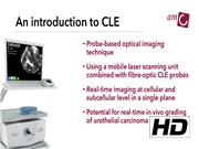
Endoscopic evaluation with postoperative histopathologic assessment of suspicious tissue forms the cornerstone for the diagnosis of urothelial carcinomas of the bladder (UCB) and of the upper urinary tract (UTUC). However, the limitation of the current diagnostic pathways is the lack of real-time in vivo histologic assessment. Confocal laser endomicroscopy (CLE) is an optical imaging technique that has the potential to overcome this limitation. This probe-based technique may enable real-time assessment of histologic grade of urothelial carcinoma during cystoscopy and ureteroscopy. 1 –5 In the bladder, real-time grade assessment could allow for immediate risk stratification of small bladder tumors during cystoscopy and therewith enable laser fulguration of identified low-risk UCB in an outpatient setting. In the upper urinary tract, real-time grade assessment of UTUC could improve intraoperative risk stratification for the selection of endoscopic treatment. Moreover, CLE may have the potential to improve the limited diagnostic yield and accuracy from endoscopic tissue biopsies for UTUC diagnosis. 6 In this video, we introduce the principles of CLE, illustrate its use during cystoscopy and ureteroscopy, and present the evaluation of CLE images for the diagnosis of UCB and UTUC.
Materials and Methods:
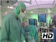
We performed CLE imaging during rigid cystoscopy and flexible ureteroscopy in patients with suspicious lesions for UCB or UTUC within the scope of two ongoing clinical trials (
Results:
Placing the CLE probe in contact with the tissue of interest enables real-time imaging at cellular resolution. Tumor diagnosis and grade differentiation are based on visual evaluation of the cellular microarchitecture. After CLE imaging, the standard endoscopic procedure may be resumed.
Conclusions:
CLE is an optical imaging technique that enables real-time in vivo observation of the cellular microarchitecture. This probe-based technique may be used during standard cystoscopy and ureteroscopy for intraoperative risk stratification of UCB and UTUC by histologic grading. This could lead to an advancement in personalized care with improved intraoperative decision-making. The results of the ongoing studies to evaluate the diagnostic value of CLE are awaited.
No competing financial interests exist.
The authors have received and archived patient consent for recording/publication of the video recording procedure.
Runtime of video: 7 mins 24 secs
Parts of the video were used for abstract presentations at the AUA 2017 and the Challenges in Endourology 2016 and 2017.
Keywords
Get full access to this article
View all access options for this article.