
Abstract
Introduction and Objective:
Management of the obstructive megaureter is one of the most difficult problems in urological surgery, especially in the neonate and young infant. 1 It can be managed using a number of operative techniques with the primary goal being to minimize the potencial for further injury to the affected kidney. However, the commonly used methods of providing drainage have their limitations. A method for internal drainage would provide a more acceptable means of relieving obstruction in infants, 2 especially with uncontrolled urinary tract infection or deteriorating renal function, until a time at which these patients could undergo a more definitive surgical repair. Our aim is to present the minimally invasive treatment of ectopic obstructive megaureter 3 as an initial and temporizing approach in duplicated ectopic megaureters with preserved moiety function. The video describes the technique in detail with the results and has a run time of 5 mins.
Materials and Methods:
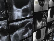
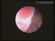
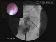
We have treated with this technique, eight infants with the unilateral duplicated system and ectopic obstructive upper ureter. All patients were symptomatic. The indications for treatment were urinary tract infections with increasing hydronephrosis (2p), pyonephros (2p), and pyelonephritis (4p). In all cases, dimercaptosuccinic acid (DMSA) renal scan showed good functioning upper moiety. In all cases, the urethrocystoscopy failed to show the meatus of the ectopic ureter. During the cystoscopy, the dilated distal end of the ureter was identified with ultrasound. Under ultrasound scan and direct cystoscopic vision, the retrovesical ectopic ureter was punctured transvesically. The puncture was done with a 5F needle and contrast was instilled in the ectopic ureter and retrograde pyelogram obtained. Once assessed the puncture into the ectopic ureter, a 0.014″ was negotiated into the upper moiety through the needle. The punctured site was then dilated with a 5 mm high-pressure balloon and a 3F double-J stent inserted.
Results and Conclusions:
There were not perioperative or postoperative complications. Median follow-up is 74 months (31–90). Urinary tract infections disappeared in all cases after the procedure. The assessment done 6 months postoperatively demonstrated a significant decrease in the grade of the hydronephrosis in all cases. No further surgical treatment has been required in three patients free of infections and with preserved function in the upper moiety. In three cases, an open ureteric reimplant was required and in two cases, an upper pole hemynephrectomy. Endoscopic transvesical puncture of ectopic megaureter and ureteral neomeatus creation is a minimally invasive technique that is successful as initial and temporizing management to avoid urinary tract infections and, in some cases, also decrease the dilatation and preserve the parenchyma function.
No competing financial interests exist.
Runtime of video: 5 mins
Keywords
Get full access to this article
View all access options for this article.