
Abstract
Introduction:
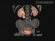
A retrocaval ureter (RCU) is a rare clinical entity that results from the abnormal persistence of the subcardinal vein. It is more common among males and usually presents in the 3rd or 4th decade of life.1,2 Whereas not all cases result in clinical obstruction, patients can present with obstruction-related problems: flank pain, upper urinary tract infections, urolithiasis, and hypertension. We present a video-enhanced description of the robot-assisted surgical management of a 25-year-old male with a symptomatic RCU and ipsilateral renal stone. The video outlines the initial presentation, provides all related radiologic investigations, details surgical techniques, and provides outcome data.
Methods:
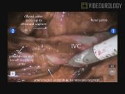
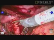
All preoperative imaging studies confirmed the diagnosis of a right RCU with ipsilateral 8-mm renal stone. Despite attempts at conservative medical management before surgery, the patient's flank pain was recalcitrant. Unfortunately, the patient did not tolerate placement of an indwelling stent and, as such, it was removed in favor of a nephrostomy tube (NT). After appropriate preparation of the patient, pneumoperitoneum was obtained using a Veress needle technique and the da Vinci© robot was docked after placement of all ports: 12-mm camera, two 8-mm robotic, 10-mm assistant, and 5-mm liver retractor. The NT was also prepped into the sterile field to permit intraoperative antegrade fluid instillation. First, the right colon and liver were mobilized to expose the retroperitoneum. The duodenum was then mobilized medially to expose the inferior vena cava (IVC) and interaortocaval space. The abnormally located ureter was identified anterior to the IVC and dissected cephalad until it coursed posterior to the IVC. At this point, dissection was initiated lateral to the IVC, where the distended proximal ureter was identified. After mobilizing a short portion of the ureter proximally, it was followed caudad until it too coursed posterior to the IVC. During mobilization of the ureter, attention was paid to minimize ischemic injury; sufficient periureteric tissue was maintained and limited cautery was used. The ureter was sharply transected at its most posterior, retrocaval point. Without removal of the retrocaval segment, the proximal and distal ends of the ureter were spatulated widely. Using the 10-mm assistant port, a flexible cystoscope was inserted into the proximal ureter. A stone basket was utilized to grasp and remove the renal stone through the same port. Over an antegradely placed ureteric stent, a water-tight, tension-free ureteroureterostomy was performed in a running fashion using a 4-0 monocryl-modified van Velthoven suture.3 At the end of the procedure, a Jackson-Pratt drain was placed and the NT left open to drain.
Results:
The surgical time was 135 minutes and there were no postoperative complications. The patient was fit for discharge on post-operative day (POD) #2, but remained in hospital until POD #4 due to social issues. The NT was removed 5 weeks postoperatively and the ureteric stent was removed the following week. A follow-up renal scintigraphy demonstrated no evidence of obstruction and the patient was pain free at 9 months.
Conclusions:
Robot-assisted surgical management of an RCU and ispilateral renal stone is a safe and feasible option.
No competing financial interests exist.
Runtime of video: 6.58 mins
Keywords
Get full access to this article
View all access options for this article.