
Abstract
Introduction:
Vena caval injury is a rare but serious intraoperative complication that may occur during laparoscopic urologic surgery. 1 Traditional teaching would suggest emergency laparotomy and open repair. 2,3 With advanced laparoscopic and robotic instruments, laparoscopic management of inferior vena cava (IVC) injury is now possible. We reviewed our experience in laparoscopic management of vena caval injury and demonstrated a case of robot-assisted repair in a video.
Materials and Methods:
We reviewed all cases of laparoscopic renal and adrenal surgery performed by a single surgeon (C.P.S.) from 2002 to 2011. Cases with intraoperative vena caval injury were retrieved and compared with their corresponding video archives.
Results:
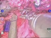
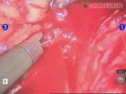
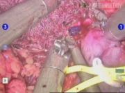
Five cases of vena caval injury were identified, all occurring during right-sided surgery. In two patients, hemorrhage occurred during laparoscopic nephrectomy. Bleeding stopped after direct compression using a 5-mm aspiration-irrigator device and application of gelatin-thrombin hemostatic matrix (Floseal; Baxter). Laparoscopic nephrectomy was completed uneventfully. During laparoscopic adrenalectomy in another patient, bleeding was stopped with intracorporal suturing using 3/0 polygalactin suture with a Lapra-Ty clip (Ethicon Endosurgery). The fourth patient had massive hemorrhage during laparoscopic donor nephrectomy after the graft was retrieved. Emergency conversion to laparotomy was required to repair the IVC. The fifth patient shown in this video developed brisk bleeding from vena caval injury during robot-assisted laparoscopic partial nephrectomy. The injury was repaired with robotic assistance using 3/0 polygalactin suture with Lapra-Ty clips. Proximal control and distal control of the vena cava were provided by the patient-side assistants using two 5-mm aspiration-irrigator devices. Partial nephrectomy was completed without further complication. Blood loss was 900 mL.
Conclusions:
In most patients, major vena caval injury may be managed without emergency laparotomy. Management of each injury had to be individualized. The key steps for securing hemostasis included increasing intra-abdominal pressure, direct tamponade, achieving proximal and distal control with additional instruments, and intracorporal suturing with or without robotic assistance.
The authors have nothing to disclose
.
Runtime of video: 7 mins 40 secs
Get full access to this article
View all access options for this article.