
Abstract
Purpose:
Major vascular injuries, which are life threatening, may occur during laparoscopic procedures. 1 The aim of this video is to illustrate the key points to avoid complications and to manage them during renal vein dissection.
Method:
Three cases are presented in the video; right transperitoneal laparoscopic nephrectomy of a 59-year-old male patient for a nonfunctioning kidney related to renal stone disease, laparoscopic right retroperitoneal radical nephrectomy of a 59-year-old male patient for a 3.5 cm renal mass, and laparoscopic left transperitoneal radical nephrectomy of a 63-year-old male patient for a 5 cm renal mass, respectively. All procedures were completed laparoscopically. The operation time was 220, 130, 190 minutes, respectively, and the estimated blood loss was 110, 25, 125 cc, respectively. Patients were discharged after removal of drain on the 3rd day of surgery.
Results:
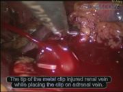
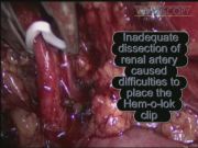
In the first case, the Hem-o-lok® clip applicator injured the renal vein during clip placing on the renal artery. Bleeding was decreased by traction of the renal vein, but the surgeon was unable to place clips on the artery and vein together. Thereafter, Hem-o-lok clips were placed on the artery and vein separately. In the second case, inadequate dissection of the renal artery caused difficulties to place the Hem-o-lok clip. Because the tip of the clip was placed on the junction of the renal vein, segmental vein, and vena cava inferior (VCI), more dissection of the renal artery was performed and a second clip was placed under the first one. In the last case, the tip of the metal clip injured the renal vein while placing the clip on the adrenal vein. Bleeding was decreased by traction of the renal vein and the Hem-o-lok clip was placed on the proximal side. Retrograde bleeding due to an unsecured renal artery was controlled by placing the metal clips on the distal side of the renal vein. Afterward, the Hem-o-lok clips were placed on the renal artery.
Conclusion:
Complete and clear identification of the renal vein, the artery with their branches and delicate dissection of renal hilum should be performed. The locking tip of the Hem-o-lok clip must be clearly visualized at the back of the vessel before closure to avoid injuries. The renal vein can be injured easily by a part of sharp instruments like the tip of a clip applicator especially when applicator removal after clip placement as it was shown in the first case. In the second case, tissue at the clicking point of the Hem-o-lok clip may cause displacement. Therefore, it was mandatory to place a second secure clip proximal to first one even it is close to VCI. At the third case, adequate dissection of the renal vein and its branches should be performed. In case of any bleeding, adequate traction of the renal vein and increasing the abdominal pressure may decrease bleeding, until obtaining a reliable clip placement. To ensure correct clip placement during bleeding, suction from an additional port should be used for providing better visualization in the surgical area.
Runtime of video: 8 mins 30 secs

Keywords
Get full access to this article
View all access options for this article.
References
Supplementary Material
Please find the following supplemental material available below.
For Open Access articles published under a Creative Commons License, all supplemental material carries the same license as the article it is associated with.
For non-Open Access articles published, all supplemental material carries a non-exclusive license, and permission requests for re-use of supplemental material or any part of supplemental material shall be sent directly to the copyright owner as specified in the copyright notice associated with the article.