
Introduction
Despite decades of research, the mechanism of regulating blood flow and oxygen metabolism associated with neural activation still remains unclear. To study that issue, reliable measurement of cerebral oxygen metabolic rate (CMRO2), as well as cerebral blood flow (CBF), is essential. Of the imaging tools, the blood oxygenation level dependent MRI technique based on the hypercapnia calibration 1 has been extensively used, although it estimates only the relative changes. However, the PET-based absolute quantification technique has been hardly applied for small animals due to technical difficulties. Here, we report novel methods overcoming existing difficulties for absolute quantification of CBF and CMRO2 in rats using a high-resolution dedicated animal PET (microPET). No arterial blood sampling procedure for the determination of an arterial input function (AIF) was performed.
Methods
Rats were imaged using the O-15 labeled tracers. An intravenous bolus injection of H215O 2 and a bolus inhalation of O15O 3 were used for CBF and CMRO2 measurements, respectively. The AIF required for absolute quantification was obtained using a time-activity curve over the heart after necessary corrections. We have validated our method in two ways. The first was to validate our intravenous injection (H215O) technique based on non-invasive AIF determination against the intracarotid injection technique which does not require AIF measurement. The second was to validate overall CBF and CMRO2 techniques using a hypothermia challenge to see hypothermic suppression effect on both measurements. For each rat, the right external carotid artery and a femoral vein were catheterized for injecting O-15 water for CBF measurements and a tracheotomy was performed for administering O-15 oxygen for CMRO2 measurement. During imaging procedure, alpha-chloralose (30 mg/kg/hr, intravenous infusion) was used for anesthetic. CBF and CMRO2 measurements were done in hypothermia (rectal temperature: 32 °C) and normothermia (37 °C) successively for each rat.
Results
Our data for 10 rats shows that the CBF (ml/100g/min) values obtained using the intravenous injection based on non-invasive AIF determination (normothermia: 54. 62±5.08, hypothermia: 45.23±6.05) agreed well with the reference values obtained using the intracarotid injection (normothermia: 54.37±4.60, hypothermia: 47.41±8.64). In addition, the CMRO2 (ml/100g/min) was reduced by 26.7% by the hypothermia (normothermia: 5.00±0.36, hypothermia: 3.66±0.58), as expected. A CMRO2 comparison map between hypothermia and normothermia obtained for a rat is shown in Fig. 1: Axial, coronal and sagittal slices from the left. Hypothermic suppression effect on the CMRO2 is clearly shown.
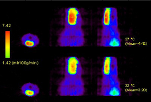
Axial, coronal and sagittal slices from the left. Hypothermic suppression effect on the CMRO2 is clearly shown.
Discussion and conclusion
Quantitative CBF and CMRO2 measurement techniques using microPET without requiring arterial blood sampling for small animals were proposed and validated for the first time. Our technique would be of great use for any CBF/CMRO2 measurement study using small animals (e.g. brain activation studies) as well as for validating other techniques (e.g. MRI-based techniques).