




Introduction
Magnetic resonance imaging (MRI) of pathophysiological vascular events requires MR contrast agents (MRCA) linked to bioactive molecules. Here we describe a simple method to prepare a large MRCA by covalently linking albumin to the gadolinium-diethylenetriaminepentaacetic acid (Gd-DTPA), a commonly used MRCA. We employed a novel, Arsenazo III-based reaction to detect unbound gadolinium in the reaction mixture. The preparation was evaluated against a large, commercial MRCA, Gadomer17 (Schering AG, Germany) in a rat 9L gliosarcoma brain tumor.
Methods
The coupling of DTPA and gadolinium to bovine serum albumin (BSA) was performed using published procedures1, 2. To 15 ml of 10 μmol BSA in HEPES buffer (pH 7.5), a suspension 0.714 g DTPA anhydride in DMSO was slowly added with constant stirring. The solution was maintained between pH 6–7 with the addition of 3 M NaOH. After stirring for 1 hr at room temperature, 0.742 g of gadolinium chloride dissolved in water was slowly added to the reaction mixture, until the very first indication of unchelated gadolinium was observed with 10 μmol Arsenazo III in acetate buffer at pH 4.0 3 . The mixture was stirred cold overnight, dialyzed x3 each v/s 0.1 M citrate buffer (pH 6.5) and water and then lyophylized. This preparation and Gadomer17 were tested for contrast enhancement using the rat tumor model. After MRI, the rats were injected with the plasma albumin binding dye Evans blue for histological verification of leakiness of tumor vasculature.
Results
The product was free of unbound Gd when assayed 4 ; molecular weight by electrophoresis was ∼90 kDA, similar to other reported values 2 . The molar labeling ratio of Gd:BSA was about 30:1 3 . During MRI it gave a similar, but not identical, pattern of enhancement as Gadomer17, which has a comparable molecular size (Figure 1A & B). Most notable was the similarity in the ring of contrast enhancement of both and their close alignment to the highly vascular edge of the tumor (Figure 1C). The image of Gd-Albumin resembled closely that of Evans blue leakage (Figure 1B & C); the spread of Evans blue-albumin toward the midline and cortex was well matched by Gd-albumin contrast pattern.
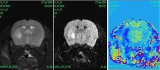
MR images of a 9L gliosarcoma tumor from the same rat after contrast enhancement with Gadomer17 (A) and Gd-Albumin (B). Note the ring of enhancement (arrows) with both MRCA and their close agreement with tumor histology and Evans blue leakage (outlined in C). (Inset in A: a T1 pre-contrast image)
Conclusion
Arsenazo III reaction provides a simple, sensitive test for instantly detecting unbound Gd in MRCA. Excess Gd can precipitate the protein in the reaction mixture. Repetitive testing with Arsenazo III can prevent such negative outcomes during preparations. This method may also be applicable in preparation of Gd-chelates with other bioactive molecules. The tumor data suggests that Gd-albumin is useful for MR imaging of albumin distribution in experimental and human studies.
Footnotes
Acknowledgements
Grant support: NIH-RO1HL70023.