
An important function of lithium as a neuroprotective agent has been documented previously. Significant reduction in infarct size and DNA damages with the corresponding improvement of neurological scores were reported as results of post stroke lithium administration. In the current study, therapeutic efficacy of chronic lithium treatment was assessed by various MRI-derived parameters (i.e., apparent diffusion coefficient (ADC), fractional anisotropy (FA), and vessel size index (VSI)) and quantified with fMRI activity using electrical stimulation of rat forelimbs. The fMRI responses were examined by measuring the local hemodynamic MRI signal intensity based on blood oxygenation level dependence (BOLD) and cerebral blood volume weighted (CBVw) responses. The ipsilesional fMRI activations in the somatosensory (SS) cortex of lithium-treated (n=6) and saline-treated rats (n=6) two weeks after middle cerebral artery occlusion (MCAO) were compared. ADC and FA values of local brain tissues were significantly correlated with the magnitude of fMRI responses for the lithium-treated rats while no such correlations were seen in the saline controls. The ipsilesional/contralesional BOLD signal intensity response ratios of lithium-treated rats were larger than those of saline-treated control rats. In contrast, the CBVw response ratios were similar between two groups. These results demonstrated that the lithium-treatment of post-stroke animal models positively enhanced BOLD fMRI response in the lesion hemisphere. Overall activation volume and magnitude were larger for saline-treated rats than the lithium-treated group in both contra and ipsilesional somatosensory cortices. However, for both BOLD and CBVw responses, the mean activated volume ratio (ipsilesional/contralesional) was higher for lithium-treated rats than controls. The CBVw activation signal magnitude ratios (Figure 1 upper panel) were significantly higher than BOLD ratios for the control animals; however, similar for the lithium-treated rats. As shown in Figure 1 (bottom panel), functional status index (FSI) which represents the mean ratio between BOLD and CBVw ipsi/contralesional activation signal intensity ratio for the individual somatosensory cortex was significantly higher for lithium-treated rats than the control group. From the acquired ADC maps, ex vacuo dilation of the ipsilesional lateral ventricles was identified for most of rats while other structural damages (e.g., FA) were also evident at this late stage (2 weeks) of recovery. Among the acquired structural parameters, ADC and FA were significantly correlated with CBVw fMRI activation magnitude (i.e., area under the curve) for the lithium-treated rats while no such correlations were found for the saline controls. When compared to the contralesional values, the ipsilesional mean VSI ratio (ipsi/contra) was larger for the lithium-treated rats; however, the micro vascular volume (i.e., Delta-R2) ratio (ipsi/contra) was larger for control rats. These results imply that possible vascular transformation affects fMRI characteristics.
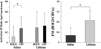
activation SI ratio and functional status index in somatosensory cortex.