
Introduction
Dramatic and rapidly reversible structural changes are observed in dendritic spine morphology following excitotoxic or hypoxic stress 1 . However, morphological and biochemical alterations in post-synaptic densities persist for up to 24 hours following transient ischemia. This suggests that the synapse may be the origin of signals that propagate toward the cell body and instigate delayed post-ischemic neuronal death. Synaptically localized cell adhesion molecules are important regulators of synaptic plasticity and synaptogenesis. Regulation of cell adhesion and cytoskeletal structure in dendritic spines is necessary for the maintenance of mature synaptic connections. Therefore, we examined the effects of transient cerebral ischemia on the expression of synaptic proteins 20 hours post-ischemia.
Methods
Adult male C57 mice were subjected to a transient (60 min) middle cerebral artery occlusion (MCAO) followed by 20 h of reperfusion. Synaptosomes were prepared from both the contralateral and ipsilateral hemispheres using the density gradient centrifugation method and characterized by electron microscopy. Synaptosomal cell adhesion molecule expression was determined using western blots. Synaptosomal proteins were ICAT-labeled, digested and analyzed by nanoLC-MS mass spectrometry. In-house software was used to identify differentially expressed paired ICAT peaks, which were then sequenced by nanoLC-MS/MS.
Results
Using western blot, it was found that Agrin and Catna2, but not GluR2, were significantly reduced 20 hours after MCAO (Figure 1). ICAT protein profiling experiments (Table 1) revealed the prominent over-expression of the actin binding protein dystrophin as well as various forms of tubulin. Several mitochondrial proteins were prominently under-expressed indicating substantial mitochondrial damage.
Focal cerebral ischemia induces alterations in synaptosomal protein expression (partial list) as determined by ICAT nanoLC-MS/MS.
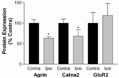
Cerbral ischemia alters synaptosomal protein expression as determined by western blot.
Conclusions
Cerebral ischemia has a lasting effect on the expression of proteins that regulate synaptic plasticity, in particular cytoskeletal proteins including the actin interacting proteins Catna2 and Dmd and the tubulin subunits. These findings indicate that a prominent re-organization of the synaptic cytoskeletal architecture occurs following cerebral ischemia. This is accompanied by a decrease in the metabolic capacity of the perisynaptic area, as evidenced by the decrease in mitochondrial enzymes. This study demonstrates that the synaptic proteome is dramatically affected by cerebral ischemia and indicates the occurrence of extensive synaptic remodeling.