




Introduction
Positron emission tomography (PET) is a powerful method that enables the simultaneous analysis of multiple hemodynamic factors. In the analysis of occlusive cerebrovascular diseases, various combinations of cerebral blood flow (CBF), metabolism, and blood volume (CBV) are observed depending on the severity of hemodynamic stress. Nonetheless, there have been no appropriate analyses to interpret the combination of factors in multivariate space. In this report, we differentiated the distribution of multiple PET-measured hemodynamic factors of patients with moyamoya disease, as a representative of chronic occlusive cerebrovascular disease, to examine if such analysis is useful to clarify the pathophysiology of the disease.
Methods
Data of 100 patients with moyamoya disease (mean of age 31.3,. range 12–58) that were obtained from 1991–2004 were retrospectively analyzed. PET measurement of CBF, CBV, cerebral metabolic rate for oxygen (CMRO2), and oxygen extraction fraction (OEF) were performed with inhalation of 15O labeled gases and with continuous arterial blood sampling. Patients were classified into 5 groups depending on clinical presentation as presented previously (1);non-symptomatic (NS) patients, patients presenting transient ischemic attack (TIA group), those with infarction associated with TIA (I/TIA), those with a permanent deficit with infarction (PD), and those with hemorrhagic onset (H group). Values of patients groups were compared with a normal group (Norm).
Results
1. In earlier 57 patients, multivariate analysis of covariance to test the distribution of three dimensional (CBF, CBV, OEF) vector was performed, indicating that the significant difference of distribution existed between every possible pair out of six groups except NS vs. H, and H vs. PD among the frontal cortex.(figure 1).
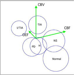
2. Using the data of these earlier patients, a multivariate discriminant was obtained and applied for the prediction of clinical presentation of latter patients using three factors (CBF, CBV, OEF) in frontal cortex. In more than 85 percent of patients, clinical type was correctly determined by this method, but such prediction was impossible by using single factor.
3. Significant alteration of factors in three dimensional space was detected before and after surgical revascularization in each patient.
Conclusions
Muti-variate statistical differentiation of hemodynamic factors could provide useful information that cannot be obtained in single factor analysis. This method was even useful in the determination of homodynamic stage in single patient.