
Abstract
Cerebral ischemia induces a complex transcriptional response with global changes in gene expression. It is essentially regulated by transcription factors as well as epigenetic players. While it is well known that the inhibition of transcriptionally repressive histone deacetylases leads to neuroprotection, the role of histone methyltransferases in the postischemic transcriptional response remains elusive. We investigated the effects of inhibition of the repressive H3K9 histone methyltransferases SUV39H1 and G9a on neuronal survival, H3K9 promoter signatures and gene expression. Their inhibition either with the specific blocker chaetocin or by use of RNA interference promoted neuronal survival in oxygen glucose deprivation (OGD). Brain-derived neurotrophic factor (BDNF) was upregulated and BDNF promoter regions showed an increase in histone marks characteristic for active transcription. The BDNF blockade with K252a abrogated the protective effect of chaetocin treatment. In conclusion, inhibition of histone methyltransferases SUV39H1 and G9a confers neuroprotection in a model of hypoxic metabolic stress, which is at least in part mediated by BDNF.
INTRODUCTION
Cerebral ischemia induces large changes in gene expression with a general tendency toward gene repression.1–3 Accordingly, repressive epigenetic marks accumulate: DNA methylation levels are reported to rise 4 and vast histone deacetylation processes take place. 5 Inhibiting epigenetic enzymes that function as transcriptional repressors, such as the DNA methyltransferase DNMT1, or the family of histone deacetylases (HDACs), leads to maintenance of activating epigenetic marks after ischemia. This, in turn, promotes the upregulation of genes, including key mediators of neuroprotection and regeneration and hence results in improved outcome after stroke.6,7 Epigenetic drugs, such as pan-HDAC inhibitors, have been identified as promising treatment option for diverse neurologic diseases that involve epigenetic dysregulation including ischemia. 8
In contrast to DNA methylation and histone acetylation, the role of histone methylation is poorly understood. Histone methylation is a posttranslational modification that can be found at lysine and arginine residues of histones (H). Methylation at lysine residues (K) occurs in three different states: mono-, di-, and tri-methylation (me 1/2/3). Different methylation states are associated with gene activation or repression. Tri- and di-methylation on histone 3, lysine 9 (H3K9) is linked to transcriptional repression. In contrast, monomethylation and the unmethylated state of H3K9, that is often acetylated (ac), are found in transcriptionally active sites of the genome. 9 Enzymes involved in the establishment of repressive H3K9 methylation marks are, among others, the methyltransferases G9a, catalyzing H3K9 me0 to me1 and me1 to me2 steps, 10 as well as SUV39H1 that catalyzes H3K9 di- to tri-methylation. 11 Both SUV39H1 and G9a are found in diverse repressive complexes and act in concert with other epigenetic players, such as DNA methyltransferases 12 and diverse HDACs. 13 As such they contribute to the induction and maintenance of transcriptional repression in the genome.
The aim of our study was to elucidate the role of histone methylation and selected associated enzymes SUV38H1 and G9a in the context of cerebral ischemia. We hypothesized that the inhibition of repressive histone methyltransferases could induce gene activation and confer neuroprotection in ischemia. We hence employed oxygen glucose deprivation (OGD), a widely used
MATERIALS AND METHODS
Isolation of Rat Cortical Neurons and Neuronal Cell Culture
Primary rat cortical neurons were derived from embryos (E17) of Wistar rats (Bundesinstitut für gesundheitlichen Verbraucherschutz und Veterinärmedizin, Berlin, Germany). Cultivation took place in supplemented neurobasal medium in 24-well, or 6-well plates as described in detail in Meisel
Chemicals and Drug Administration
Chaetocin (C9492), BIX-01294 (B9311), and K252a (K1639) were purchased from Sigma Aldrich (Taufkirchen, Germany). All were solved in dimethyl sulfoxide in 3 mmol/L stock solutions. After toxicity testing in neuronal cultures, chaetocin was used at a 30-nmol/L concentration, BIX-01294 at 100 nmol/L, and K252a at 50 nmol/L. In all cases, higher concentrations evoked signs of toxicity. Drug administration to neuronal cultures occurred as pretreatment on day
Lentivirus Production, Titration, and Application
Interfering RNA target sequences were designed using the internet applications of Invitrogen (http://www5.invitrogen.com/custom-genomic-products/tools/mirna/). Selected sequences (SUV39H1: A GGA CAA GAA AGC TTG GCT AG and G9a: T AGA GCT TCG ACT TCA GAC TT) were BLAST-searched against rat and mouse genome sequences to ensure target match and to exclude unspecific targets. A nontargeting ‘scrambled’ construct was used as a control (A AAT GTA CTG CGC GTG GAG AC). With the BLOCK-iT Pol II miR Expression Vector Kit (Invitrogen, Darmstadt, Germany) microRNA-embedded short hairpin RNA (miR-shRNA) constructs were generated containing the respective sequences. The miR-shRNA constructs were finally cloned into a third-generation lentiviral vector based on Addgene plasmid 27232 (ref 15) with the following modifications: microRNA delivery was driven by a ubiquitin promoter and the initial reporter protein was exchanged with a myc-tagged red fluorescent protein (RFP). HEK 293FT cells (Invitrogen, Darmstadt, Germany) were used as a producer cell line. Cotransfection with the packaging vectors psPAX (Addgene 12259) and 7.5pMD2G (Addgene 12260) was performed as described in Dull
Oxygen Glucose Deprivation
Primary neuronal cultures were subjected to OGD on DIV 9. In experiments establishing the OGD model it was shown that 100 μmol/L glutamate or 100 μmol/L NMDA induces cell death on DIV 9 already (data not shown). In OGD experiments, medium was removed from cells and preserved. Cells were once rinsed in PBS and subjected to OGD for 135 ± 5 minutes in a balanced salt solution at
Evaluation of Cell Survival and Damage: Lactate Dehydrogenase Assay
Neuronal injury was assessed by measuring lactate dehydrogenase release (LDH) in the supernatant 24 hours after the injury paradigm on DIV 10. Further, a second measurement followed after total lysis of cells using 0.5% Triton-X for 30 minutes at 37°C. Data are presented as percentage of cell death calculated from the ratios between the unit of LDH activity per mL per well and the maximum LDH activity per well after total lysis.
Evaluation of Cell Survival and Damage: Cell Counts
Fluorescent microscopic images of neuronal cultures were taken on DIV 9 directly pre OGD, as well as on DIV 10, 24 hours after OGD, with a Leica DFC360 FX microscope combined with a Leica DFC360 FX camera (Leica, Wetzlar, Germany) and a computerized software program (Leica Application Suite V3.3.0, Heerbrugg, Switzerland). Ten regions of interest (ROIs) per well were preselected and repeatedly analyzed at x400 magnification, maintaining identical settings for all experiments. Red fluorescent protein-positive cells were counted in a blinded manner (around 60 cells per ROI pre OGD). Mean values of the 10 ROIs were calculated per well and counted as
Immunoblots
After whole cell protein harvest and electrophoretic separation, proteins were transferred onto cellulose membranes and probed with the following antibodies purchased from Cell Signaling (Frankfurt am Main, Germany): G9a: CS 3306; SUV39H1: CS 8729; Myc: CS 2272; ß-actin: CS 4967; H3K9me3: CS 9754; H3K9me2: CS 4658; and H3K9ac: CS 9671. Primary antibodies were applied in a 5% bovine serum albumin tris-buffered saline solution with 0.1% Tween-20 (TBST). Blocking and second antibody incubation were performed in 5% milk TBST.
Chromatin Immunoprecipitation and Sequencing
Around 50 million rat cortical neurons per condition (chaetocin versus vehicle-treated cells, both pre and post OGD) were cultivated, pretreated with 30 nmol/L chaetocin/vehicle on DIV 8 and harvested on DIV 9, 1 hour after OGD. Cells were crosslinked in 1% formaldehyde in growth medium for 10 minutes at 4°C, and the reaction stopped with 125 mmol/L glycine. Cultures were rinsed in cold PBS, scraped, centrifuged, and washed twice with PBS, and the pellet snap frozen in liquid nitrogen. Resuspension took place in a first lysis buffer (50 mmol/L HEPES-KOH, pH 7.5, 140 mmol/L NaCl, 1 mmol/L EDTA, 10% glycerol, 0.5% NP-40, 0.25% Triton X-100, freshly added protease inhibitors) for 10 minutes at 4°C followed by centrifugation and uptake in a second lysis buffer (10 mmol/L Tris-HCl, pH 8.0, 200 mmol/L NaCl, 1 mmol/L EDTA, 0.5 mmol/L EGTA) for 10 minutes, RT. For nuclear lysis, the pelleted chromatin was solubilized (10 mmol/L Tris-HCl, pH 8.0, 100 mmol/L NaCl, 1 mmol/L EDTA, 0.5 mmol/L EGTA, 0.1% Na-Deoxycholat, 0.5% N-Laroylsarcosine) and sheared by pulsed sonication for 40 minutes at 4°C in a bioruptor (Diagenode, Seraing, Belgium) to a bulk DNA reduction to less than 400 bp. To get rid of cell debris, the sheared chromatin was clarified by centrifugation at 20 000
Expression Analysis and Polymerase Chain Reaction
For mRNA expression analysis, real-time quantitative reverse transcription PCR (qRT-PCR) was performed using LightCycler 2.0 (Roche, Mannheim, Germany). Briefly, whole-cell mRNA was harvested in Trizol (1 mL per 3 Mio cells). After chloroform extraction, RNA containing pellets were solved in diethylpyrocarbonate-treated water. After DNase digest, PCR inhibitors were removed with the help of NucleoSpin RNA clean-up KIT (Macherey Nagel, Düren, Germany). Equal amounts of RNA were reverse transcribed with M-MLV reverse transcriptase and random hexamers. For qRT-PCR, the LightCycler FastStart DNA Master SYBR Green I Kit (Roche) was used. All PCRs were performed in duplicate. The relative expression of each gene of interest (GOI) was calculated compared with a reference gene (ref) by the delta Cp (crossing point) method with efficiency correction using the equation E−Cp/GOI)(GOI)/E−Cp (ref)(ref). Mean values of duplicates were determined. The reference genes ß-actin and reep5 with the most stable expression patterns in OGD were chosen in a previously performed methodological study using NormFinder Software.
20
In all experiments, PCRs for both reference genes were performed and similar results obtained with either reference. Amplification efficiencies (
Thermal cycling conditions and amplification efficiencies for each primer pair are listed in Table 1.
PCR primer pairs
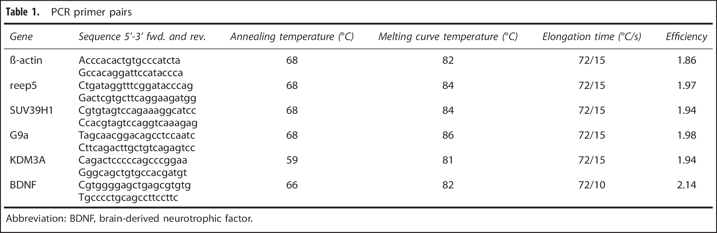
Abbreviation: BDNF, brain-derived neurotrophic factor.
Statistical Analysis
Data are presented as box/scatter plots and mean ± 95% confidence interval as well as bars with mean ± s.d.
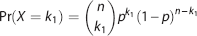
To calculate significant differences between sequence reads in the ChIP-seq experiments the following assumptions were made: Given are two datasets for two conditions C1 and C2 consisting of N1 and N2 uniquely mapped reads. In a given genomic region for C1 and C2 we observe k1 and k2 mapped reads, respectively. The probability is modelled as a series of n = k1 + k2 Bernoulli experiments with probability of success of p = N1/(N1 + N2). The probabilities follow the Binomial distribution:
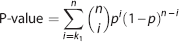
For the calculation of the P-values the probabilities were summed up:
RESULTS
Pharmacological Inhibition of the Histone Methyltransferases SUV39H1 and G9a with Chaetocin Promotes Neuronal Survival after Oxygen Glucose Deprivation
To test the hypothesis whether inhibition of the transcriptional repressors SUV39H1 and G9a induced neuroprotection in OGD, two pharmacological inhibitors were employed: chaetocin, an inhibitor of members of the SUV39 family of histone methyltransferases with inhibitory effects against SUV39H1 and G9a22,23 and second, BIX-01294, an inhibitor of G9a. 24 Primary rat cortical neuron cultures were pretreated with chaetocin or BIX-01294 24 hours before OGD. Cell death was assessed by LDH measurements 24 hours after OGD. Cell death was significantly reduced in the chaetocin-treated group compared with the vehicle-treated group in OGD (Figure 1A). In contrast, BIX-01294 pretreatment had no significant effect on neuronal viability post OGD (Figure 1B).
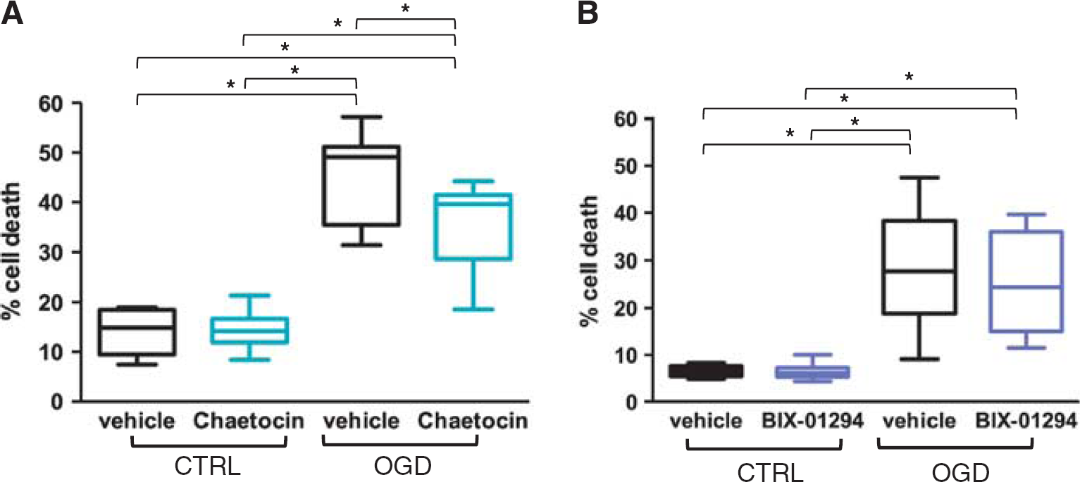
Chaetocin but not BIX-01294 protects neurons subjected to oxygen glucose deprivation (OGD). Percentage of cell death upon inhibitor/vehicle pretreatment in neurons subjected to OGD or control (CTRL) conditions as assessed by lactate dehydrogenase (LDH) measurement. A two-way analysis of variance (ANOVA) was conducted that examined the effect of OGD and treatment on cell survival followed by a Tukey
Knockdown of the Histone Methyltransferases SUV39H1 or G9a Protects Rat Cortical Neurons in Oxygen Glucose Deprivation
The miR-shRNA constructs targeting SUV39H1 or G9a were delivered to cortical neurons
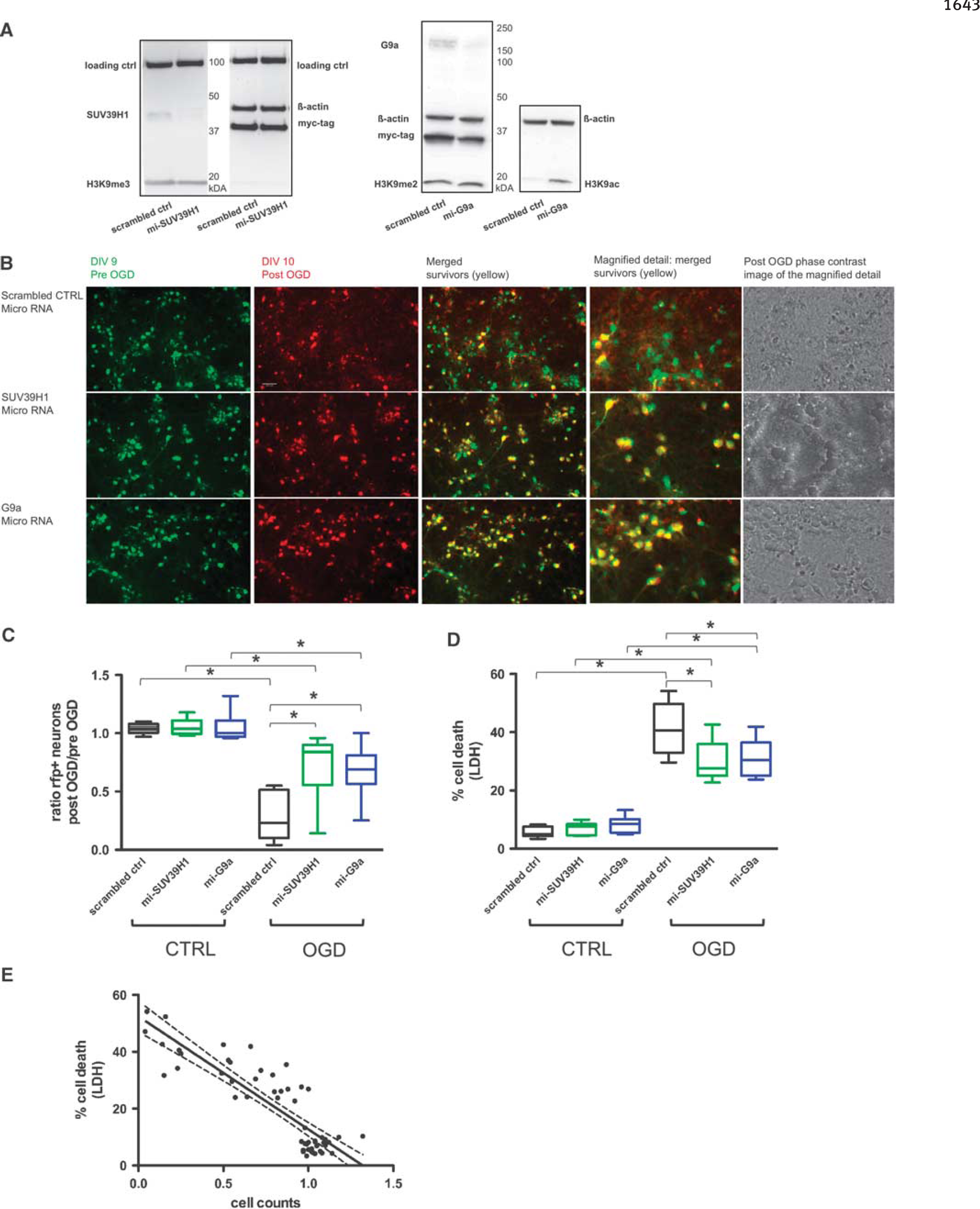
Specific miRNA-based knockdown of histone methyltransferases SUV39H1 or G9a protects neurons subjected to oxygen glucose deprivation (OGD). (
The neuroprotective effect of SUV39H1 and G9a knockdown in OGD assessed by cell counts was confirmed by LDH measurement of cell death (Figure 2D). Lactate dehydrogenase measurements and cell counts correlated significantly — as previously shown in a similar experimental setting17,25 — which corroborated the robust neuroprotective effect upon SUV39H1 and G9a knockdown (Figure 2E).
SUV39H1 and G9a Expression Is Not Induced by Desferrioxamine
As the transcription factor hypoxia inducible factor 1 (HIF-1) regulates a large number of genes in response to hypoxia, we tested, whether the mRNA expression of either SUV39H1 or G9a was induced by 48 hours treatment with the iron chelator DFO, a known activator of HIF. The histone demethylase KDM3A, shown to be HIF-1 induced in hypoxic cancer cells 26 was upregulated in primary rat cortical neurons upon DFO treatment and served as a positive control. However, neither SUV39H1 nor G9a mRNA expression was affected by DFO (Figure 3).
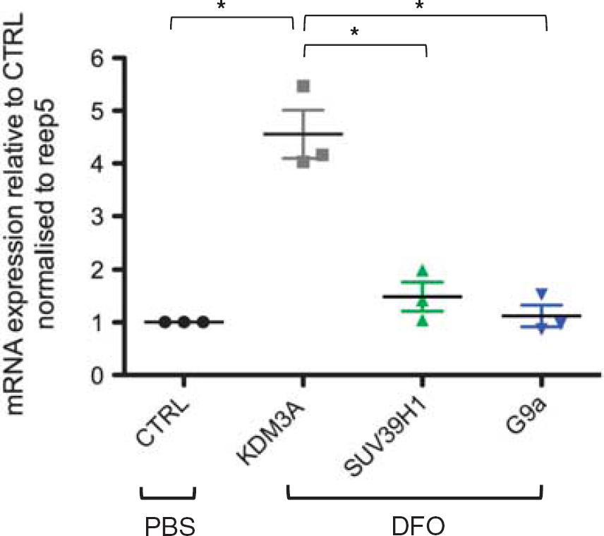
KDM3A but not SUV39H1 or G9a mRNA expression is upregulated by desferrioxamine (DFO). Expression of KDM3a, SUV39H1 and G9a mRNA after 48 hours DFO or phosphate-buffered saline (PBS) (= CTRL) treatment was determined by quantitative reverse transcription PCR (qRT-PCR) and normalized to reference gene reep5. A one-way analysis of variance (ANOVA) was used to determine mRNA expression difference upon DFO treatment followed by Tukey's multiple comparison test to isolate differences among groups
Chaetocin Treatment Increases Activating H3K9 Marks at Brain-Derived Neurotrophic Factor Promoter
To capture alterations in epigenetic profiles upon SUV39H1 and G9a inhibition by chaetocin, promoter regions of genes involved in neuroprotection and regeneration were analyzed by chromatin immunoprecipitation followed by sequencing (ChIP-seq) 1 hour after OGD. Chromatin immunoprecipitation was performed using an antibody against unmethylated, but acetylated H3K9, assuming that the inhibition of SUV39H1 and G9a would induce a shift from repressive (methylated) to activating (unmethylated) H3K9 posttranslational modifications around target genes. Brain-derived neurotrophic factor, which is known to have a crucial role in promoting postischemic survival,
27
was chosen for closer analysis. Several TSS were analyzed (
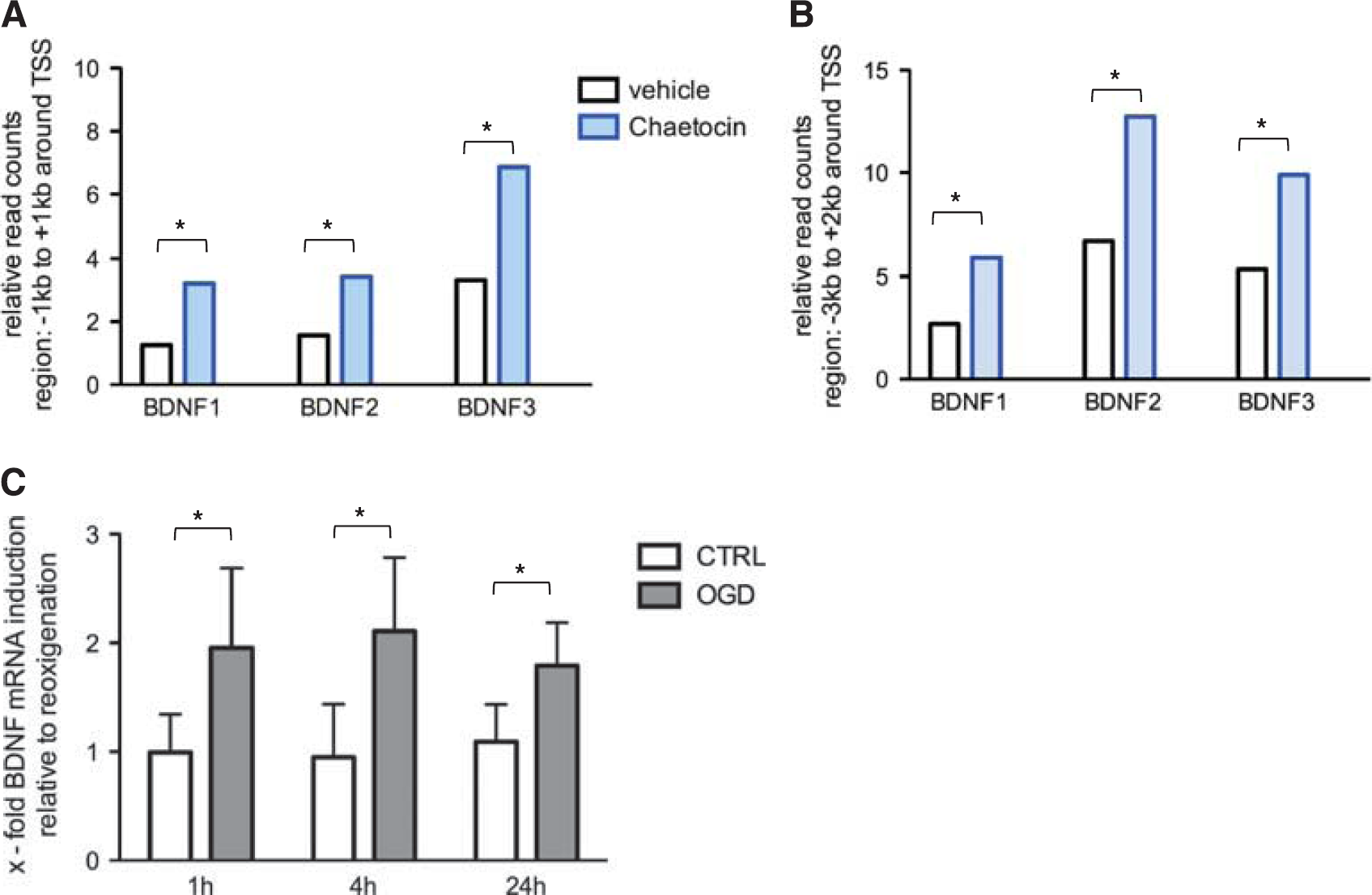
Chaetocin induces H3K9 promoter acetylation and transcription of brain-derived neurotrophic factor (BDNF) post oxygen glucose deprivation (OGD). (
Chaetocin Inhibition of SUV39H1 and G9a during Oxygen Glucose Deprivation Increases Brain-Derived Neurotrophic Factor mRNA
After SUV39H1 and G9a inhibition
Brain-Derived Neurotrophic Factor Upregulation Is Essential for Chaetocin-Induced Neuroprotection in Oxygen Glucose Deprivation
To investigate whether chaetocin exerted its neuroprotective effect by inducing BDNF, BDNF-TrkB signaling was blocked by applying K252a, a selective inhibitor of tyrosine kinases (TrK). Rat cortical neurons were pretreated with chaetocin or vehicle or chaetocin+K252a or K252a alone on DIV 8 and subjected to OGD on DIV 9 followed by cell death assessment on DIV 10. While chaetocin treatment again significantly protected neurons against OGD, the additional administration of K252a attenuated the protective effect of chaetocin. BDNF-TrkB blockade by K252a administration alone did not significantly alter cellular survival upon OGD (Figure 5).
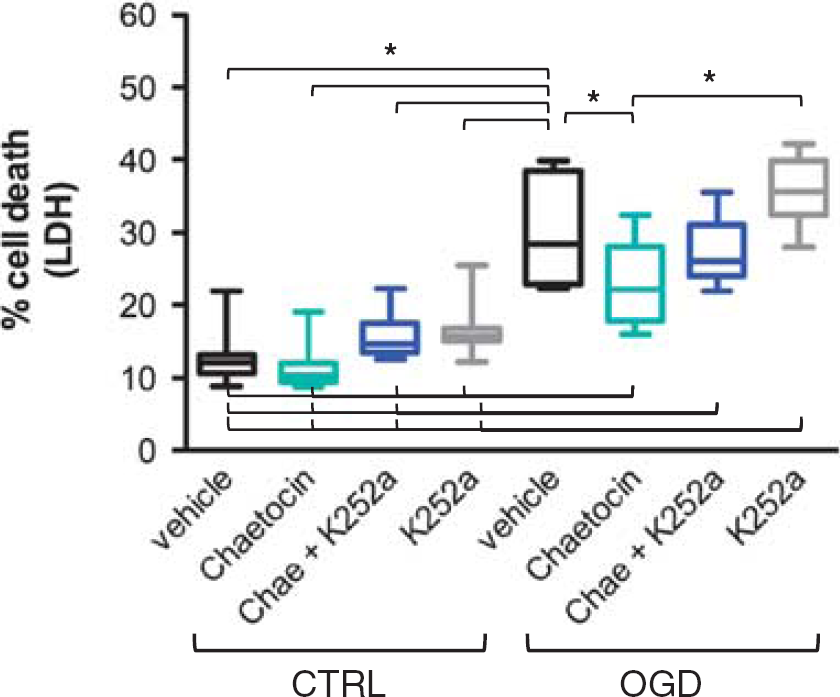
BDNF-TrκB blockade attenuates chaetocin-induced protection in oxygen glucose deprivation (OGD). Blockade of BDNF-TrkB with K252a and SUV39H1 and G9a inhibition with chaetocin. Evaluation of cell death by lactate dehydrogenase (LDH) measurement. Included are 10 independent experiments, consisting of 3 wells/condition each. A two-way analysis of variance (ANOVA) was conducted that examined the effect of OGD and treatment on cell survival. There was a significant interaction between effects of OGD and treatment, F(3,72) = 3.546 with
DISCUSSION
We identified the histone methyltransferases SUV39H1 and G9a as crucial players in OGD-induced cell damage. Inhibition of SUV39H1 and G9a, by knockdown or pharmacological inhibition, conferred significant protection to neurons subjected to OGD. SUV39H1 and G9a inhibition increased H3K9ac in promoter regions of BDNF post OGD — a sign for increased transcriptional activity — and resulted in elevated BDNF mRNA levels after experimental ischemia. Blockade of BDNF-TrkB signaling attenuated chaetocin-induced protection of rat cortical neurons subjected to OGD.
From DNA methylation and histone acetylation studies in stroke models, it is known that postischemic gene repression can be attenuated through epigenetic remodeling and that a subsequent neuroprotective effect is based on transcriptional activation of cytoprotective genes.5–7 With the current study we could for the first time demonstrate that inhibition of transcriptional repressors on the level of histone methylation can effectively promote neuronal survival in an
Recently, it was also shown in an
For actual changes in gene transcription, an orchestrated interplay between different epigenetic levels, such as histone methylation and acetylation, is necessary. 9 Both SUV39H1 and G9a act in multiple repressive complexes and interact with regulatory elements to silence transcription.12,13,29 The inhibition of these two histone methyltransferases can induce a shift from repressive to activating posttranslational histone modifications beyond H3 demethylation. Our observation that the knockdown of the transcriptional repressor G9a alone increases global H3K9 acetylation levels (Figure 2A) — a sign for transcriptional derepression — is consistent with findings of others. A reduction of global H3K9me3 or H3K9me2 levels is not visible in the immunoblots upon SUV39H1 or G9a knockdown. Other enzymes known to be involved in the establishment or eradication of H3K9 methylation such as ESET or members of the KDM4 family that are not blocked by SUV39H1 or G9a knockdown might account for this finding. Moreover, the lack of alterations of the H3K9 methylation status on the global level does not exclude changes at single gene promoters. Discrepancies between histone lysine modifications on the global level and locally at promoters of single genes have been described. For example, ischemic preconditioning induced a global decrease of activating H3K4me2 and H3K4me1 compared with controls, while H3K4me3 levels remained unchanged. Repressive global H3K9me2 levels increased by almost 60%. 30 However, subsequent analysis of histone methylation at promoters of induced neuroprotective genes showed methylation changes contrary to those assessed upon global analysis, e.g., locally increased H3K4me2 and decreased H3K9me2 levels. The authors explained this discrepancy by the fact that preconditioning led to decreased H3K4me2 and increased H3K9me2 in noncoding regions of frequent long interspersed nuclear element sequences, correlating with the globally found histone methylation pattern. 31
Histone deacetylases 1 and 2 interact with G9a in a repressor complex assembled around the REST (repressor-element-1-transcription-factor), which is induced upon experimental stroke. The REST depletion was shown to prevent epigenetic silencing and rescue neurons. 29 Thus, the neuroprotective effect by G9a knockdown in our paradigm might be mediated by inhibiting the REST complex, which links histone deacetylation to histone methylation, subsequently followed by transcriptional derepression of key cytoprotective players.
Interestingly, blocking G9a with the inhibitor BIX-01294 did not yield neuroprotection in OGD in contrast to both pharmacological blockade with chaetocin and G9a knockdown. This discrepancy might be due to the concentrations of BIX-01294 we used in our primary neuronal culture. To avoid neurotoxicity we used concentrations that might be too low for induction of neuroprotection. Initially, BIX-01294 was tested in much less sensitive cells (murine fibroblasts and stem cells) 24 and later found to display a poor separation of toxicity and functional potency. 32 A number of new G9a inhibitors are currently being designed and constantly improved. 32 Cellular application, however, still seems to be a challenge with the compound BIX-01294 as well as with more recent products. 33
The exclusive inhibitory specificity toward SUV39 family members as well as the exact mechanism of action of chaetocin are a matter of debate; its inhibitory potency against SUV39H1 and G9a, however, has been confirmed by several research groups.22,23,34,35 In the present study, chaetocin was for the first time employed in neurons. We could hence identify it as a novel neuroprotective agent, which might promote survival not only in models of cerebral ischemia but also in other models of neurodegeneration.
As observed with HDAC inhibition in experimental ischemia36,37 the neuroprotective effect of chaetocin seems to rely on the induction of important neuroprotective and regenerative players, such as BDNF. The neurotrophin BDNF has an important role in neuronal plasticity and survival. It exerts its function through binding to its receptor tyrosine kinase B (TrkB) and subsequent induction of downstream signaling cascades. In the ischemic brain, BDNF induction promotes protective and neurogenic developments. The crucial role of BDNF-TrkB signaling in neuroprotection was observed in diverse models of ischemia. In a model of glutamate excitotoxicity, BDNF-TrkB blockade partially inhibited the protective effect of lithium. 38 Neuroprotection provided by PBI-05204, a plant extract from Nerium oleander, is blocked by BDNF-TrkB inhibition with K252a in ischemic brain slices. 39 Furthermore, neurogenic effects of HDAC inhibitors were attenuated upon BDNF blockade with BDNF antibodies in neurons 40 as well as with K252a in a rodent model of ischemia: sodium butyrate-induced neurogenesis in various brain regions 41 as well as oligodendrogenesis and reduction of white-matter injury were demonstrated to depend on BDNF-TrkB signaling. 42 Here, we showed that chaetocin-induced neuroprotection in OGD was diminished by BDNF-TrkB blockade with K252a (Figure 5). This finding identifies BDNF as one crucial mediator of chaetocin-induced regeneration and protection in experimental ischemia.
CONCLUSION
Pharmacological as well as genetic inhibition of the histone methyltransferases SUV39H1 and G9a significantly promoted neuronal survival post OGD. The protective effect was based on a shift in promoter signatures toward transcriptional activity and mediated by upregulation of protective genes such as BDNF.
Footnotes
SS contributed to experimental design, data acquisition and analysis, writing article and approval of current version; CH contributed to RNA interference experiments: miRNA design, data acquisition and analysis, revising article and approval of current version; HL contributed to BDNF experiments, PCR data acquisition and analysis, revising article and approval of current version; JF contributed to experimental design of miRNAs, revising article and approval of current version; JH contributed to ChIP-data acquisition and analysis, revising article and approval of current version; FY and AM contributed to study design, revising article and approval of current version. SM contributed to experimental design and analysis, revising article and approval of current version.
The authors declare no conflict of interest.