[11C]befloxatone is a positron emission tomography radioligand to image monoamine oxidase A (MAO-A) in the brain, which has been used in preclinical studies and in clinical protocols. However, a recent study found that [11C]befloxatone binding potential (k3/k4) has a poor correlation with MAO-A protein levels measured in the human brain. We here show that this poor correlation only depends on the choice of the parameter when performing kinetic modeling. In particular, the total volume of distribution of [11C]befloxatone shows a tight correlation with both protein and mRNA levels of MAO-A in the human brain.
In a recent work, Tong et al reported the in vitro region-wise distribution of protein levels of monoamine oxidase A (MAO-A) in the human brain.1 These results have important implications for brain imaging studies. Using data from published positron emission tomography (PET) studies, they found good correlations between in vitro MAO-A levels and PET measures of MAO-A density/activity using [11C]harmine (r=0.86, P = 0.003) and [11C]clorgyline (r=0.82, P = 0.007), but not with [11C]befloxatone (r=0.61, P = 0.15), essentially because of the low [11C]befloxatone binding potential reported in the thalamus.2 Thus, the authors suggested caution in interpretation of the data obtained with this radioligand. Two hypotheses can be put forward to explain this poor correlation: (1) the in vivo kinetics of [11C]befloxatone poorly reflect the actual MAO-A distribution, and therefore [11C]befloxatone should be abandoned; (2) the selected quantitative parameter, (k3/k4), is not the optimal one, and thus a different outcome parameter should be chosen.
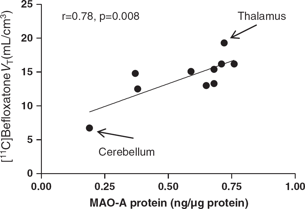
The full kinetic modeling of [11C]befloxatone has been recently published.3 This study showed that a two-tissue compartment model can reliably quantify total distribution volume (VT) values. In agreement with in vitro data, the lowest [11C]befloxatone VT values were found in the cerebellum (6.7 ± 0.9 ml/cm3), while the highest were in the thalamus (19.3 ± 1.4 ml/cm3).3 Importantly, [11C]befloxatone VT values are well correlated with the in vitro MAO-A protein levels published by Tong et al.,1 to a degree similar to that found for the other MAO-A radioligands (r = 0.78, P = 0.008) (Figure 1). Notably, the thalamus/cerebellum VT ratio is slightly higher for [11C]befloxatone (2.9), than for [11C]harmine VS (2.1) and [11C]clorgyline Λλk3 (2.4), thus indicating a slightly better, although still imperfect, quantitative proportionality with in vitro data (3.9).1 Furthermore, [11C]befloxatone compartmental VT values are well correlated not only with protein levels, but also with mRNA expression of MAO-A,4 which suggests that MAO-A undergo little in vivo post-transcriptional modifications.
One possible reason why (k3/k4) showed a poor correlation with protein levels might be due to the very slow wash-out from the brain of [11C]befloxatone,3 which may introduce errors in the estimation of microparameters, especially k4. In addition, a K1/k2 constrained model presupposes a constant non-displaceable compartment across the brain. If this condition is not verified, a regional quantification error may ensue.
Correlation between [11C]befloxatone VT values and in vitro MAO-A protein levels. Please compare this figure to Figure 6 in the paper by Tong and colleagues1.
Finally, in our kinetic modeling paper, we also showed that spectral analysis provided the most reliable quantification of [11C]befloxatone at the voxel level.3 However, the correlation between protein levels and voxel-wise spectral analysis VT values is weaker than with compartmental modeling (r = 0.67, P = 0.033). Therefore, compartmental modeling at the regional level should be the method of choice to quantify [11C]befloxatone binding.
In summary, [11C]befloxatone can reliably quantify MAO-A concentrations in human PET studies, and its preferred outcome measure should be VT estimated by compartmental modeling.
Footnotes
The authors declare no conflict of interest.
ACKNOWLEDGMENTS
The authors would like to thank Drs Joanna Fowler and Junchao Tong for useful discussions and suggestions.
References
1.
TongJMeyerJHFurukawaYBoileauIChangLJWilsonAA. Distribution of monoamine oxidase proteins in human brain: implications for brain imaging studies. J Cereb Blood Flow Metab (2013); 33(6):863–871
2.
LeroyCBragulatVBerlinIGregoireMCBottlaender MRoumenovD. Cerebral monoamine oxidase A inhibition in tobacco smokers confirmed with PET and [11C]befloxatone. J Clin Psychopharmacol (2009); 29(1):86–88
3.
Zanotti-FregonaraPLeroyCRoumenovDTrichardCMartinotJLBottlaenderM. Kinetic analysis of [11C]befloxatone in the human brain, a selective radioligand to image monoamine oxidase A. EJNMMI Res (2013); 3(1):78
4.
Zanotti FregonaraPLeroyCRizzoGRoumenovDTrichard CMartinotJL. Imaging of monoamine-oxidase A in the human brain with [11C]befloxatone: quantification strategies and correlation with mRNA transcription maps. Nucl. Med. Commun. (2014) In press