23. Use of [11C]-(+)-A-dihydrotetrabenazine (DTBZ) to monitor disease progression in a transgenic mouse model of Parkinson's disease
I. Guenther1, D. Rubins1, K. Schlingmann1, X. Meng1, S.E. Brown2, S. Sanabria1 and J.J. Cook1
1Imaging Department; 2Parkinson's Disease Department, Merck & Co, West Point, Pennsylvania, USA
Objectives: The aim of this study was to evaluate the use of the vesicular monoamine transporter 2 (VMAT2) ligand (+)-[11C]dihydrotetrabenazine (DTBZ) as a potential biomarker for the in vivo quantification of disease progression in the transgenic ‘MitoPark’ (MP) mouse model of Parkinson disease (PD). MP mice express a conditional knockout of mitochondrial transcription factor A (Tfam) targeted to dopaminergic neurons. This results in a parkinsonism phenotype, with adult onset of slowly progressing impairment in motor function accompanied by loss of dopaminergic neurons.1
Methods: A series of PET studies was performed in 8 MP mice and 8 age-matched littermate wild type (WT) controls (age 6 to 23 weeks). Animals were anesthetized with isofluorane (1% to 2%) and were administered ∼0.2 mCi [11C]DTBZ via tail vein. Dynamic PET images were acquired for 30 min in a microPET Focus 220. PET images were reconstructed using a 3-D maximum a posteriori reconstruction (MAP) algorithm. Reconstructed images were aligned with a standard mouse brain regional template and time-activity curves were obtained in the striatum and cerebellum. From the resulting image data, [11C]DTBZ binding potential (BP) in striatum was calculated using the Logan Plot with cerebellum as reference region.
Results: MitoPark mice show progressive bradykinesia, akinesia, and tremor phenotypes reminiscent of PD, before dying prematurely at 26 to 33 weeks of age.2 A full characterization of this strain is currently underway by the Merck PD team. Figure 1 summarizes measured BP of all [11C]DTBZ scans (21 scans on WT and 20 scans on PD mice with 1 to 5 scans per animal). MP mice exhibit a significant reduction in [11C]DTBZ BP compared to WT mice at 13 weeks of age. BP in WT slightly increased with age, while longitudinal imaging of MP mice showed a progressive reduction of BP with increased age. These results demonstrate that the degeneration of the striatal dopaminergic neurons in this transgenic animal model of PD can be followed in vivo by imaging the progressive loss of VMAT2 using the PET tracer [11C]DTBZ. This is consistent with findings previously reported for other animal models of PD such as the 6-OHDA lesioned rat and the MPTP lesioned monkey3,4 and in PD patients.5
Conclusions:In vivo imaging using the PET tracer [11C]DTBZ shows a progressive loss of striatal VMAT2 in the MitoPark mouse model of PD. This parallels the progressive degeneration of the striatal dopaminergic system in this model.1 Thus, VMAT2 imaging in this model may be useful for evaluating potential neuroprotective therapeutics for PD.
49. What to do with all that data?!
L. Martens
EMBL-EBI, Hinxton, Cambridge, UK
Novel technologies in different OMICS fields are generating vast amounts of data when applied to samples such as brain tissue biopsies, autopsies, or CSF fluid. Once the analysis is complete, the difficult task of interpreting the output data begins. Here we aim to provide some pointers into possible analyses that can be performed based on a large amount of across-experiments proteomics data from central nervous system tissues, as well as some analyses we have done for human plasma and serum. Taken together, these tools indicate that large amounts of data can be a blessing, and that they can enable powerful analyses that can inform the use of technology in analysing a specific sample, as well as the understanding of the underlying biology.
111. Machanisms associated with Alzheimer's disease in ischemic hippocampus degeneration
M. Ułamek1, M. Jabłoński2 and R. Pluta1
1Neurodegenerative Disorders, Mossakowski Medical Research Centre, Polish Academy of Sciences, Warsaw; 2Orthopedic and Rehabilitation, Skubiszewski Medical University of Lublin, Lublin, Poland
Objectives: For several reasons the favored brain sector for the investigation of ischemic degeneration is the hippocampus. First of all the hippocampus is the region which displays the characteristic neuropathology in ischemic and Alzheimer's brain. Second the hippocampus is implicated in spatial learning and memory. Third the hippocampus especially its sector CA1 is one of the brain areas very sensitive to ischemic and Alzheimer's injury. Finally the relationship of etiology and between ischemic injury dementia and Alzheimer's disease type dementia are recently much debated. Moreover the characterization of the neuronal cells death in ischemic hippocampus is crucial for the development of new effective therapies for both diseases. Additionally brain ischemia is well known for its ability to influence the function of the blood-brain barrier. We assessed blood-brain barrier permeability for horseradish peroxidase and different parts of amyloid precursor protein in animal hippocampus exposed to global brain ischemia.
Methods: Using rats (n = 10) blood-brain barrier and amyloid precursor protein changes were studied after 10 mins brain ischemia1 with 6 months survival. As controls sham-operated animals (n = 6) were sacrificed in due time. Rats were perfusion fixed for these investigations. Five brains were cut at 60 μm slices in the coronal plane by a vibratome for horseradish peroxidase staining.2 Next after putting slices on microscope slides they were investigated. Paraffin sections from other 5 brains were selected for amyloid precursor protein staining3 and structural studies. Control brains went through the same procedures as ischemic.
Results: The blood-brain barrier leakage especially in the medial part of the hippocampus was associated with increased staining of C-terminal of amyloid precursor protein/β-amyloid peptide in perivascular space. At 6 months after ischemia in CA1 area complete pyramidal neurons loss was noted in 93% of brains. At 7% of brains we observed incomplete neuronal loss with degenerating neurons in a trace of the hippocampal CA1 pyramidal cell layer. Additionally acute and chronic neuronal degeneration appeared in CA2, CA3 and CA4 areas and dentate gyrus that were not involved in early pathology. Atrophy of the hippocampus was found after ischemia. Generally atrophic hippocampus is indicative of an active progressing neurodegenerative processes.
Conclusions: These data indicate that late neurotoxic C-terminal of amyloid precursor protein and β-amyloid peptide accumulation around blood-brain barrier vessels in hippocampus postischemia may represent a secondary injuring process that could exacerbate stroke outcome by additional delayed neuronal death with development dementia.4 Prolongation of survival time results in neuronal changes within hippocampal areas of non-selective vulnerability. These data also suggest that gradual accumulation of above proteins and the amount accumulation correlate very well with extent of pathology in hippocampus and survival time after ischemia.4 It is looking that amyloid precursor protein products control neurodegeneration in ischemic hippocampus.
197. Proton (1H) magnetic resonance spectroscopy of autosomal recessive cerebellar ataxias
I. Iltis1, D. Hutter1, K. Bushara2, C. Gomez3 and G. Öz1
1Center for Magnetic Resonance Research; 2University of Minnesota, Minneapolis, MN; 3University of Chicago, Chicago, Illinois, USA
Objective: Friedreich's ataxia (FRDA) and ataxia with oculomotor apraxia type 2 (AOA2), the most frequent forms of autosomal recessive ataxias,1,2 exemplify the two main mechanisms that underlie these conditions.3 Therefore it is of interest to assess the metabolic and cellular alterations in these two diseases that represent different molecular pathways to neurodegeneration. Here, we investigated the neurochemical alterations in the brains of patients with FRDA and AOA2 by high field 1H MRS.
Methods: Two patient populations and their respective age-matched control groups participated in this study: eight patients with FRDA (5 F/3 M, average age ±s.d., 27±9 years) and their controls (10 F/10 M, 26±5 years), 8 patients with AOA2 (4 F/4 M, 38±9 years) and their controls (7 F/12 M, 36±11 years). Neurochemical profiles of cerebellar hemispheres (volume, 4.9 mL) and vermis (6.3 mL) were measured at 4T using previously described data acquisition and analysis methods.5 Metabolite concentrations were corrected for the amount of cerebrospinal fluid (CSF) in each voxel.
Results: In the vermis, both patient groups exhibited a higher concentration of myo-inositol (AOA2 +44%, P<0.0001, FRDA +12%, P = 0.023) and a higher glutamine/glutamate ratio (AOA2 +35%, P = 0.001, FRDA +31%, P = 0.002) (Figure). Disease-specific alterations were also identified: N-acetylaspartate (NAA) was significantly lower in the vermis in FRDA, while choline, creatine and glucose concentrations were increased in AOA2. In the cerebellar hemispheres, NAA concentrations were significantly lower in both patient groups, and glutamine/glutamate ratio was higher only in patients with AOA2. Significant atrophy was observed in the vermis of both patient groups, characterized by a higher CSF content (AOA2 versus controls, 52%±13% versus 11%±4%; FRDA versus controls, 18%±5% versus 11%±3%).
Quantification of metabolites.
Conclusion: High level of myo-inositol is indicative of fibrous gliosis, described in post-mortem studies.6 Neuroaxonal loss is likely to be responsible for the lower NAA level observed in these patients, while an altered glutamine/glutamate ratio possibly indicates an alteration in neurotransmission. Disease- and region-specific alterations in the metabolic profile of the vermis and cerebellar hemispheres were identified in two populations of patients with cerebellar ataxias using 1H MRS at 4T.
Supported by Kory and Scott Tabor Ataxia Research Fund, NIH R21 NS056172, P41 RR08079, P30 NS057091, M01RR00400 and MN Medical Foundation.
260. Using PET to study the effects of deep brain stimulation of the subthalamic nucleus in the treatment of Parkinsons disease
J. Sidtis1,2, M. Tagliati3, D. Sidtis1,4, V. Dhawan5 and D. Eidelberg5
1Geriatrics, Nathan Kline Institute, Orangeburg; 2Psychiatry, New York University Medical School; 3Neurology, Mount Sinai School of Medicine; 4Speech Language Pathology and Audiology, New York University, New York; 5Center for Neuroscience, Feinstein Institute for Medical Research, Manhasset, New York, USA
Background and aims: Bilateral high-frequency deep brain stimulation of the subthalamic nucleus (STN-DBS) has become a significant approach to the treatment of Parkinsons disease (PD). However, the mechanism of action is still not well understood.
Methods: Seven subjects with bilateral STN-DBS underwent two sets of 12 H20 PET scans on separate days, once with the stimulators on at therapeutic settings, once with the stimulators off. For each session, there were 2 replications of a rest state and 5 speech tasks. In addition to the 12 scans, composite images were created from the initial 6 (first half) and the subsequent 6 scans (second half). Regions of interest encompassing each slice were applied to 59 slices. CBF was measured at 3 thresholds: maximum CBF, mean CBF of voxels at the upper 10% of activity, and mean CBF of all voxels (100%). Total CBF was calculated at 10% (mean CBF*10% voxels) and at 100% (mean CBF*100% voxels).
Purpose: The determination of global effects is critical to characterizing regional changes associated with specific tasks and specific neurological disorders. This study examined global effects of bilateral STN-DBS on CBF in subjects with ideopathic PD. This research is part of a larger study of the effects of PD and STN-DBS on regional CBF and speech.
Results: The effects of STN-DBS and time (first versus second half) were evaluated using nonparametric statistics to avoid assumptions about underlying distributions. In the ON condition, there were no significant effects of time. However, in the OFF condition, there were significant decreases from the first to second half of the scanning session for CBF max, and for total flow at 10% but not 100% (Wilcoxon Signed Ranks Test, P<0.03). There were no significant ON-OFF effects for the first half of the study, but in the second half of the study OFF was significantly lower than ON for each measure (Wilcoxon Signed Ranks Test, P<0.03). The time course of these declines was further examined by ordering the scans by their sequence during the scan session rather than task. As expected, there was no significant effect of sequence in the ON condition. In the OFF condition, there were significant declines for both CBF max (Chi-square = 23.64; P = 0.014) and CBF 10% (Chi-square = 26.03; P = 0.006).
Conclusions: CBF was stable when STN-DBS was ON, but CBF decreased over time in the OFF condition. The effects of STN-DBS have been considered in terms of specific changes in the basal ganglia, and on local field effects. These results demonstrate a third mechanism of action in STN-DBS: a global effect consisting of higher peak CBF. The changes in CBF associated with STN-DBS may reflect generalized increases in cortical excitability. This mechanism may play a role in therapeutic response, undesired side effects, or both. PET should play a significant role in understanding these effects (Supported by R01 DC007658).
351. Multimodal imaging of the hippocampus in early Alzheimer's disease
I. Yakushev1, A. Gerhard2, M. Lorscheider1, I. Schermuly1, H.-G. Buchholz3, J. Albrecht1, A. Hammers4, M. Schreckenberger3 and A. Fellgiebel1
1Department of Psychiatry and Psychotherapy, University of Mainz, Mainz, Germany; 2Wolfson Molecular Imaging Centre, The University of Manchester, Manchester, UK; 3Department of Nuclear Medicine, University of Mainz, Mainz, Germany; 4Division of Neurosciences and Mental Health, MRC Clinical Sciences Centre, Imperial College, London, UK
Objectives: Structural and functional disturbances of the hippocampus in early Alzheimer's disease (AD) have been consistently detected using various imaging modalities. Recently, functional magnetic resonance imaging (MRI) and neuroanatomical studies have provided evidence for a functional differentiation along the longitudinal axis of the hippocampus with the hippocampal head playing a crucial role in the formation of memories. Thus focussing on the hippocampal subdivisions, the objective of the present study was to investigate associations between different structural and functional imaging markers of hippocampal integrity and memory performance in early AD.
Methods: Global and regional hippocampal volumes, diffusivity, and glucose metabolism were quantified in 20 patients with early AD (MMSE 24.5±2.9, mean±SD) using structural MRI, diffusion-tensor imaging (DTI), and positron emission tomography with 18fluorodeoxyglucose (FDG-PET). The study group included 11 subjects with mild Alzheimer's dementia and 9 subjects with amnestic mild cognitive impairment (MCI), who had an ‘Alzheimer-typical’ FDG-PET finding. Such individuals were repeatedly shown to deteriorate to Alzheimer's dementia within 1 to 2 years.1,2 Regions-of-interest (ROIs) were placed manually onto the hippocampal head (HH), body (HB), and the whole hippocampus bilaterally. PET images were carefully corrected for partial volume effects taking into account both spill-in and spill-out effects. The measures from the different modalities were correlated for each ROI followed by correlations between the ROI values and scores on memory performance tests controlling for age. Finally, stepwise regression analyses with memory tests as dependent and ROI measures as independent variables were performed.
Results: In the whole group, the verbal delayed recall score correlated with mean diffusivity (MD) of the left HH (r = −0.64, P = 0.003) and with the volume of the left HB (r = −0.61, P = 0.006). Neither correlation retained the statistical significance when the analyses were restricted to the subgroup of MCI subjects. In the regression analyses, MD of the left HH was the only predictor of memory performance explaining 30.6% of the variance (model P = 0.007). Glucose metabolism correlated with MD in the left HH only (r = −0.48, P = 0.038). When the analyses were restricted to the subgroup of MCI subjects, the correlation appeared highly significant (r = −0.90, P = 0.003).
Conclusions: The main finding of our study is the inverse correlation between performance on the verbal delayed recall test and diffusivity of the left HH. While the memory performance was also associated with the left HB volume, among the measures of three imaging modalities diffusivity of the left HH was the only predictor of verbal memory impairment in early AD. Furthermore, the correlation of the left HH diffusivity and glucose metabolism, which was especially pronounced in the subgroup of MCI subjects, indicates a close relationship between PET and DTI measures as early imaging markers of non-specific tissue damage. The lack of the association between hippocampal metabolism and memory performance is somewhat surprising, but is consistent with a recent report.3 We continue to work on the present study, introducing new variables and increasing the sample size.
494. The role of molecular interactions in the pathogenesis of Alzheimer's disease: could nanotechnology put a stop to it?
A. Nazem1 and G.A. Mansoori2
1Mashhad University of Medical Sciences, Mashhad, Iran; 2Chemical and Bio Engineering & Physics, University of Illinois at Chicago, Chicago, Illinois, USA
Background and aims: The current medical approach toward Alzheimer's disease (AD) is to lower the symptoms associated with it. However, the need for therapeutic agents to halt the irreversible progression of the disease process is completely palpable through scientific community. Designing such therapeutic agents is not attainable without a complete knowledge of the underlying molecular mechanisms and the chemical structures of the involved molecules in the pathologies under survey.
Nanosciences and technology are a new area of scientific revolution, promising drastic breakthroughs in medical diagnostic and therapeutic approaches. The potentials of this new field, however, are not clear for most of clinical scientists. In this project we aim to show these potentials for Alzheimeŕs disease treatment on a strong foundation of AD molecular mechanisms.
Methods: We first reviewed the molecular mechanisms involved in the pathogenesis of AD. For this purpose we include original research articles published between years 2000 and 2008, from journals with impact factor greater than 3. We then classify the various steps into three major categories: predisposing processes, pathogenetic processes and protecting mechanisms. Afterwards we scrutinize the potential of nanotechnology building blocks and complex systems in ceasing the pathogenetic processes.
Purpose: Our fiest objective in this article was to reach a unifying picture of the various molecular mechanisms involved in AD pathogenesis. Based on these mechanisms, we present the applications of nanotechnology building blocks and complex nano-systems that are potentially capable of stopping the AD pathology.
Results: Some nanotechnology building blocks, nanoparticles and complex nanosystems, are suggesting shortcuts to the treatment of AD. These nanoscience based systems are biologically functional. Designing these nanoscale agents have been recently provided by nanotechnology.
Conclusions: Through nanotechnology it would be possible to design targeted drug delivery systems, against different culprit molecules in AD pathogenesis. However, further research is needed to examine the biological compatibility of nanotechnology products.
631. Cortical amyloid load and glucose metabolism in Alzheimer's disease and mild cognitive impairment with [11C]PIB- and [18F]FDG-PET imaging
S. Hatashita and H. Yamasaki
Shonan Atsugi Hospital & Clinic, Atsugi, Japan
Objective: The aim is to investigate the deposition of amyloid plaques by carbon 11-labeled Pittsburgh Compound B ([11C]-PIB) in the brains with dementia (Alzheimer's type, AD) and mild cognitive impairment (MCI). We clarify the relationship between cerebral amyloid load and glucose metabolism.
Method: Forty-three patients with AD, 49 with MCI and 80 healthy control (HC) were studied. All 172 patients underwent 60-mins dynamic [11C]-PIB PET and 15-mins static [18F]-FDG PET. [11C]-PIB data was acquired from 35 to 60 mins after injection. Regions of interest (ROI) were defined on co-registered MRI and used in the analysis of the PET data. Distribution volume ratios (DVR) of PIB retention were determined using Logan graphical analysis (cerebellar gray as reference region). [18F]-FDG PET images were extracted using 3 dimensional stereotactic surface projections (3D-SSP) by a Z-score on a pixel-by-pixel basis. Quantitative analysis for [18F]-FDG used the standardized uptake value ratio (SUVR) values of cortical regions.
Result: All of 43 AD patients (MMSE: 19.6±4.6, CDR: 0.87±0.57) had a robust increase of PIB binding in the anterior and posterior cingulate, precuneus, frontal, parietal, lateral temporal cortical areas (typical PIB AD-pattern). The mean value of DVR in whole cortical areas was the highest among all groups (2.43±0.56, P<0.01). On FDG PET 3D-SSP images, twenty (46%) of 43 AD patients with PIB AD pattern showed a significant reduction of cortical glucose metabolism in temporo-parietal, frontal, and precuneus cortices (classic metabolic AD-pattern). The mean value of FDG SUVR in cortical areas was the smallest (0.91±0.05, P<0.01). However, increased PIB bindings did not correlate to comparable metabolic decreases in any of cortical areas. Of the 49 MCI patients (MMSE: 27.3±1.8, CDR: 0.5), twenty-seven showed AD like patterns of amyloid deposition similar to AD (DVR: 2.16±0.30, P<0.01) whereas the remaining 22 patients had no PIB retention in cortical areas. Three (11%) of 27 MCI PIB positive patients had the hypometabolic AD-pattern and five (18%) showed hypometabolism only in posteior cingulate gyrus and precuneus on FDG images. The values of DVR in cortical areas were not correlated with FDG SUVR. In the precuneus, however, increased PIB retention was significantly related to the decreased glucose metabolism in MCI group, but not in the AD group (r = −0.23, P<0.05). Seven of 84 HC had the PIB retention in cortical areas (DVR: 1.92±0.27, P<0.01), but did not show the hypometabolic AD-pattern or hypometabolism in the precuneus.
Conclusion: The [11C]-PIB PET determines cortical amyloid load at different stage of AD, but the pattern of cortical amyloid deposition is not identical to hypometabolic pattern that seen in the [11C]-PIB PET. This amyloid plaque formation is not directly responsible for cerebral glucose metabolism in cortical regions.
761. Mapping progressive losses in [18F]FPCIT binding in early stage Parkinson's disease: a longitudinal PET study
Y. Ma, S. Peng, P. Spetsieris, V. Dhawan and D. Eidelberg
Center for Neurosciences, The Feinstein Institute for Medical Research, Manhasset, New York, USA
Objectives: PET imaging with radioligand [18F]FPCIT provides a valuable means for quantifying regional abnormality and clinical correlate in dopamine transporter (DAT) binding associated with Parkinson's disease (PD). We have previously shown bilateral losses of DAT binding in the posterior putamen in hemi-PD subjects. In this study, we sought to delineate the topographic distribution of striatal DAT binding reductions in PD patients and its longitudinal change during early phases of the disease progression.
Methods: We performed dynamic [18F]FPCIT PET studies over 100 mins in eight parkinsonian patients (age = 59.2±8.0 yr; Hoehn & Yahr stages 1 to 2.5; UPDRS = 8.5±5.3) at baseline and at approximately four years. Ten age-matched normal subjects (age = 62±10.4 yr) served as controls. Volumetric maps of [18F]FPCIT binding were calculated on a voxel-by-voxel basis at 95 mins post-injection and spatially normalized based on corresponding mean images generated over early frames. Statistical parametric mapping (SPM) was then used to localize binding reductions in PD relative to controls as well as to detect interval changes between the two time points in PD patients. The analysis was restricted within a striatal brain mask due to the existence of a prior hypothesis. Local changes in FPCIT binding was assessed post-hoc over spherical volumes (8 mm in diameter) centered at the peak coordinates of interest.
Results: Relative to the normal mean, [18F]FPCIT binding decreased significantly in the bilateral putamen of the PD group at baseline (contralateral: −26 −8 4, Zmax = 6.36, cluster = 5.5 mL; ipsilateral: 34 −16 2, Zmax = 5.33, cluster = 5.1 mL; P<0.001; Figure 1A). The decrease was larger in extent at the follow-up time point (contralateral: −28 −8 4, Zmax = 6.70, cluster = 7.1 mL; ipsilateral: 32 −10 4, Zmax = 5.67, cluster = 5.9 mL; P<0.001). The cluster in the ipsilateral putamen was smaller in size than that in the contralateral side, but localized slightly more posterior. A significant reduction was also seen between the two time points only at a lower threshold of P<0.01, with more DAT binding loss in the ipsilateral putamen. Progressive losses of [18F]FPCIT binding were evident in the post-hoc analysis (e.g. Figure 1B). By contrast, there were no increases in DAT binding within the striatum of PD patients.
SPM maps (A) and post-hoc VOI plot (B).
Conclusions: Brain mapping analysis of [18F]FPCIT PET data is capable of quantifying and mapping anatomically specific abnormalities in straital DAT activity in patients with PD. Our data seem to suggest different rates of decline in striatal DAT binding in the contralateral and ipsilateral putamen. This method offers a useful tool to track the onset and progression of PD at its early stages.
773. A progressive increase in brain microhemorrhages correlates with sporadic late-onset dementia development
W.M. Kirsch1, M. Schrag1, G. Mcauley1, J.P. Larsen2, S. Peterson2, W. Britt III3, F. Petersen4, C. Dickson1, D. Kido5, M. Ayaz6, E.M. Haacke6, W.J. Pearce7, L. Liotta8 and H.V. Vinters9
1Neurosurgery Center for Research, Training, and Education; 2Department of Medicine, Geriatric Medicine; 3Department of Psychiatry; 4Health Research Consulting Group; 5Department of Radiology, Loma Linda University, Loma Linda, California; 6MRI Institute for Biomedical Research, Detroit, Michigan; 7Center for Perinatal Biology, Division of Physiology, Loma Linda University, Loma Linda, California; 8Department of Molecular and Microbiology, George Mason University, Manassas, Virginia; 9Division of Neuropathology, UCLA Medical Center, Los Angeles, California, USA
Objectives: Our goal is to define the pathogenesis of sporadic late-onset dementias studying brain microvasculature with new minimally invasive technologies.
Methods: For the past 5.5 years we have monitored the cognitive course of 76 mildly cognitively impaired (MCI) and 28 cognitively normal elderly individuals applying new MR contrast (Susceptibility Weighted Imaging, SWI) and proteomic tools (‘carrier protein stripping’). The protein discovery tools are novel and are yielding previously unknown blood proteins and peptides considered to be of very low abundance. Low molecular weight fragments in the range of 40 kDa or less are being detected. Validation of these biomarkers has been conducted using antibodies raised against the candidates. The SWI sequence at 1.5T is at least fourfold more sensitive than conventional 1.5T GE-T2* for the detection of brain microbleeds (BMB). SWI is a 3-dimensional velocity compensated gradient echo sequence combining magnitude information with phase information that enhances contrast of magnetic field inhomogeneities combing phase and magnitude information permits shorter echo times, improves signal to noise ratio and detection of subvoxel susceptibility scores.
Results: SWI at 1.5T revealed an unanticipated association of cognitive loss with increasing BMB typical for ‘cerebral amyloid angiopathy (CAA).’ SWI is superior to conventional gradient echo T2* (GE-T2*) for BMB detection. Nine of 26 MCI participants followed to dementia have significant MH and unique serum proteins, peptides from the heme degradation pathway. Our human experiment is the first prospective evidence for CAA microvasculopathy in the pathogenesis of late onset dementia.
Conclusions: As a result of the association between cerebral MH and progression to dementia, 3 key endpoints have been added to our study:
quantifying the location of MH by 3T SWI during the course of dementia,
proteomic studies of peripheral blood to complement SWI BMB detection to develop a clinical test for CAA and
determine the role of CAA hypoxic and apoptotic mechanisms responsible for neuronal and cognitive loss in an appropriate transgenic mouse model.
Defining the role of microvasculopathies in the pathogenesis of sporadic late-onset dementia may result in new therapeutic strategies.
This research was funded by NIH grant AG20948.
785. Whole-brain PET study of Parkinson's patients reveals a complex pattern of rCBF changes associated with deep brain stimulation
A. Tabesh1, B. Ardekani1,2, M. Tagliati3, V. Dhawan4, D. Eidelberg4 and J. Sidtis1,2
1The Nathan S. Kline Institute for Psychiatric Research, Orangeburg; 2Department of Psychiatry, New York University School of Medicine; 3Department of Neurology, Mount Sinai School of Medicine, New York; 4Center for Neuroscience, Feinstein Institute for Medical Research, Manhasset, New York, USA
Objective: To investigate changes in the whole-brain multivariate pattern of regional cerebral blood flow (rCBF) quantified using positron emission tomography (PET) in Parkinson's patients with deep brain stimulation (DBS) on (ON) and off (OFF) during rest and speech motor tasks.
Methods: Six subjects with Parkinson's disease and bilateral DBS implants underwent PET scans in two sessions. In each session, PET images were acquired at rest (2 images) and while performing five speech motor tasks (10 images), with both the ON and OFF conditions. A total of 142 (72 ON and 70 OFF) images were obtained (two missing OFF images). All images were spatially normalized to a template using SPM1 and their non-brain voxels were masked out. The remaining voxels (2 × 2 × 2 mm3) were used for multivariate classification using the linear Gaussian classifier (LGC).2 Classification accuracy was estimated using the leave-one-out scheme.2
Results: The classification accuracy of the LGC was 96.5% (sensitivity = 97.2%; specificity = 95.7%). Figure 1 shows the discriminant pattern of the LGC superimposed on the SPM PET template in neurological convention. Coefficients of the discriminant function with the largest 5% absolute values are shown. Blue and red scales indicate negative and positive coefficients, respectively. Slices are in the inferior to superior order and are 10 mm apart. Regions with increased rCBF due to DBS (in red) were thalamus, globus pallidus, caudate, and putamen. Regions with decreased rCBF were supplementary motor area and cerebellum. Increased and decreased rCBF were both observed in different regions of the frontal and temporal lobes.
Conclusions: Using a whole-brain multivariate classification approach, we showed that PET images corresponding to DBS OFF and ON conditions can be reliably differentiated. The high accuracy of classification confirms the reproducibility of the observed differences in the sense that they accurately predict the OFF or ON status of a previously unseen PET image. The whole-brain approach enables analysis without the need for specific a priori hypotheses regarding regions of interest. Previous PET studies had applied voxel-wise analysis techniques to investigate rCBF differences between the OFF and ON conditions.3,4 The multivariate classification approach offers the additional benefit of accounting for potential correlations between rCBF in different brain regions overlooked by voxel-wise techniques. The multivariate approach also allows for assigning a significance level to each brain region analogous to p-values in voxel-wise analysis.5
Acknowledgment: Supported by R01 DC007658.
860. Brain arachidonic acid metabolic pathway is altered during cuprizone-induced demyelination
S. Palumbo1, C.D. Toscano2, A. Silva3 and F. Bosetti1
1BPMS, NIA/NIH, Bethesda; 2Center for Drug Evaluation and Research, Office of New Drugs, Office of Drug Evaluation I, Division of Neurology Products, Food and Drug Administration, Silver Sprig; 3NINDS, NIH, Bethesda, Missouri, USA
Objectives: Multiple sclerosis (MS) is characterized by recurrent and progressive demyelination/remyelination cycles which result in development of scleroses (scars) in white and gray matter of CNS, axonal damage and neuronal loss. There is no cure for MS, and only symptomatic treatments are available.1 Chronic cuprizone exposure produces progressive demyelination in mice brain which reverses upon its removal from the diet.2 Since there is limited evidence of altered arachidonic acid (AA) metabolism in MS, we hypothesized that cuprizone exposure would alter the expression of genes involved in AA metabolism, and that these genes represent potential therapeutic targets.
Methods: Adult C57B16 mice were exposed to cuprizone for 6 weeks and then returned to a normal diet. Demyelination was assessed by histochemistry and magnetic resonance imaging. Real-Time PCR and western blotting were used to determine gene and protein expression, respectively, of critical enzymes involved in AA metabolism.
Results: We demonstrated that the Magnetization Prepared Rapid Gradient Echo (MPRAGE) is a useful MRI technique to assess quantitatively structural changes in myelin rate in the corpus callosum during cuprizone exposure. Histochemistry with the myelin stains Black Gold and Fluoromyelin demonstrated that frank demyelination and influx of glial cells into the corpus callosum begins at week 3 and peaks at week 5. However, a decrease in myelin and oligodendrocyte markers, myelin basic protein (MBP) and 2,3-cyclic nucleotide 3-phosphodiesterase (CNPase), was evident at week 1. Increased expression of CD11b and glial acidic fibrillary protein (GFAP), evidence of activated microglia and astrocytes, was also observed at week 1. Coincident with these early changes, we found an increase in cyclooxygenase (COX)-2 that persisted throughout the demyelination process as well as lipoxygenase 15-LO which was increased at week 1 and peaked in the height of frank demyelination suggesting that these AA metabolism genes are either involved in or respond to the earliest sign of demyelination. Moreover, western blotting analysis showed that 15-LO protein levels, peaked in the height of frank demyelination. Both gene and protein expression of 5-LO was not significantly changed during the early stages of demyelination but it peaked during week 5, when glial markers and frank demyelination also reached their peak of expression. While expression of 12-LO was not consistently increased during demyelination, increased 12-LO expression was observed during the remyelination, suggesting a role for this isoform in the recovery process. The expression cPLA2 and sPLA2 also increased at the peak of frank demyelination, while Ca2+-independent iPLA2 expression was not changed.
Conclusions: Multiple enzymes involved in AA metabolism are altered in the cuprizone model of MS suggesting that COX- and LO- derived AA metabolites are involved in demyelination and remyelination. Moreover, MPRAGE is a sensitive and non invasive method that allows to arrange longitudinal studies and to create a bridge between animal and human studies. These data may help to develop new biomarkers and therapeutic targets to treat demyelinating diseases.
862. Perfusion angiography: a novel technique for characterization of perfusion in cerebral ischemia
D. Liebeskind1, G. Szilagyi2, S. Black2 and B. Buck3
1University California Los Angeles, Los Angeles, California, USA; 2Neurology, University of Toronto, Toronto, ON; 3Neurology, University of Alberta, Edmonton, Alberta, Canada
Objectives: Most perfusion imaging modalities for acute stroke or cerebral ischemia depend on contrast bolus tracking, yet the anatomy of specific flow routes remains obscure. Conversely, conventional angiography is increasingly used for endovascular procedures in acute stroke; however, standard perfusion parameters may be difficult to ascertain. We developed a novel post-processing technique that allows for rapid determination of various perfusion parameters from digital subtraction angiography (DSA).
Methods: Angiodensitometry of DSA data acquired in acute stroke was utilized to estimate perfusion. A standalone computer software algorithm was developed to iteratively process temporal changes in contrast intensity, yielding concentration-time curves based on the known arterial inflow within each pixel. Cerebral blood volume (CBV) is calculated at each pixel by numerical integration over the entire corresponding concentrationtime curve. Cerebral blood flow (CBF) is determined by deconvolving the tissue concentration-time curve with the arterial input function (AIF) using singular-value decomposition with a block-circulant deconvolution matrix.
Subsequent generation of multiparametric perfusion maps allowed for region of interest analyses.
Results: Perfusion angiography images were processed in 20 cases of acute ischemic stroke, exhibiting various types of occlusive lesions and degrees of collateral circulation. Perfusion maps were generated to display cerebral blood flow (CBF), cerebral blood volume (CBV), mean transit time (MTT) and cerebral perfusion pressure (CPP).
Simultaneous visualization of anatomic structures allowed the user to identify specific flow routes. Perfusion images were generated in frontal, lateral, and oblique planes. Software capability included use of DSA data with variable frame rates and spatial resolution. Validation was performed on multicenter datasets and correlation assessed with noninvasive perfusion imaging modalities.
Conclusions: Perfusion angiography provides a novel means to rapidly assess numerous perfusion parameters at the time of endovascular procedures. Serial changes in perfusion associated with treatment may be evaluated with this software in a multicenter setting.
876. Diagnostic accuracy of visual versus statistical PET image analysis for distinguishing between normal controls and Alzheimer's disease
J. Mountz1, A. Muthukrishnan1, J. Price2, E. Deeb1, M. Rudolph2, C. Mathis2 and W. Klunk2
1University of Pittsburgh Medical Center; 2University of Pittsburgh, Pittsburgh, Pennsylvania, USA
Objectives: Visual assessment of F-18 FDG brain PET scans for diagnosis of Alzheimer's disease (AD) is widely used. Software programs for image analysis compare FDG PET scans to normal controls (NC) and generate statistical maps of hypometabolic regions. We examined NC and dementia patients to compare the accuracy of visual assessment to 3D-SSP and PMOD.
Methods: Patients were screened through the University of Pittsburgh Alzheimer's Disease Research Center using the NINCDS-ADRDA criteria. Nineteen NC, 17 MCI, and 16 AD subjects underwent F-18 FDG brain PET scans on the Siemens HR+ PET scanner after i.v. injection of approximately 7 mCi F-18 FDG. Analysis was performed by two expert brain PET scan interpreters (JM and AM). Scans were randomly sampled for visual interpretation and rated using the 3D-SSP and PMOD algorithms. Raters scored each scan from 1 to 4 where 1 = definitely normal, 2 = questionably normal, 3 = questionably abnormal, and 4 = definitely abnormal. The same rating scale was used for assessment of AD where 1 was definitely not AD and 4 was definitely AD. Agreement by both readers was necessary to score as either normal or AD, and differences were resolved by consensus.
Results: Figure shows two representative F-18 FDG PET scans. Normal control (left) and Alzheimer's disease patient (right) are represented. Image shows the format by which each scan was interpreted: Visual Assessment (top), 3D-SSP (middle), and Pmod (bottom). Table shows sensitivity and specificity for diagnosis of Alzheimer's disease for each test: Visual Interpretation, 3D-SSP, and Pmod. Sensitivity (top left) and specificity (bottom right) are shown bolded as percentages. False positive percent (top right) and false negative percent (bottom left) are also shown. AD was correctly classified as 56% visual, 50% by 3D SSP and 75% by PMOD. Normal controls were correctly classified as 79% visual, 95% by 3D-SSP and 89% by PMOD. PMOD was more sensitive to identify AD, but with a mild loss in specificity compared to 3D-SSP.
Visual interpretation of PET
3D-SSP
Pmod
AD patient
Control patient
AD patient
Control patient
AD patient
Control patient
Scan +
56%
21%
50%
5%
75%
11%
Scan −
44%
79%
50%
95%
25%
89%
Conclusions: Visual assessment provides fair accuracy in discriminating NC from AD, but when combined with a statistical method the accuracy significantly increases. Caution should be exercised when using visual or statistical methods alone to diagnosis AD.
893. Blockade of adenosine A2a receptor by antagonist increases [Ca2+]l in HEK cells: possible relevance to neuroprotective interventions in Parkinson's disease
P.M. Luthra and S.K. Barodia
Dr. B.R. Ambedkar Center for Biomedical Research, University of Delhi, Delhi, India
Objective: A2a receptor antagonists showed neuroprotective effects in models of diseases, such as brain ischemia,1 and MPTP-induced neuronal damage.2 Hence, adenosine A2a receptor antagonists may possess neuroprotective effects in neurodegenerative diseases such as Parkinson's disease, although the mechanisms responsible for such effects are primarily unknown.3 In the nervous system, Ca2+ acts as an important second messenger in regulating neurotransmitter release, synaptic activity, and even neuronal survival.4 It has been reported that large elevations in [Ca2+]i do not always correlate with neurotoxicity but may instead have neuroprotective signaling effects.5 In the present work modulation of [Ca2+]i concentration was studied in the presence of A2a receptor antagonist SCH 58261 and agonist NECA using stably transfected HEK 293 cells with human A2a receptor.
Methods: HEK 293 cells were stably transfected with pcDNA3.1 (+) containing human A2a receptor cDNA using CaPO4 method. Western blot analysis (using anti-HA mouse antibody) indicated the presence of moderate levels of A2a receptor proteins in stably transfected HEK 293 cells. Modulation of free cytosolic Ca2+ concentration in human embryonic kidney (HEK 293) cell line was studied with A2a receptor antagonist SCH 58261 and agonist NECA using the fluorescent Ca2+ indicator fura-2AM.
Results: Activation of adenosine A2a receptor in transfected HEK 293 cell line with A2a receptor antagonist SCH 58261 leads to 20% increase in intracellular Ca2+ i.e. [Ca2+]i level. The present study on the human A2a receptor transfected HEK 293 cells activation lead to inhibition of cAMP-dependent protein kinase A (PKA) activity.
Conclusion: The studies illustrate that the elevation of intracellular Ca2+ i.e. [Ca2+]i level by A2a receptor antagonists inhibited the cAMP-dependent PKA activity. A2a receptor antagonists induced enhancement of [Ca2+]i may be related to striatal neuroprotection.
In crease in [Ca2+]i by A2a receptor antagonist.
962. Extradural motor cortex stimulation and dopamine transporter imaging in advanced Parkinsońs disease: a pilot study
D. Di Giuda1, I. Bruno1, B. Cioni2, A.R. Bentivoglio3, D.P. Dambra1, R. Mazza1, F. Cocciolillo1 and A. Giordano1
1Nuclear Medicine Institute; 2Neurosurgery Institute; 3Neurology Institute, Catholic University of the Sacred Heart, Rome, Italy
Objectives: Extradural motor cortex stimulation (EMCS) has recently been proposed as a possible alternative to deep brain stimulation in the treatment of advanced Parkinson's disease (PD). Aim of this pilot study was to investigate long-term effects of EMCS on striatal Dopamine Transporter (DAT) availability in PD patients using 123I-Ioflupane SPECT.
Methods: Six advanced PD patients (5 f, 1 m; mean age: 63.2±5.6 yrs) were enrolled in the study. A quadripolar electrode strip was placed epidurally over the motor cortex, unilaterally in 2 and bilaterally in 4 patients. Therapeutic stimulation during one year of follow-up was applied through the electrode contralateral to the worst clinical side. Neurological assessment (UPDRS) and 123I-Ioflupane SPECT were performed before implant and after 6 and 12 months of continuous EMCS. Caudate-to-occipital cortex and putamen-to-occipital cortex radiotracer uptake ratios were calculated using a region of interest method.
Results: Five patients showed clinical improvement during EMCS, while 1 was unresponsive. Mean total UPDRS score was reduced by 22% at 6 months and 16% at 12 months; UPDRS III in ‘off-med’ showed a mean decrease of 20% at 6 months and 10% at 12 months. No side effects because of surgery or stimulation were reported at follow-up. At baseline SPECT, all patients showed severe nigrostriatal impairment. Changes in specific to non-specific 123I-Ioflupane uptake ratios were observed between pre-operative and follow-up investigations but did not reach statistical significance. However, a progressive decrease in radiotracer uptake was observed in the striatum contralateral to the implant side, reflecting a loss of DATs from baseline to the one-year follow-up scan. On the contrary, a tendency towards stability of uptake ratios was detected in the striatum ipsilateral to the implant. Semi-quantitative analysis results for striatal subregions showed a trend towards reduction of DAT availability in the caudate contralateral to the implant side. On the other hand, 123I-Ioflupane uptake ratios remained stable in bilateral putamen during the observation period. In particular, a trend towards increase was detected in the putamen, more evident after 6 months and ipsilaterally to the implant.
Conclusions: This preliminary study showed that clinical improvement in patients with advanced PD undergoing continuous unilateral EMCS was paralleled by stable DAT availability in the putamen, especially on the side ipsilateral to the implant. On the contrary, the loss of DATs measured with 123I-Ioflupane SPECT continued to progress in the caudate, particularly on the side contralateral to the implant. Given the absence of progressive DAT decline in bilateral putamen, it might be hypothesised that chronic EMCS could act as a ‘protective’ mechanism against further putaminal neuronal degeneration. However, a possible pharmacological effect of long-term treatments and changes in medication dosage on DAT expression or affinity cannot be excluded.
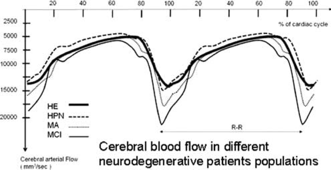
1074. Cerebrospinal fluid and blood flows in mild cognitive impairment, Alzheimer disease and normal pressure hydrocephalus
S. Stoquart-El Sankari1,2, C. Gondry-Jouet3, D. Mbayo3, A. Fichten4, O. Godefroy2, M.-E. Meyer1 and O. Balédent1
1Department of Imaging and Biophysics; 2Department of Neurology; 3Department of Radiology; 4Department of Neurosurgery, Amiens University Hospital, Amiens Cedex, France
Objectives: Phase-Contrast MRI (MRI) is a non invasive reliable technique which enables rapid quantification of cerebrospinal fluid (CSF) and cerebral blood (CBF) flows, and is helpful for aetiological diagnosis of Normal Pressure Hydrocephalus (NPH). In some patients, differential diagnosis with neurodegenerative pathologies (such as Alzheimer disease: AD) may be difficult. Our purpose was to study the effects of AD or Mild Cognitive Impairment (MCI) on intracranial flows.
Methods: Nine MCI and 6 mild AD patients were identified after neuropsychological assessment and neurological examination. They underwent cerebral MRI using 3T scanner. PC-MRI pulse sequence was performed, and dynamic flow images were analyzed with home-made processing software. Results were compared to normal values recently published in age matched elderly healthy (EH) population (n = 12), and to NPH patients, using multivariate analysis.
Results: Arterial flows didn't show statistical difference between AD patients (569±69 mL/mins) and EH (509±103). Although not statistical, we found increased CBF in MCI patients (647±124). Cervical CSF stroke volumes analysis showed similar values in the 3 populations (EH: 457±147 μL; AD: 600±194; MCI: 560±146). Similarly, aqueductal CSF stroke volumes were comparable in EH (34±16 μL) and AD (35±17). Interestingly, aqueductal CSF was hyperdynamic in MCI patients (71±24), but less than in NPH patients (196±100).
Conclusion: These preliminary data suggest increased cerebral perfusion and ventricular CSF pulsations in predemential MCI patients. Surprisingly, we didn't find a significant difference between AD patients and healthy elderly. This may be due to small studied populations. PC-MRI enables reliable, safe and rapid measurements of CSF and blood flows. It provides complementary data helpful for the differential diagnosis between NPH and AD patients.
1075. Choroids plexus and CSF alteration in mild cognitive impairment?
O. Balédent1,2, M. Haddad-Rmeilly1,2, S. Stoquart-El Sankari1,3, J.-M. Serot4, O. Godefroy3, P. Bailly2 and M.-E. Meyer1,2
1Department of Imaging and Biophysics; 2Department of Nuclear Medicine; 3Department of Neurology; 4Department of Geriatrics, Amiens University Hospital, Amiens Cedex, France
Objectives: Cerebrospinal fluid (CSF) plays a fundamental role in brain pathophysiology. Recent research has underlined the CSF's involvement in neurodegenerative processes, such as Alzheimer's disease (AD). The CSF is produced by the choroid plexus (CP) but the latter's functional activity has only been studied in vitro. We present a new approach for studying these targets with [18F]fluorodeoxy-D-glucose (18F-FDG) positron emission tomography (PET) in Mild cognitive impairment popualtion.
Methods: The first population is constituted of 10 subjects (7 males and 3 females) aged of more than 65 years (73±6 years) with a malignant pathology not affecting the brain. A neurological exam has been done to every patient to exclude all neurological pathology. The second population is constituted of 8 subjects (5 males and 3 females) identified as MCIa in the neurological department. a 45 mins dyn-PET acquisition was scheduled in 34 time frames for each patient. CSF and CP regions of interest were manually selected on new reconstructed parametric map presented in other abstract of this congress. Mean raw uptake curves were reconstructed and FDG fixation was measured.
FDG fixation in choroid plexi presents a linear relationship with FDG fixation found in the CSF (R2 = 0,602). This preliminary result shows the ability of PET for observation of CSF secretion.
Results: The results show for the first time that it is possible to monitor the kinetics of FDG uptake by the CP and the CSF during a PET scan. The FDG kinetics in the CP differ from those seen in the CSF and all other brain tissues. CSF and CP FDG fixations were significantly decrease in MCI population.
Conclusion: This work is an initial step towards using PET to evaluate CSF production alteration in MCI population.
References
1.
Ekstrand. PNAS2007;104(4):1325–30.
2.
Renzi. Soc. Neurosciences2008; Abstracts 741.2.
3.
Collantes. Rev Esp Med Nucl, 2008;27(2):103–11.
4.
Doudet. NeuroImage2006;30:26–35.
5.
Bohnen. J Cereb Blood Flow Metab2006;26:1198–212.
6.
PlutaR. Acta Neuropathol1991;83:1–11.
7.
PlutaR. Brain Res1994;633:41–52.
8.
PlutaR. Brain Res1994;649:323–328.
9.
PlutaR. Ischemia-reperfusion pathways in Alzheimer's disease. Nova Science Publishers, New York. 2007.
10.
Le BerI. Brain2004;127:759–67.
11.
ViauM. Brain Res.2005;1049:191–202.
12.
TaroniFDiDonatoS. Nat Rev Neurosci2004;5:641.
13.
MascalchiM. Radiology2002;223:371–8.
14.
TkáčI. Appl Magn Reson2005;29:139–57.
15.
CriscuoloC. Neurology2006;66:1207–10.
16.
Chételat. Neurology2003;60:1374–7.
17.
Mosconi. Neurology2004;63:2332–40.
18.
Walhovd. Neuroimage (in press).
19.
MaY. Parametric mapping of [18F]FPCIT binding in early stage Parkinson's disease: A PET study. Synapse2002;45:125–33.
20.
KirschWMMcAuleyGPetersenFChenLHKimIBaqaiWSchragMHaackeEMAyazMKhanABrittWIIILarsenJPetersonSDicksonCOyoyoUHolshouserBMuellerCVintersHAKidoD. Serial Susceptibility Weighted MRI measures brain iron and microbleeds in dementia. Journal of Alzheimer's Disease, accepted January 2009.
LudwinSK. Ann Neurol1994;36(Suppl):S143–5. Review.
28.
MonopoliALozzaGForlaniAMattavelliAOnginiE. Blockade of adenosine A2a receptors results in neuroprotective effects in cerebral ischaemia in rats. NeuroReport1998b;9:3955–9.
29.
ChenJFXuKPetzerJPStaalRXuYHBelsteinMSonsallaPKCastagnoliKCastagnoliNJrSchwarzschildMA. Neuroprotection by caffeine and A2A adenosine receptor inactivation in a model of Parkinson's disease. J Neurosci2001;21:RC143 (1–6).
30.
ImpagnatielloFBastiaEOnginiEMonopoliA. Adenosine receptors in neurological disorders. Emerg Ther Targets2000;4:635–64.
31.
FangBAPangPeter KTBenishinChristina G. The role of Ca2+ channel modulation in the neuroprotective actions of estrogen in -amyloid protein and 1-methyl-4- phenyl-1,2,3,6-tetrahydropyridine (MPTP) cytotoxic models. Neurochemistry International2004;45:31–8.
32.
FriedmanLK. Calcium: A role for neuroprotection and sustained adaptation. Molecular Interventions2006;6:315–29.
33.
PrioriALefaucheurJP. Chronic epidural motor cortical stimulation for movement disorders. Lancet Neurol2007;6(3):279–86.
34.
CioniB. Motor cortex stimulation for Parkinsońs disease. Acta NeurochirSuppl2007;97(Pt 2):233–8.
35.
BaledentOHenry-FeugeasMCIdy-PerettiI. Cerebro-spinal fluid dynamics and relation with blood flow: A magnetic resonance study with semiautomated cerebrospinal fluid segmentation. Invest Radiol2001;36:368–77.
36.
Stoquart-ElsankariSBaledentOGondry-JouetCMakkiMGodefroyOMeyerME. Aging effects on cerebral blood and cerebrospinal fluid flows. J Cereb Blood Flow Metab, 2007.
37.
SerotJMBeneMCFoliguetBFaureGC. Morphological alterations of the choroid plexus in late-onset Alzheimeŕs disease. Acta Neuropathol2000;99:105–8.