125. P-glycoprotein function in the blood-brain barrier of epileptic rats
S. Syvänen1, G. Luurtsema2, C.F.M. Molthoff2, A.D. Windhorst2, M.C. Huisman2, A.A. Lammertsma2, R.A. Voskuyl1 and E.C.M. de Lange1
1Division of Pharmacology, LACDR, Leiden University, Leiden; 2Nuclear Medicine and PET Research, VU University Medical Center, Amsterdam, The Netherlands
Objectives: It has been hypothesized that P-glycoprotein (P-gp) is responsible for drug resistance in epilepsy by hindering anti-epileptic drugs from penetrating the blood-brain barrier and reaching their targets inside the brain.1,2 The objective of this study was to compare the brain distribution of (R)-[11C]verapamil, a frequently used PET tracer for studying P-gp function,3–5 in naïve and epileptic rats and thus to investigate possible differences in P-gp function between these two groups.
Methods: Male Sprague-Dawley rats were treated with kainic acid to induce epilepsy (n = 20) or with saline (n = 20). At 1 week after treatment, each rat underwent a PET study with (R)-[11C]verapamil. In half of the rats of each group, the P-gp inhibitor tariquidar was administered 20 to 30 mins prior to the start of the (R)-[11C]verapamil scan. Blood samples were withdrawn during the PET scan and analyzed with regard to tracer metabolism in plasma. Brain radioactivity concentrations, expressed as standardized uptake values (SUV) were compared between naïve and epileptic rats as well as between tariquidar treated and untreated rats.
Results: Differences in brain SUV uptake between naïve and epileptic rats were small. In both groups uptake of (R)-[11C]verapamil in the brain was very low with SUV values ranging from 0.60 at the start of the 60 mins scan to 0.15 at the end of the scan. Pretreatment with tariquidar resulted in an up to 10-fold increase in brain SUV in both naïve and epileptic rats. In the tariquidar treated rats there was a small tendency towards slower wash-out of verapamil from epileptic rat brains than from naïve rat brains. Plasma kinetics and metabolism of (R)-[11C]verapamil were unaltered by both tariquidar treatment and epilepsy.
Conclusions: P-gp function in naïve and epileptic rats appeared to be similar. Therefore, this study did not confirm the hypothesis that P-gp plays a major role in limiting brain uptake of anti-epileptic drugs that are P-gp substrates. P-gp function, however, was investigated 1 week after induction of epilepsy. It cannot be ruled out that P-gp function may be altered at other time points after induction of epilepsy.
222. An improved method to measure cerebral P-glycoprotein function reveals changes in brain uptake of (R)-[11C]-verapamil following status epilepticus in rats
J.P. Bankstahl1, C. Kuntner2, M. Bankstahl1, T. Wanek2, J. Stanek2, M. Müller3, O. Langer2,3 and W. Löscher1
1Institute for Pharmacology, Toxicology, and Pharmacy, University for Veterinary Medicine Hannover, Hannover, Germany; 2Department of Radiochemicals and microPET Imaging, Austrian Research Centers, Seibersdorf; 3Department of Clinical Pharmacology, Medical University of Vienna, Vienna, Austria
Objective: Multidrug efflux transporters like P-glycoprotein (Pgp) at the blood-brain barrier (BBB) are believed to play an important role in resistance to antiepileptic drug treatment. (R)-[11C]verapamil (VPM) positron emission tomography (PET) can be used to measure Pgp function, but low brain uptake of VPM hampers the mapping of Pgp function in different brain regions. We investigated if this limitation can be overcome by Pgp modulation prior to PET imaging and if this method is suitable to quantify differences in Pgp expression after status epilepticus in rats, which we found in a previous study.1
Methods: Two groups of naïve female Sprague–Dawley rats (n = 12) underwent single VPM PET scans 2 h after intravenous administration of different doses of the third generation Pgp inhibitors elacridar or tariquidar. Radioactivity uptake in brain was expressed as the area under the time activity curve (AUC) normalized for injected 11C-activity. A sigmoidal dose-response curve was fitted to AUC values measured after different inhibitor doses. To study regional differences of cerebral Pgp activity, paired VPM PET scans, before and after administration of tariquidar (3 or 15 mg/kg), were performed both in naïve rats and in rats 48 h after pilocarpine-induced status epilepticus.
Results: Inhibitor administration resulted in up to 10-fold increased VPM AUCs with ED50 values of 1.16±0.12 and 3.00±0.24 mg/kg for elacridar and tariquidar, respectively. In the paired PET scans, the 15 mg/kg tariquidar dose uniformly increased radioactivity distribution across different brain regions as compared to baseline scans, both in naïve and epileptic rats. In epileptic but not in naïve rats, the 3 mg/kg tariquidar dose resulted in obvious regional differentiation of brain radioactivity distribution, with lowest uptake observed in cerebellum. Immunohistochemical staining demonstrated an almost 2-fold increase of Pgp labeling in the cerebellum of epileptic rats as compared to other brain regions.
Conclusion: Our data suggest that VPM PET in combination with administration of moderate doses of Pgp inhibitors can be used for the mapping of regional Pgp function in the brain. The current data will help to study changes in Pgp-function at the BBB in further models of epilepsy as well as in pharmacoresistant patients.
The research leading to these results has received funding from the European Community's Seventh Framework Programme under grant agreement no. 201380 (EURIPIDES).
890. Ictal SPECT during seizures pharmacologically provoked in pre-surgical evaluation of extratemporal lobe epilepsy
D. Di Giuda1, C. Caldarella1, C. Barba2, R. Mazza1, F. Cocciolillo1, D.P. Dambra1, G. Colicchio3 and A. Giordano1
1Nuclear Medicine Institute, Catholic University of the Sacred Heart, Rome; 2Pediatric Neurology Unit, Children's Hospital A. Meyer, Florence; 3Neurosurgery Department, Catholic University of the Sacred Heart, Rome, Italy
Objectives: Ictal SPECT is a reliable indicator of the seizure onset zone in patients with intractable epilepsy. Contrarily to the contribution in temporal epilepsy, the localising ability of ictal SPECT in the pre-surgical work-up of extratemporal epilepsy (ETE) is lower due to several factors affecting results. Obtaining true ictal radiotracer injections remains a challenge at most Centres, mainly for logistical reasons. Aim of the study was to assess the diagnostic value of pharmacologically provoked ictal SPECT in surgical ETE.
Methods: 26 patients (15 m, 11 f, mean age: 27.7±8.8 years) underwent the following pre-surgical protocol: neurological examination, MRI, neuropsychological and psychodynamic assessments, scalp-video-EEG monitoring, interictal and ictal SPECT with 99mTc-Ethyl-Cysteinate-Dimer (ECD) and invasive neurophysiologic recordings if necessary. Interictal SPECT was carried out after a seizure-free period of >24 h. Ictal SPECT was performed during seizure provoked with pentylenetetrazol slow injection and recorded by scalp-video-EEG and/or intracranial-EEG monitoring. Visual interpretation of images was performed blind to clinical data. SPM analysis of individual ictal SPECT in comparison with a normal brain SPECT database was available in 16/26 cases.
Results: MRI was normal in 3/26 cases. In all patients radiotracer injections were performed during provoked seizures semiologically identical to the spontaneous ones. The average delay from clinical and EEG seizure onset to radiotracer injection was 6 and 16 secs, respectively. Visual analysis of interictal and provoked ictal SPECT was concordant with the epileptogenic zone (EZ) in 22/26 (84.6%) epileptic patients: 19/22 subjects showed the typical hypo-hyperperfusion pattern, while in the remaining three cases no interictal hypoperfusion areas were detected. Fifteen of these 22 patients with concordant results underwent ablative surgery: 13 patients were seizure-free after at least 2 years of follow-up while in 2 subjects surgical follow-up was too short to be considered. Discordant results between SPECT findings and the EZ were observed in 2 patients while in 2 cases with large anatomical lesions provoked ictal SPECT failed to demonstrate focal hyperperfusion. In these two cases ablative surgery was performed with poor outcome. SPM analysis of provoked ictal SPECT was concordant with visual interpretation and final definition of the EZ in 14/16 and 12/16 cases, respectively.
Conclusions: This study demonstrated the localising value of provoked ictal SPECT in refractory ETE. The diagnostic efficacy of our procedure was supported by the satisfying results at surgical outcome, also in patients with non-localising MRI. The optimisation of 99mTc-ECD injection timing is the main advantage of our procedure, indispensable for unambiguous localisation of extratemporal foci. SPM analysis may improve localisation of the seizure onset zone and aid in distinguishing it from seizure propagation areas.
957. 1H HRMAS NMR metabolic profile of mouse brain intoxicated by an organophosphorus compound
F Fauvelle, G. Testylier, F. Dorandeu, P. Arvers, A. Foquin and P. Carpentier
CRSSA, La Tronche, France
Soman is an organophosphorus neurotoxic. It induces epileptic seizures that can last for several hours leading to brain oedema and neuronal lesions, mainly in the piriform cortex and the hippocampus. Limited amount of data is available on the associated brain metabolic disturbances. The purpose of this study was to analyse by HRMAS 1H-NMR spectroscopy the brain metabolic profile in piriform cortex (Pir) and cerebellum (Cvl) from the initial phase (1 h) to 7 days after soman intoxication.
Methods: Mice were intoxicated with soman (172 μg/kg SC) and sacrificed 1, 4, 24, 48, 72 h and 7 days after. Pir and Cvl biopsies were immediately stored in liquid nitrogen after dissection.
The HRMAS 1H-NMR experiments were performed on a Bruker DRX Avance spectrometer at 9.4 Tesla, at 4°C and 4 KHz spinning. A spin-echo sequence with a 30 ms total echo time was used.
In a first analyse, 17 metabolites have been quantitated using jMRUI-sofware.1 Then the Projection to Latent Structure by means of partial least squares Discriminant Analysis (PLS-DA) was applied to rough NMR data. All spectral region between 4.65 and 0.5 ppm were segmented in 0.02 ppm rectangular buckets and loaded in the SIMCA-P software version 10 (Umetrics, Umea, Sweden) for PLS-DA analysis.
Results: Metabolite variations:
In the initial phase of intoxication (1 and 4 h), epileptic seizures have evolved into a severe status epilepticus. Alanine and GABA in Pir reached their maximum level at 1 h while Choline and Acetate were maximum at 4 h.
24 h after soman injection, seizures are not totally arrested but are now discontinuous. Pir biopsies were characterized by a strong increase of Lactate and a strong decrease of NAA. Myo-inositol exhibited a significant decrease at 24 h and 48 h in Pir. Glutamine strongly increased not only in Pir but also in Cvl.
Seven days after intoxication only lactate and NAA in Pir remained at an abnormal level.
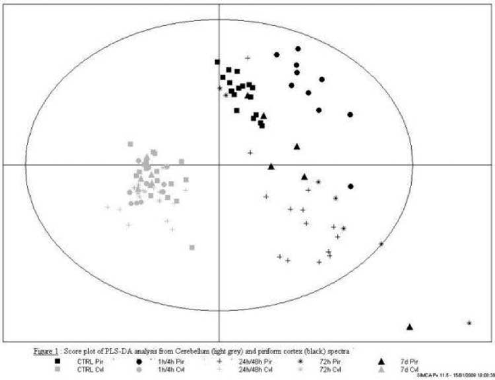
Multivariate analysis: PLS-DA score plot showed the very good separation between samples based upon brain structure, even for samples of intoxicated animals (Figure 1). However, a clear difference appear between the two strutures: cortex samples are much more dispersed than cerebellum samples. The second effect that is superimposed on structural effect for the class separation is the kinetic effect; samples appear separated according to their metabolic profile at different phases after intoxication.
Conclusion: This study enabled the acute observation of metabolic disturbances at different time points after soman intoxication. The metabolites identified reflected the different diseases occurring during soman intoxication from epileptic activity, brain oedema, neuro inflammation to neuronal death and brain adaptation.
1069. Role of oxidative and nitrosative stress during theophylline induced seizures in experimental animals
K. Gulati, A. Ray and V. Vijayan
Department of Pharmacology, University of Delhi, Vallabhbhai Patel Chest Institute, Delhi, India
Theophylline, a methylxanthine, is a very effective bronchodilator in the treatment of obstructive airway disease. However, its narrow therapeutic index and propensity to induce neuroexcitatory effects are a major concern for this pharmaco-economically viable drug. Seizures can be problem during its use particularly in extremes of age and in epileptics, and identification of therapeutic strategies to counteract such iatrogenic seizures could lead to the development of an effective antidote for theophylline toxicity. Current therapeutic tools utilizing conventional anti-seizure approaches are ineffective in antagonizing such seizures, and thus the present study evaluated some novel mechanisms involved in theophylline induced convulsions in mice. Theophylline is metabolized by the xanthine-xanthine oxidase pathway, which is an important source of free radical generation, and thus the possible involvement of oxidative stress was studied in this phenomenon. Aminophylline (theophylline et ethylene diamine) induced seizures and mortality in mice in a dose related manner, which could not be replicated by phosphodiesterase inhibitors or the cerebral blood flow enhancer, pentoxyfylline. Further, such seizures were not consistently antagonized by conventional anticonvulsants or adenosine agonists. Interestingly, pretreatment with antioxidants (alpha tocopherol, ascorbic acid and melatonin) and the NO synthase inhibitor, L-NAME induced differential degrees of attenuations in aminophylline-induced seizures and mortality. The most potent effects were seen with the combination treatment of melatonin and L-NAME. Such seizures were paralleled by elevations in brain malondialdehyde (MDA), NO metabolites (NOx), and lowered reduced glutathione (GSH) levels, which were reversed by the antioxidant pretreatments. On the other hand, a combination of sub threshold doses of the methylxanthine and the NO precursor, L-arginine, had synergistic seizurogenic effects. Further, sub effective doses of aminophylline when combined with sub threshold intensities of electroshock also induced tonic clonic seizures which were also prevented by pretreatments with antioxidants, NO synthase inhibitors and their combination therapy. Biochemical assay of brain homogenates in these mice also showed elevations in MDA and NOx and reductions in GSH activity, which were normalized by antioxidant treatments. These results indicate that oxidative stress and ROS-RNS interactions may be involved in theophylline induced seizures and mortality, and antioxidants, alone or in combination with NO synthase inhibitors could form useful treatment strategies in such situations. Further, this hypothesis could provide a good nidus for translational research in co morbid situation involving asthma and epilepsy, and also other iatrogenic convulsive states.
BankstahlJPLöscherW. Resistance to antiepileptic drugs and expression of P-glycoprotein in two rat models of status epilepticus. Epilepsy Research2008;82:70–85.
7.
BarbaCDi GiudaDPolicicchioDBrunoIPapacciFColicchioG. Correlation between provoked ictal SPECT and depth recordings in adult drug-resistant epilepsy patients. Epilepsia2007;48:278–85.
8.
CalcagniMLGiordanoABrunoIParbonettiGDi GiudaDDe RossiGTronconeLColicchioG. Ictal brain SPET during seizures pharmacologically provoked with pentylenetetrazol: A new diagnostic procedure in drug-resistant epileptic patients. Eur J Nucl Med Mol Imaging2002;29:1298–306.