9. Is cerebral oxygenation negatively affected by infusion of noradrenaline in healthy subjects?
P. Brassard, T.S. Larsen and N.H. Secher
Department of Anaesthesia, The Copenhagen Muscle Research Centre, Rigshospitalet, Faculty of Health Sciences, University of Copenhagen, Copenhagen, Denmark
Introduction: One approach of increasing mean arterial pressure (MAP), in order to secure a pressure gradient for vital organs, is provided by the use of vasopressor agents. The consequence of augmenting MAP by elevating vascular resistance or cardiac output (CO) on blood flow and oxygenation of the brain is not clear. More specifically, the influence of noradrenaline (NA), which is an α-agonist causing vasoconstriction, on cerebral hemodynamics is ambiguous. Furthermore, the influence of NA on cerebral oxygenation has not been investigated.
Aim: The aim of this study was to evaluate the impact of the infusion of NA on cerebral oxygenation in healthy subjects.
Methods: Three doses of NA (0.05, 0.1 and 0.15 mg kg−1 mins−1 for 20 mins each) were infused in nine healthy subjects (6 males; 26±6 years old, mean±s.d.). MAP, cerebral oxygenation characterized by frontal lobe oxygenation (ScO2) and jugular venous oxygen saturation (SjvO2), middle cerebral artery mean flow velocity (MCA Vmean), cardiac output (CO) and arterial pressure for carbon dioxide (PaCO2) were evaluated.
Results: MAP increased from 88 (22) [median (range)] to 115 (30) mm Hg with increasing doses of NA (P<0.05 for 0.1 and 0.15 mg kg−1 mins−1 of NA) and reflected an increase in total peripheral resistance (20.3 (13.7) to 25.2 (12.1) mm Hg 1/L mins−1; P<0.05) as CO remained at a baseline value (Table). ScO2 and SjvO2 lowered with increasing doses of NA, reaching statistical significance with NA infused at 0.1 mg kg−1 mins−1 and higher (ScO2: 78 (19)% to 69 (22)%; P<0.05; SjvO2: 67%±8% to 64%±7%; P<0.01). MCA Vmean was reduced with each doses of NA (56.9±11.2 to 55.0±11.7 cm secs−1; P<0.05). Finally, PaCO2 lowered with increasing doses of NA (5.4±0.4 to 5.1±0.4 kPa; P<0.001).
Data are mean±s.d. or median (range). Infusions of NA are in mg kg−1 min−1;
P<0.05,
P<0.01, and
P<0.001 versus saline.
Conclusion: This study suggests that infusion of 0.1 mg kg−1 mins−1 and higher doses of NA negatively affects cerebral oxygenation as it increases MAP by peripheral vasoconstriction.
64. Rapid cerebral hemodynamic modulation during set shifting: evidence of time-locked associations with cognitive control in females
D. Schuepbach1, S. Duschek2 and D. Hell1
1Psychiatric University Hospital, University of Zürich, Zürich, Switzerland; 2Department of Psychology, University of Munich, Munich, Germany
Objectives: Set shifting provokes specific alterations of cerebral hemodynamics in large basal cerebral arteries.1 However, no clear gender differences have been reported so far. In the following functional transcranial Doppler (fTCD) study, we introduced cerebral hemodynamic modulation, a means that assesses rapid changes of brain perfusion, to gender aspects of set shifting during Wisconsin Card Sorting Test.
Methods: Male and female participants underwent the WCST during measurements of the middle and anterior cerebral arteries. Parameters of task performance and cerebral hemodynamic modulation during set maintenance and set shifting were investigated by means of multi- and univariate analyses of variance. Correlation analyses examined the linkage between task performance and rapid cerebral hemodynamic modulation.
Results: Males showed mostly positive values of cerebral hemodynamic modulation during set shifting whereas in females, maximum modulation was restricted to the behaviorally relevant time point of set shifting. Further, we observed time-locked associations between slowing during set shifting and early rapid cerebral hemodynamic modulation of the left and the right MCA in females (R = −0.82, P = 0.0036 and R = −0.90, P = 0.0004, respectively), but not in males.
Conclusions: This study provides evidence of gender related cerebral hemodynamic modulation during set shifting and of time-locked brain behavior relationship during cognitive control in females, and it emphasizes the importance of temporal dynamics of brain perfusion during cognitive control in both genders. Future fTCD studies are urgently needed to examine neurovascular mechanisms that mediate rapid cerebral hemodynamic modulation and time-locked associations during cognitive control.
140. Depth-resolved microscopy of cerebral blood flow and volume during somatosensory stimulation with doppler optical coherence tomography
V. Srinivasan1, S. Sakadzic1, I. Gorczynska2, A. Yaseen1, J. Fujimoto2 and D. Boas1
1Photon Migration Imaging Laboratory, MGH/MIT/HMS Athinuola A. Martinos Center for Biomedical Imaging, Massachusetts General Hospital/Harvard Medical School, Charlestown; 2Department of Electrical Engineering and Research Laboratory of Electronics, Massachusetts Institute of Technology, Cambridge, Massachusetts, USA
Objectives: Investigation of laminar differences in cerebral blood flow and volume during functional activation has been difficult due to the limited spatial resolution of techniques such as MRI and diffuse optical imaging. Recently two-photon microscopy has emerged as a useful tool for depth-resolved measurements of microvasculature during functional activation. However, in conventional two-photon microscopy penetration depths are limited to ∼500 microns, simultaneous measurements at multiple cortical depths are difficult, and quantification of high velocities in diving arterioles can be challenging. Doppler Optical Coherence Tomography (OCT) is a new microscopy technique that potentially addresses these limitations. The objective of this study is to investigate Doppler OCT for measuring changes in cerebral blood flow and volume during functional activation.
Methods: Sprague Dawley rats (N = 5) were prepared with cranial windows, identically to our previous preparations for two-photon microscopy.1 Imaging was performed during forepaw stimulation (300 μs pulses at 3 Hz for 4 secs with 0.5 to 1.1 mA amplitude) while rats were under alpha-chloralose anesthesia. Rats were first imaged during forepaw stimulation using 2D optical intrinsic signal imaging (OISI) to determine the response area and to confirm physiological stability of the animal.
A spectral/Fourier domain OCT microscope was constructed for in vivo imaging of the rat cerebral cortex. Briefly the light source consisted of a Mai Tai Ti:Sapphire laser (Spectra Physics) operating at ∼830 nm and spectrally broadened in a nonlinear UHNA3 fiber (manufactured by Nufern, distributed by Thorlabs). The axial (depth) resolution was approximately 4.5 microns in air (3.3 microns in tissue). A home-built spectrometer enabled an imaging speed of 22,000 axial scans per second and an axial imaging range of 2.5 mm in air. The transverse resolution was ∼11 microns. OCT cross-sectional images were acquired repeatedly at a rate of 4 frames per second across 2 mm of cortical tissue for 8 secs (1 secs pre-stimulus, 4 secs stimulus, and 3 secs post-stimulus). Doppler OCT signals were extracted by high-pass filtering each OCT image along the transverse dimension, calculating a windowed autocorrelation function at each position, and performing a linear fit to the phase of the autocorrelation function. 25 to 30 repetitions were performed for the contralateral and ipsilateral forepaw, with 20 secs between stimuli.
Results:Figure 1A shows fractional flow changes and Figure 1B shows fractional diameter changes of a single representative diving arteriole in the somatosensory cortex during contralateral (blue) and ipsilateral (red) stimulation.
Diving arteriole during functional activation.
Conclusions: Doppler OCT can simultaneously measure spatiotemporal characteristics of blood flow and volume over cortical depths of 1 mm. Therefore, Doppler OCT has potential as a research tool to understand neurovascular coupling mechanisms and elucidate the BOLD signal.
161. Predictive factors of cerebral hyperperfusion syndrome before and immediately after carotid angioplasty and stent placement
T. Iwata, T. Mori, H. Tajiri and M. Nakazaki
Shonan Kamakura General Hospital, Kamakura City, Japan
Objectives: To anticipate high risk of hyperperfusion syndrome (HPS) following carotid angioplasty and stent placement (CAS) is useful for perioperative management. The purpose of our retrospective study was to find predictors of HPS before and immediately after CAS and to investigate the utility of single-photon emission computed tomography (SPECT) and transcranial color-coded real-time sonography (TCCS) as predictors.
Methods: Included for analysis were patients:
who underwent elective CAS in our institution from July 2005 to March 2008,
with unilateral carotid stenosis,
who underwent 99mTc-ethyl cysteinate dimmer (99mTc-ECD) SPECT study before and immediately after CAS,
who underwent acetazolamide (ACZ) challenge test of 99mTc-ECD SPECT study before CAS and
who underwent TCCS study before and immediately after CAS.
Regional cerebral blood flow (rCBF) by using 99mTc-ECD SPECT and mean blood flow velocity (mBFV) in the middle cerebral artery (MCA) by using TCCS were examined before and immediately after CAS. Clinical HPS was defined as symptoms as follows:
throbbing ipsilateral frontotemporal or periorbital headache and
at least one symptom of temporary deterioration of consciousness level, focal seizure and hemiparesis.
Age, sex, degree of stenosis, hypertension (HT), hyperlipidemia (HL), diabetes mellitus (DM), asymmetry index (AI) = (rCBF in the affected hemisphere coupled with a carotid stenosis/rCBF in the contralateral hemisphere) before and immediately after CAS, AI change = (AI immediately after CAS−AI before CAS), AI ratio = (AI change/AI before CAS), regional activity-to-cerebellar activity (R/CE) ratio = (rCBF in the affected hemisphere coupled with a carotid stenosis/rCBF in the ipsilateral cerebellum hemisphere) before and immediately after CAS, R/CE ratio-change = (R/CE immediately after CAS−R/CE before CAS), R/CE ratio-ratio = (R/CE ratio-change)/(R/CE before CAS), CVR = [(post-ACZ rCBF−resting rCBF)/resting rCBF], MCA mBFV in the affected hemisphere before CAS and MCA mBFV ratio = (MCA mBFV immediately after CAS in the affected hemisphere/MCA mBFV before CAS in the affected hemisphere) were assessed.
Results: Eighty consecutive patients underwent CAS and ten of them presented HPS after CAS. In TCCS study, the dropout rate due to an insufficient acoustic temporal bone window was 20% (16/80). Between HPS and non_HPS groups, there were significant differences in severe carotid stenosis, CVR and MCA mBFV in the affected hemisphere (P<0.05: Mann–Whitney U test) in the preoperative items, and significant differences in AI after CAS, AI change, AI ratio, R/CE ratio after CAS, R/CE ratio-change, R/CE ratio-ratio and MCA mBFV ratio (P<0.05: Mann–Whitney U test) in the postoperative items, although there were no significant differences in age, sex, HT, HL, DM, AI before CAS and R/CE before CAS. Logistic regression analysis showed that CVR [Odds ratio (95%CI); 0.674 (0.492 to 0.926), P = 0.015] was the significant predictor among the preoperative items, and that MCA mBFV ratio [Odds ratio (95%CI); 9.696 (1.550 to 60.657), P = 0.015] and R/CE ratio-change [Odds ratio (95%CI); 1.169 (1.018 to 1.342), P = 0.027] were the significant predictors among the postoperative items.
Conclusions: Significant predictors of HPS were CVR before CAS, and MCA mBFV ratio and R/CE ratio-change immediately after CAS. SPECT and TCCS studies are useful to predict HPS.
176. Anatomical study of normal variations of circle of willis in 132 fetuses, infants and adults
S. Ansari1, M. Dadmehr2, B. Eftekhar3, F. Nejat2, S. Kamali Ardakani2, S.M. Ghodsi2, J.E. Heavner4, B. Vidic4 and J. Mocco1
1Department of Neurosurgery, University of Florida, College of Medicine, Gainesville, Florida, USA; 2Department of Neurosurgery, Tehran University of Medical Sciences, Tehran, Iran; 3Royal Hobart Hospital, Tasmania, TAS, Australia; 4Health Science Center, Texas Tech University, Lubbock, Texas, USA
Objective: Several studies have investigated the variations in the anatomy of each segment of circle of Willis whereas a few have addressed the variations of this arterial circle as a whole. In this study the entire circle of Willis and its variations were studied in a cohort of Iranian people and compared with previous reports.
Material and method: 132 brains of recently deceased Iranian people (102 adults and 30 fetuses and infants) were dissected and anatomic variations of the circle of Willis were observed. The dissection process was digitized for further studies. Using computer software (Osiris) the external diameters and length of the vessels were measured and the circle variations were classified. The variations of the circle as whole and segmental variations were compared with previous studies.
Results: Nearby 41% of the circles were symmetric. Uni-and bilateral hypoplasia of posterior communicating arteries (PcoAs) constituted the most common variation in our study which was similar to previous works. Aplasia of the precommunicating part of the anterior cerebral artery (A1) and the precommunicating part of the posterior cerebral artery (P1) were not observed. In 3.3% of fetuses and infants and 3% of adult instances both right and left posterior communicating arteries were absent. There was one case of anterior communicating artery (AcoA) aplasia in adult group which was not seen in fetus and infant instances. As a whole, there were no two identical circles regarding to their dimensions.
Conclusion: The anatomical variations discovered in Iranian circle of Willis in this current study were not significantly different to those of more diverse populations reported in the literature. On the whole, the frequencies of the different variants of the entire cerebral arterial circle and segmental variations were comparable with previous studies. The main differences between the fetal and adult disposition are the diameter of the PcoA and the circular part of the posterior cerebral artery.
182. Rapid cerebral hemodynamic modulation during complex cognitive functions: evidence of time-locked associations with age
D. Schuepbach and D. Hell
Psychiatric University Hospital, University of Zürich, Zürich, Switzerland
Objectives: There is evidence that age affects complex cognitive functions from a neuropsychological and neurophysiological perspective. Age related changes in neurovascular coupling have been also described.1 We recently introduced cerebral hemodynamic modulation,2 a means that assesses rapid changes of brain perfusion, to executive functions, and we found evidence of time-locked associations between rapid cerebral hemodynamic modulation and performance during those tasks. In the following functional transcranial Doppler (fTCD) investigation, we correlated rapid cerebral hemodynamic modulation during a variety of executive functions with age.
Methods: Subjects underwent executive tasks, namely the Wisconsin Card Sorting Test, a means of abstraction and cognitive flexibility, the Tower of Hanoi puzzle and the Stockings of Cambridge, which are considered as measures of planning. Bilateral fTCD measurements of the middle (MCA) and anterior (ACA) cerebral arteries were carried out during the tasks. Partial correlation analyses examined the linkage between rapid cerebral hemodynamic modulation and age during early phases of those paradigms, with gender and test performance as covariates.
Results: There were significant and inverse associations between early phases of rapid cerebral hemodynamic modulation in the MCA and age in executive tasks (P<0.05), which were mostly observed during time intervals of 1 to 2 secs and 2 to 3 secs after the start of the paradigms. Older subjects showed significantly less increase of brain perfusion due to cognitive stimuli than younger participants. However, task performance was a significant cofounder in some tests.
Conclusions: These results suggest that there is a pattern of age related associations with cerebral hemodynamic modulation in the lateral hemispheres during complex cognitive functions suggestive of higher modulatory qualities in young age. Unsurprisingly, task performance attenuated some associations. Since there is evidence of time-locked associations between rapid cerebral hemodynamic modulation and performance during complex cognitive functions, the finding of age-related linkage with cerebral perfusion could be used as a marker of cognitive decline and as a tool for the development of cognitive enhancers for neuropsychiatric disorders such as schizophrenia or Alzheimer's disease.
189. Lipid-soluble cigarette smoke particles upregulate contractile endothelin type B and thromboxane A2 receptors of rat middle cerebral arteries
H. Sandhu1, C.B. Xu1,2 and L. Edvinsson1,2
1Department of Clinical Experimental Research, Glostrup Research Institute, Glostrup University Hospital, Glostrup, Denmark; 2Division of Experimental Vascular Research, Institute of Clinical Sciences in Lund, University Hospital of Lund, Lund, Sweden
Objective: Cerebrovascular smooth muscle cells (SMCs) are associated with enhanced expression of endothelin type B (ETB) receptor expression. Stroke is strongly associated with several cardiovascular risk factors; e.g. cigarette smoking, increase in low density lipoproteins (LDL), hypertension, inter alia.1 We examined the question if lipid-soluble cigarette smoke particles affect the expression of the contractile ETB and thromboxane A2 (TP) receptors during organ culture; a method which mimics vascular receptor upregulation seen in cerebral ischemia.2
Method: Kentucky Reference cigarettes are smoked with aid of vacuum and the smoke particles retrieved on a filter. These were dissolved in 1 mL dimethyl sulfoxide (DMSO) or in water. Fresh cotton filters soaked in DMSO or water were used as controls. Middle cerebral arteries (MCAs)—removed from adult male Sprague Dawley rats (body weight approximately 250 g)—are incubated for 24 h with 0.15 μL DMSO-soluble smoke particles (DSP), water-soluble cigarette smoke particles, DMSO or water. The MCAs are mounted in myographs and contractile responses to the ETB specific receptor agonist sarafotoxin (S6c) or the TP specific receptor agonist U46619 are studied. Real time PCR and immunohistochemistry are used to semi-quantify the ETB and TP receptor mRNA and protein levels, respectively.
Results: S6c does not elicit significant contraction of fresh artery segments; however, after organ culture for 24 h S6c induces marked vasoconstriction which does not differ if a small volume of DMSO or water is present during the organ culture.2 Addition of DSP to the organ culture increases the maximum of the S6c response; Emax from 60%±7% in control DMSO or water, to 105%±10% in DSP (P<0.05). The water soluble smoke particles did not enhance the contraction. The Emax of the U46619 response also increased; from 112%±5% in control to 148%±12% in DSP (P<0.05). Real time PCR showed that DSP upregulates both ETB and TP receptor mRNA expression; thus suggesting an effect at the transcriptional level. Immunohistochemistry showed that the ETB and the TP receptors are upregulated by the organ culture in the SMCs and not in the endothelium. Addition of DSP to the organ culture further upregulates this ETB and TP receptor expression at the same location.
Conclusion: We demonstrate for the first time that lipid-soluble cigarette smoke particles but not water-soluble cigarette smoke particles or nicotine per se in smoking dosage (date not shown), enhance the SMC expression of ETB and TP receptors. The ETB and TP receptor mediated contractions are upregulated in the SMC and this occurs in parallel with enhanced expression of their mRNA levels. The in vitro organ culture model can be used to study the molecular signal pathways leading to upregulation of the contractile ETB and TP receptors.
192. Cerebral blood flow simulation using a vascular graph model
J. Reichold1, M. Stampanoni2,3, A.L. Keller4, A. Buck5, P. Jenny1 and B. Weber6
1Institute of Fluid Dynamics, ETH Zurich, Zurich; 2Swiss Light Source, Paul Scherrer Institut, Villigen; 3Institute for Biomedical Engineering, University and ETH Zurich, Zurich, Switzerland; 4Max Planck Institute for Biological Cybernetics, Tübingen, Germany; 5Nuclear Medicine, University Hospital Zurich; 6Institute for Pharmacology & Toxicology, University of Zurich, Zurich, Switzerland
Background and aims: This work aims at constructing a computational model that can faithfully predict cerebral blood flow (CBF) in realistic vascular networks. We propose a vascular graph (VG) model, based on simple fluid dynamic principles, that comprises an upscaling algorithm, which both reduces the computational cost and allows CBF simulations even when the discrete topology of the capillary bed is unknown.
Methods: The cerebral vasculature can be represented by a graph of resistive elements. Each vessel bifurcation or end-point is a vertex, the blood vessels themselves designate the graph's edges that connect pairs of vertices. By requiring mass conservation at the vertices and providing appropriate boundary conditions, the resulting linear system of equations can be solved to yield pressure and flow values for the entire graph.
This procedure has been thoroughly described for the analogous problem in electrical circuits (Weinberg, 1962), was adapted to vascular networks by Lipowsky and Zweifach (Lipowsky et al, 1974), and was recently extended by Boas and coworkers (Boas et al, 2008). We have further enhanced the model with focus on three-dimensionality and applicability to complex realistic vascular networks. By embedding the vasculature in a computational grid representative of brain tissue, the interaction between the two compartments can be captured in a truly three-dimensional fashion.
The proposed upscaling algorithm replaces the discrete topology of the capillary bed by a coarser-scale network with similar fluid dynamical properties. This is achieved by dividing the computational domain into sub-volumes for each of which effective conductance values of the capillary bed are computed.
Results: The VG model was applied to high-resolution angiography data of the rat cortex obtained with synchrotron radiation based x-ray microscopy (Weber et al, 2006). Our simulations reproduce the well-established finding that the dilation of an arteriole leads to an increase in CBF in the feeding vessel, as well as the capillary bed it irrigates and the corresponding draining veins. We also find that flow decreases in all other arterioles that supply that same region of the capillary bed. This decrease, however, is not as pronounced as the flow increase in the dilated vessel. Our simulations further suggest that the spatial specificity of a local dilation depends both on the cortical depth of the dilation site as well as the type of vessel (Duvernoy et al, 1981) whose diameter is modified.
Our simulations of occluded penetrating arterioles demonstrate that CBF recovers downstream of the occlusion with increasing number of bifurcations. Approximately 350 micron away from the occlusion site, the effect of the constriction is negligible. This distance is consistent with the spatial distribution of the penetrating arterioles and compares well with the recent findings of Nishimura and coworkers (Nishimura et al, 2007).
Conclusions: We have presented a modeling framework that can compute blood pressure and flow, as well as scalar transport and exchange between vasculature and tissue. The introduction of an upscaling algorithm extends the model's applicability to very large networks and eliminates the need for detailed knowledge of the capillary bed topology.
206. Evaluating crosstalk between epoxyeicosatrienoic acids (EETS) and neuropeptide signaling in neurogenic vasodilation
J. Iliff1,2 and N. Alkayed1,2
1Department of Anesthesiology and Peri-Operative Medicine; 2Department of Physiology and Pharmacology, Oregon Health and Science University, Portland, Oregon, USA
Objectives: Epoxyeicosatrienoic acids (EETs) are vasodilator eicosanoids synthesized by P450 epoxygenases and metabolized by soluble epoxide hydrolase (sEH).1 We recently reported that the machinery for EETs synthesis and metabolism is present in perivascular vasodilator nerve fibers and associated ganglia innervating the rat middle cerebral artery. We have also found that EETs play a functional role in the regulation of cerebral blood flow (CBF) by these nerves.2,3 The vasomotor actions of these fibers have been attributed to the release of the neuropeptides vasoactive intestinal peptide (VIP) and calcitonin gene-related peptide (CGRP).4 We therefore set out to determine if EETs and neuropeptide signaling interact in the neurogenic regulation of CBF. In the present study, we evaluated the following potential modes of interaction between EETs and the vasodilator action of VIP and CGRP:
post-junctional potentiation of neuropeptide vasomotor action by EETs,
parallel and additive vasomotor effects of EETs and neuropeptides,
intracellular regulation of pre-junctional neuropeptide release by EETs, and
dependence of neuropeptide-evoked vasodilation upon endothelium-derived EETs.
Methods: CBF was monitored over front-parietal cerebral cortex in adult male Wistar rats by laser Doppler flowmetry (LDF). The neuropeptides VIP or CGRP (1 nmol/L to 1 μmol/L) were perfused over the pial surface via a closed cranial window and the LDF responses were measured in the presence and absence of 14,15-EET (100 nmol/L).
Results: VIP (n = 5) and CGRP (n = 5) evoked a concentration-dependent hyperemia in the cortex with EC50 values of 0.475 μmol/L and 6.64 μmol/L and with maximal values of 26%±7% and 22%±3% at 1 μmol/L concentration, respectively. In the presence of 14,15-EET, neither the EC50 nor the maximal CBF responses to VIP or CGRP were significantly altered.
Conclusions: These data suggest that neurogenic EETs do not act post-junctionally to potentiate the vasomotor action of the neuropeptides VIP or CGRP. Further studies will evaluate the other modes of interaction between neurogenic EETs and neuropeptide signaling. Resolving the interactions between these cerebral vasodilator signaling pathways may provide novel therapeutic options for the treatment of such pathological states as migraine, stroke and vasospasm after subarachnoid hemorrhage. Supported by NS44313 to NJA and a predoctoral NRSA to JJI.
229. Impact of cytokines and growth factors on contractile endothelin receptors in rat cerebral arteries
E. Stenman, H. Ahnstedt, M. Henriksson and L. Edvinsson
Clinical Sciences, Experimental Vascular Research, Lund University, Lund, Sweden
Objective: Cerebral ischemia induces an increased endothelin receptor mediated contraction in rat cerebral arteries via enhanced expression of endothelin ETB receptors in the smooth muscle cells.1 The aim of the present study was to examine if growth factors and cytokines, well-known to be activated in cerebral ischemia,2 can influence the function and expression of cerebrovascular endothelin receptors.
Methods: Rat middle cerebral arteries (MCAs) were cultured for 24 h with or without tumor necrosis factor-α (TNF-α; 10 and 100 ng/mL), interleukin-1β (Il-1β; 10 and 100 ng/mL), platelet-derived growth factor (PDGF 10 and 100 ng/mL), epidermal growth factor (EGF; 20 and 100 ng/mL) or basic fibroblast growth factor (bFGF; 10 and 100 ng/mL). The MCAs were mounted in sensitive myographs and contractile responses were obtained for sarafotoxin 6c (ETB receptor agonist) and endothelin-1 (in this case ETA receptor agonist, due to ETB receptor desensitization). The receptor mRNA levels were examined by real-time PCR.
Results: Culture of isolated MCA segments results in enhanced expression of ETB receptors in the smooth muscle cells; a response also seen after cerebral ischemia. Here we found that TNF-α (100 ng/mL) and EGF (20 ng/mL) further enhanced the culture-induced expression of the ETB receptor mediated contraction, while EGF (100 ng/mL) decreased the ETB receptor mediated contraction. On the other hand bFGF induced an enhanced ETA receptor mediated contraction while PDGF and Il-1β did not affect the endothelin receptor mediated contraction. Real-time PCR showed that neither TNF-α (100 ng/mL) nor EGF (20 ng/mL) influenced ETB receptor mRNA after 24 h of organ culture. Instead bFGF (10 ng/mL) increased the amount of ETB receptor mRNA, which is in accordance with previous studies on cultured MCA smooth muscle cells.3 The ETA receptor mRNA levels were not affected by TNF-α (100 ng/mL), EGF (20 ng/mL) or bFGF (10 ng/mL).
Conclusion: The results suggest that TNF-α and bFGF may contribute to the up-regulated endothelin receptor mediated response seen in cerebral arteries after cerebral ischemia, while EGF has different impact on cerebrovascular endothelin receptors, depending on concentration. This mechanism could participate in the final formation of the brain infarct after cerebral ischemia.
282. Cerebrovascular reactivity and cerebral autoregulation: distinct physiological properties?
E. Carrera1, L. Lee2, S. Giannopoulos1 and R. Marshall1
1Neurology; 2Radiology, Columbia University Medical Center, New York, New York, USA
Background: Cerebrovascular reactivity testing (CVR) with CO2 challenge is used clinically as a measure of cerebrovascular reserve. Spontaneous, short time-frame, cerebral autoregulation (CA) can also be measured by determining correlations between simultaneous fluctuations of blood pressure and cerebral blood flow velocities. It is unknown, however, whether CVR measures the same physiological process as CA.
Methods: We prospectively studied CVR and CA in 20 healthy volunteers (mean age 32±8, 15 men). CBFV was monitored continuously with transcranial Doppler over both middle cerebral arteries at a depth of 50 to 56 mm using a standard headframe. Arterial blood pressure was continuously monitored using a non-invasive finger cuff. After 10 m monitoring at normocapnea, 5% CO2 was administered via facemask for 10 m. Using continuous, in-line capnometry, CVR was calculated as percent increase in MCA mean flow velocity (MFV) per mm Hg pCO2 during the inhalation period. Cerebral CA was determined by transfer function analysis to derive the phase shift between spontaneous ABP and CBFV fluctuations at 0.1 Hz.
Results: CO2 inhalation produced an average CVR of 2.85%±1.3%. A significant average decrease in phase shift occurred in response to CO2 inhalation, from 37.6%±11.4 degrees to 21.0%±15.9 degrees (P<0.001). However, there was no evidence of a significant correlation between CVR and CA change during CO2 inhalation (Pearson correlation coefficient = −0.248, P = 0.32), nor between CVR and baseline CA (Pearson = 0.341, P = 0.17), across individuals in our group.
Conclusion: Phase shift between ABP and CBFV is reduced during CO2 challenge, demonstrating a decrease in spontaneous CA with vasodilatation. Our finding that neither magnitude of CA change nor baseline CA correlated with the degree of CVR suggests that cerebrovascular reserve and CA measure distinct physiological properties. Further investigation into the relationship between these 2 components of cerebral hemodynamics is warranted to determine how best to make use of these measurements in pathological states where these properties may dissociate.
296. Fibered confocal fluorescence microscopy for in vivo study of cerebral microcirculation in the rat: vasodilatory effects of the phytoestrogen genistein
M. Castelló-Ruiz1, J.B. Salom1,2, J.M. Pradillo3, M.C. Burguete2, V.G. Marrachelli1, G. Torregrosa1,2, I. Lizasoain3 and E. Alborch1,2
1Centro de Investigación, Hospital Universitario La Fe; 2Departamento de Fisiología, Universitat de València, Valencia; 3Departamento de Farmacología, Facultad de Medicina, Universidad Complutense de Madrid, Madrid, Spain
Objectives: Conventional intravital fluorescence microscopy and confocal laser-scanning microscopy involve the positioning of a microscope objective on the cerebral microvascular field. A new image system combining confocal fluorescence microscopy with fiber optics, namely fibered confocal fluorescence microscopy (FCFM), replaces the microscope objective with a mini-optical probe.1 We sought to apply FCFM for the study of brain microcirculation in the rat and to assess the vasoactive effects of the phytoestrogen genistein, which effects relaxing isolated cerebral arteries and increasing laser-Doppler measured cerebral perfusion have been reported.2,3
Methods: Male Wistar rats (300 g) were anesthetized and the head fixed in a stereotaxic. A cranial window covering all the right parietal bone was drilled and the dura was removed. A ProFlex Z probe connected to the Laser Scanning Unit of a FCFM system (Cell-vizio, Mauna Kea Technologies) was positioned above the cortex. A bolus (700 μL) of 5 to 10 mg/mL FITC-dextran was injected through the left femoral vein to label plasma. Saline washed erythrocytes from a donor rat were labeled by incubating (2 h, 25°C) with 10 mg/mL FITC isomer I-Celite and injected into a receptor rat. Sodium nitroprusside (SNP, 10 μmol/L) and endothelin-1 (1 μmol/L) were topically administered to test for reactivity of pial vessels. Genistein (10 mg/kg) was infused directly into the brain circulation through the right carotid artery and through the femoral vein. Images were displayed, acquired and analyzed with an Apple computer fixed up with ImageCell software.
Results: Fluorescent labeling of the plasma with 10 mg/mL FITC-dextran allowed the visualization of surface corticoparietal microvessel networks including arterioles, venules and capillaries (Figure). Fluorescent labeling of erythrocytes with FITC isomer I-Celite allowed the visualization of circulating cells in the brain surface. Counterstaining of microvessels with 5 mg/mL FITC-dextran improved images of circulating erythrocytes in the brain vascular networks and their transit throughout capillaries. Topical SNP and endothelin-1 induced increases and reductions in microvessel diameter, respectively. Both intracarotid and intravenous infusion of genistein induced increases in the diameter of most but not all pial vessels. However, the intracarotid effect was immediate and higher in magnitude (70%) while the intravenous effect was delayed (6 mins) and lesser in magnitude (50%).
Conclusions: A new FCFM-based technique has been applied to in vivo study of cerebral microcirculation in the rat and it has proven appropriate to assess pial vessel vasoactivity and to observe the transit of blood cells. Vasodilatory responses to local and systemic genistein are in line with reported3 increases in cerebral perfusion induced by this phytoestrogen.
Supported by RETICS-RENEVAS from ISCIII (RD06/0026/0006).
308. Angiography and venograpy of bat intracranial vessels.adaptation to prolonged inversion
J. Ashaolu
Anatomy, University of Ilorin, Ilorin, Nigeria
Background and aims: Bat contend with prolonged inversion in their roosting colonies, and both inversion and valsalva maneuver in Man has been reported to increase intracranial pressure.1 The aim of this study is to verify if bats possess accessory drainage system(s) or arterial shunt(s) for regulating intra-cranial circulation.
Methods: 4 bats captured irrespective of sex weighing between 250 to 260 g were divided into 2 groups of 2 each (A and B). The group A and group B bats were anaesthetized with chloroform and administered with 3 mls Urografin (a radiographic contrast) through the left and right ventricles to study the venography and angiography respectively.
Purpose: To verify how bats contend with cranial hydrodynamics during inversion.
Results: X-ray images were taken and placed on a viewing box, and photographic records obtained for analysis showed that; the posterior vena cava was highly enlarged, the superior sagittal sinus communicated with the facial veins, the vertebral arteries have a midline origin that appears to emanate from the arch of aorta other than the origin from the subclavian artery, the vertebral arteries communicated by shunts with each other and, the common carotid arteries appear visually narrower compared to the vertebral arteries.
Conclusions: All these arterial and venous modifications suggest reasons for bats protection against increased intracranial pressure or increased hemodynamics when inverted in their roosting colonies.
340. Role of adenosine 2A receptor (A2AR) in autoregulation induced by acute hypotension
Y. Kusano, G. Echeverry, G. Miekisiak, T. Kulik and H.R. Winn
Neurosurgery, Mount Sinai School of Medicine, New York, New York, USA
Objective: Adenosine is a potent vasodilator and is increased in brain during hypoxia, ischemia and with neuronal activation. During hypotension, some investigators note an increase in brain adenosine concentrations even within the autoregulatory range,1 whereas other investigators note an increase in brain adenosine levels only when MABP is decreased below the autoregulatory limit.2 The aim of this study is to evaluate the role of the A2aR during induced hypotension in mice.
Methods: Anesthetized and ventilated wild type (WT) and adenosine A2aR knockout (KO) mice (C57Bl/6) were used. Catheters were placed in femoral arteries for measurement of mean arterial blood pressure (MABP) and withdrawal of arterial blood as previously described.3 MABP was lowered acutely by withdrawing arterial blood until reaching pre-determined target points within the autoregulatory range in mice (150 to 50 mm Hg). After 3 m, baseline MABP was restored. Cerebral blood flow (CBF) was measured using laser-Doppler flowmetery through the intact calvarium. Cerebral vascular resistance (CVR) was then computed from its relationship to flow and pressure. Preliminary studies measuring internal carotid artery pressure during hypotension confirmed that the changes in the femoral artery reflected the blood pressure at the base of the brain.
Results: In the A2aR KO animals (n = 9), the change in CVR (0.37±0.05, s.e.m.) was significantly (P<0.05) less than in the WT littermates (0.66±0.03). Thus, during acute hypotension, autoregulation was significantly impaired in A2aR KO mice compared to WT (Figure).
Conclusions: These results suggest that the A2a receptor plays a significant role in autoregulation during acute hypotension.
377. Long-term prognosis of unilateral cerebrovascular disease with reduced arterial vasoreactivity
M. Isozaki1, Y. Arai1, T. Kudo2, M. Kobayashi2, Y. Handa1, T. Kubota1 and H. Okazawa2
1Department of Neurosurgery; 2Biomedical Imaging Research Center, University of Fukui, Eiheiji-cho, Japan
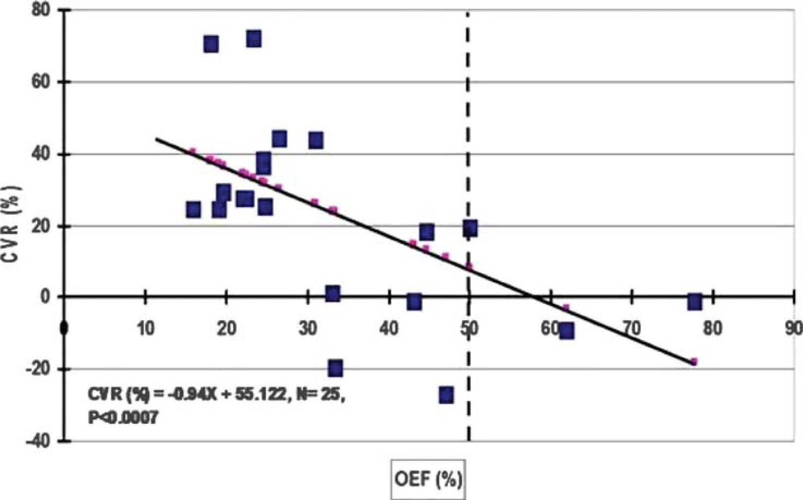
Objectives: Impaired cerebral hemodynamics like misery perfusion is a high risk factor of subsequent ischemic stroke. The acetazolamide (ACZ) challenge test, which assesses cerebral vasoreactivity (CVR), has been reported to be useful to evaluate cerebral perfusion reserve and to predict the risk of cerebral hemodynamic impairment in patients with major cerebral arterial steno-occlusive disease. There are two types of CVR reduction, with or without decrease in baseline cerebral blood flow (CBF), and the former is considered overlapping significantly with a condition of miserly perfusion. The purpose of this study was to investigate the long-term prognosis of patients with preserved baseline CBF and reduced CVR measured with PET.
Methods: Twenty-nine patients with symptomatic unilateral major cerebral arterial occlusion or severe stenosis (>70%) in the internal carotid or middle cerebral arteries were involved in this study. They suffered from ipsilateral ischemic events, including transient ischemic attack (TIA) and minor complete stroke (modified Rankin scale 1 or 2). All patients underwent O-15 gas and water PET scans to measure CBF, oxygen extraction fraction (OEF), cerebral metabolic rate for oxygen (CMRO2) and blood volume (CBV). All patients were followed at least 12 months and were medically treated for infarction and for underlying diseases such as hypertension, diabetes mellitus, and hyper-lipidemia during follow-up periods. The primary endpoint was stroke recurrence or death because of any diseases.
Results: Based on CVR values, 29 patients studied were divided into two groups of reduced CVR (N = 16, 64±7 years) and normal CVR (N = 13, 57±9 years). CVR values were 2.62%±9.56% in reduced CVR group and 25.2%±11.4% in normal CVR group. None of them showed significant decrease in baseline CBF in the ipsilateral hemisphere. There were no significant differences in CBF, CBV, OEF, CMRO2 between the groups. Patients with normal CVR were followed up for 38.9±9.3 months and those with reduced CVR were followed up for 37.5±20.5 months. There were no significant incidences of TIA, minor stroke and death during the follow-up periods. One patient with normal CVR was dead because of a heart disease at 14 months after his PET study. Kaplan–Meier analysis and Mantel-Cox log-rank statistics showed that the incidence of ipsilateral stroke or death during follow-up periods was not significantly different between the two groups.
Conclusion: Patients with CVR reduction without decrease in baseline CBF showed no ischemic events during the follow-up period in this study, indicating these patients can be treated by medication for cerebral circulation and baseline diseases. The present long-term prognosis study showed the low risk of patients with sufficient cerebral perfusion at baseline condition even though they had a poor CVR in the affected territory.
378. Autoregulation of cerebral blood flow to changes in arterial pressure in mild Alzheimer's disease
A. Zazulia1, T. Videen1, J. Morris1 and W. Powers2
1Neurology, Washington University School of Medicine, St Louis, Missouri, USA; 2Neurology, University of North Carolina, Chapel Hill, North Carolina, USA
Background: Recent studies in transgenic mice overexpressing the amyloid precursor protein indicate that impaired autoregulation of cerebral blood flow (CBF) to changes in arterial pressure may be of critical importance in the development of pathological Alzheimer's disease (AD). Given the practical relevance of such a finding in guiding treatment of hypertension in the elderly, we designed this study to determine whether autoregulation is impaired in patients with AD.
Methods: Nineteen subjects aged 74±6 years with very mild (Clinical Dementia Rating [CDR] 0.5, n = 15) or mild (CDR 1, n = 4) AD, 74% of whom had treated mild-to-moderate hypertension, underwent 15O-PET CBF measurements before and after mean arterial pressure (MAP) was lowered from 108±13 to 93±10 mm Hg with intravenous nicardipine infusion, PET imaging with the benzothiazole amyloid-imaging agent 11C-PIB, and magnetic resonance imaging. Each subject's CBF and 11C-PIB PET images were aligned, co-registered to a standard mean CBF image in Talairach atlas space, and masked to exclude non-cerebral structures. Bilateral hemispheric (global) CBF measurements were made using a mask in Talairach space restricted to brain between Z = +50 and Z = −10. CBF measurements were also obtained in regions of increased 11C-PIB uptake (>1.7 times uptake in cerebellar gray matter), in 0.56 cc spheres (10 mm diameter) in the anterior and posterior cortical borderzone regions of the middle cerebral artery territory, and in 3 to 20 cc regions of T2-weighted imaging defined leukoaraiosis.
Results: There was no significant difference in mean CBF before and after MAP reduction in the bilateral hemispheres (44.7±9.9 versus 43.1±8.9 mL × 100 g−1 × min−1, P = 0.22), regions of increased 11C-PIB uptake (48.6±14.3 versus 45.8±12.6 mL × 100 g−1 × min−1, P = 0.09), cortical borderzones (42.8±11.0 vs. 40.8±9.5 mL × 100 g−1 × min−1, P = 0.11), and regions of leukoaraiosis (20.9±7.2 versus 20.6±7.0 mL × 100 g−1 × min−1, P = 0.86). Differences in CBF among the four analyses are likely due to differences in the fraction of gray matter in the regions-of-interest.
Conclusions: The lack of significant change in CBF in bilateral hemispheres, cortical borderzones, or regions of high 11C-PIB uptake or leukoaraiosis with a 10 to 15 mm Hg reduction in MAP suggests that there is neither a generalized nor local defect of autoregulation in AD.
382. Spatial variation analysis of the plasma and red blood cell flow in rat somatosensory cortex
H. Kawaguchi1, K. Masamoto1,2, T. Obata1 and I. Kanno1
1National Institute of Radiological Sciences, Chiba; 2The University of Electro-Communications, Tokyo, Japan
Objectives: Spatiotemporal variations in the flow of fluorescently labeled plasma and red blood cells (RBCs) were characterized by calculating the locally-controlled arterio-venous transit times in the somatosensory cortex of rats from image acquired with confocal microscopy.
Methods: Sprague-Dawley rats (6 to 8 w) were anesthetized with isoflurane (5% for induction and 1.3% to 1.5% for experiments), and an area (3 × 3 mm2) on the left parietal bone over the somatosensory cortex was removed to allow access to brain surface. The respiration rate was maintained at 0.87 Hz with mechanical ventilation. For visualization of the cortical flow, a cocktail (0.02 mL) of Qdot-605 (1 μmol/L used as a plasma marker) and FITC-labeled RBCs was injected into the external carotid artery at a rate of 2.23 mL/mins. The images of RBC and plasma flow were simultaneously obtained through a band-pass filter (500 to 590 nm and 595 to 615 nm, respectively) with confocal microscopy at an excitation of 488 nm. The frame rate was 14.2 fps (interval of 70.4 ms), the total measurement time 18 secs (256 frames), and image size 512 × 512 pixels (FOV: 1.82 × 1.82 mm2). Average and standard deviation of the baseline intensity before arrival of the fluorescent markers was calculated several dozen frames from the start point of the measurements. The appearance time of the fluorescent markers was then measured by determining the earliest time point at which the intensity was greater than the average plus 2 SD on a pixel-to-pixel basis. To ensure no intensity overlap from neighboring pixels, spatial filtering was not used.
Results:Figure 1 shows a pixel-by-pixel map of the appearance time for the RBCs (A) and plasma marker (B). The pixel counts corresponding to vessels in the RBC map were 70% to 80% of that of the plasma map. The difference may be due to the rheological properties of RBCs and/or fluorescent wavelength-dependent differences in the optical properties of tissue, dyes, or detectors. The diameter of the detected vessels was observed to be greater in the plasma map than in the RBC map (1 to 2 pixels: 3.5 to 7.0 μm), which agrees well with a previous report.1 Veins of relatively large size (≥50 mm) demonstrated a laminar flow, as seen by the spatial pattern of flow distribution in the upstream veins. A small difference in the appearance time between the plasma and the RBCs was observed for the arteries (−2 to 3 frames). In contrast, the veins had a longer appearance time for the plasma marker[O1] in comparison to that of the RBCs (−2 to 9 frames). The difference varied spatially across the venous network.
Images of the appearance time of markers.
Conclusions: The spatial variation of plasma and RBC flows were successfully measured in this study. The present method can be used for the study of CBF regulation under various physiological conditions and local activations.
392. Coordinated response of arterial networks induced by local stimulation in anesthetized rat somatosensory cortex
K. Masamoto1,2, T. Obata2 and I. Kanno2
1The University of Electro-Communications, Tokyo; 2National Institute of Radiological Sciences, Chiba, Japan
Aim: To characterize the spatiotemporal dynamics of cerebrovascular networks induced by local stimulation, the cortical surface and intracortical vascular networks were imaged with confocal or multi-photon microscopy in the anesthetized rat somatosensory cortex.
Materials and methods: Sprague-Dawley rats (6 to 8 weeks) were used for the experiments following an experimental protocol approved by the Institutional Animal Care and Use Committee. The animals were anesthetized with isoflurane (5% for induction and 1.3% to 1.5% for experiments), and endotracheal intubation was performed for mechanical ventilation. Catheters were placed into the femoral vein and artery for administration of fluorescent marker and arterial blood sampling, respectively. An area (3 × 3 mm) on the left parietal bone over the somatosensory cortex was removed. Arterial blood pressure, heart rate, and respiratory parameters were monitored throughout all experiments. Rectal temperature was maintained at 37°C. For visualization of cortical vasculature, a bolus injection of Qdot 655 (1 μmol/L, 0.2 to 0.4 mL) was performed. The image of the cortical surface vessel was obtained with confocal microscopy with a 488-nm excitation, and the image of intracortical vessel was obtained with multi-photon microscopy at an excitation of 900 nm (∼2.0 W at laser output) up to a depth of 0.6 to 0.8 mm from the cortical surface with a z-step of 0.01 mm. The image resolution in a x-y plane was 512 × 512 pixels, and a sampling rate was 13 frames per second. The artery and vein emerging from the parenchyma was identified by tracking pial arterial and venous networks, respectively. The cross-section diameter was measured at the focal point. The neural stimulation in the measured region was induced with electrical pulse (1.5 mA, 1.0-ms duration at 12 Hz) to contra-lateral side of forepaw. The stimulation trial was repeated with 8 times and averaged across all trials. The CBF response and somatosensory evoked potentials were also measured with laser-Doppler flowmetry (LDF) and a surface electrode, respectively, after the vascular imaging experiments.
Results and discussion: Stimulation-induced vasodilation was observed only for arterial side, whereas no detectable changes in the vascular diameter were obtained for the venous side. The time of onset of the arterial vasodilation (∼0.5 secs) was in good agreement with the time-courses of CBF changes measured with LDF. The peak of dilation accounted for ∼10% of resting vessel diameter. The arterial dilation started at the local point along the small arterioles, and spread into large upstream arteries located on the cortical surface. About 40% of arterioles that dive into the parenchyma showed vasodilation over the somatosensory area (FOV: 1.8 × 1.8 mm2) where 15 to 20 penetrating arterioles were found. The penetrating arterioles that showed vasodilation was localized around the activated regions, although the upstream surface arteries respond globally. Further, the vasodilation of upstream arteries extended in a stimulus-dependent manner. These findings indicate that the branching point from parent surface arteries to the penetrating arterioles play a key role in controlling the spatial regulation of blood supply distribution in the intracortical regions where the energy demand locally varied depending on neural activity.
452. Involvement of calcium-calmodulin dependent protein kinase II on endothelin receptor expression in cerebral arteries of rat
R. Waldsee, S. Eftekhari and L. Edvinsson
Clinical Science, Experimental Vascular Research, Lund, Sweden
Objective: Experimental cerebral ischemia and organ culture of cerebral arteries result in enhanced expression of endothelin ETB receptors in smooth muscle cells via increased transcription.1,2 The present study was designed to evaluate the involvement of calcium-calmodulin dependent protein kinase (CAMK) on the expression of endothelin receptors after organ culture.
Methods: Rat basilar arteries were incubated for 24 h with and without the CAMK inhibitor, KN93. The contractile responses to endothelin-1 (ET-1; ETA and ETB receptor agonist) and sarafotoxin 6c (S6c; ETB receptor agonist) were studied using a sensitive myograph. The mRNA levels of ETA and ETB receptors and of CAMKII were determined with real-time polymerase chain reaction (PCR) while the protein level was evaluated by semi-quantitative immunohistochemistry.
Results: The mRNA levels of CAMKII and of the ETB receptor were increased during organ culture but there was no change in ETA receptor expression. This effect was abolished by co-incubation with KN93. In functional studies, KN93 attenuated the S6c-induced contraction and to a minor degree the ET-1 induced response. This was confirmed at the protein level by immunohistochemistry where the endothelin receptors were found co-localised with CAMKII, in the smooth muscle cells. Phosphorylated extracellular signal-regulated kinase ERK1/2 was measured by immunohistochemistry. Incubation of arteries with KN93 decreased the level of pERK1/2. In addition, pERK1/2 co-localised both the endothelin receptors and CAMKII.
Conclusion: Our results show that the CAMKII is involved in the endothelin receptor regulation and interacts with the ERK1/2 pathway, resulting in enhanced receptors expression in rat basilar artery.
523. Gaba and glutamate mediate the neurovascular coupling response to whisker stimulation partly via arachidonic acid derivatives
P. Fernandes1, A. Kocharyan1, C. Lecrux1, E. Vaucher2 and E. Hamel1
1Laboratory of Cerebrovascular Research, Montreal Neurological Institute, McGill University; 2École d'Optométrie, Université de Montréal, Montréal, QC, Canada
Background and aims: Previously, we demonstrated that increases in cortical cerebral blood flow (CBF) induced by whisker stimulation occurred concurrently with cortical activation of COX-2 pyramidal cells and specific subsets of GABA interneurons (Fernandes P et al, Brain07, Abstract #BO12–3). Here, we investigated the contribution of glutamate, GABA and neuroglial components in this evoked CBF response using pharmacological blockade of receptors and/or astroglial synthetic pathways.
Methods: Increases in cortical CBF during whisker deflection (20 secs, ∼10 Hz) were measured in the contralateral barrel cortex by laser Doppler flowmetry at baseline and after intracisternal (i.c., 3 μL of a 10−4 M, pH 7.4 buffered solution) injection of vehicles, antagonists of NMDA (MK-801), GABA-A (picrotoxin), epoxyeicosatrienoic acid (EET) (14,15-EEZE) receptors, inhibitors of prostaglandin synthetic enzyme cyclooxygenase-2 (COX-2) (NS-398) and of the EET synthetic enzyme P450 epoxygenase (MS-PPOH). The evoked CBF response was recorded in urethane-anesthetized rats following administration of these pharmacological compounds either alone or in combination, with 4 to 10 rats used per group. The femoral artery was cannulated for continuous monitoring of blood gases and blood pressure, and body temperature was measured throughout the experiments, with no differences observed among groups. Changes in CBF were compared by repeated-measures analysis of variance (ANOVA) or by one-way ANOVA for three groups.
Results: Vehicles had no effect on the CBF response evoked by whisker deflection when compared to baseline. As expected, MK-801 decreased the CBF response (−53.1%±6.8%, P<0.001) (Gsell et al, J Neurosci 2006; 26:8409–8416). Similarly, picrotoxin significantly reduced the CBF response (−39.1%±3.9%, P<0.001), and combined blockade of NMDA and GABA-A receptors demonstrated a significant additive inhibitory effect (−73.1%±5.3%, P<0.01). In agreement with a recent study (Liu et al, 2008, Am J Physiol Heart Circ Physiol, 295:H619–H631), inhibition of EETs synthesis with MS-PPOH or antagonism of the EET receptor with 14,15-EEZE diminished the perfusion response (−43.3%±4.1%, P<0.001 and −32.8%±5.6%, P<0.01, respectively). Their combined administration resulted in a significant additive effect on the CBF response (−62.4%±5.8%, P<0.01). In contrast, the combined administration of MS-PPOH with MK-801 or picrotoxin did not result in a greater effect than using MS-PPOH alone, suggesting a common route of action. The CBF response was reduced by COX-2 inhibition (−46.3%±3.9%, P<0.001), and the combined administration of NS-398 and picrotoxin had an additive inhibitory effect on the CBF response (−59.6%±5.7%, P<0.01).
Conclusions: These results show the interplay between pyramidal cells, GABA interneurons, and astrocytes in the regulation of CBF in the rat whisker barrel cortex. Together, the data indicate that:
both glutamate and GABA are involved in the perfusion response to whisker stimulation, and
they act in parallel to activate the synthesis of arachidonic acid metabolites from the EET and/or prostaglandin pathways, some of these mediators being synthesized and released by astrocytes.
Overall, the data indicate that inhibitory and excitatory cells in the somatosensory cortex contribute to the neurovascular coupling response to thalamocortical afferents, and that astrocytes can be intermediaries for both types of neurons.
Supported by CIHR grant (MOP-84209, EH).
532. Candesartan improves autoregulation of local cortical cerebral blood flow during haemorrhagic hypotension while enalaprilat does not
O.B. Paulson1,2, S.T. Sigurdsson1,3, A. Høj Nielsen3 and S. Strandgaard3
1Neurobiology Research Unit, Copenhagen University Hospital, Rigshospitalet; 2Danish Research Center for Magnetic Resonance, Copenhagen University Hospital, Hvidovre; 3Department of Nephrology, Copenhagen University Hospital, Herlev, Copenhagen, Denmark
Objectives: The renin-angiotensin system (RAS) maintains a tone in the cerebral resistance vessels, which can be influenced by RAS blockers. Several ACE-inhibitors (ACEi) have this effect. The beneficial effects of angiotensin II receptor blockers (ARBs) on stroke incidence and outcome in clinical trials might be related to this haemodynamic effect. Bradykinin is a potent endothelium dependent vasodilator which is broken down by ACE. Treatment with ACEi thus increases bradykinin levels. The present study investigated the effect of the ARB candesartan, the ACEi enalaprilat and the bradykinin antagonist HOE 140 on the lower limit of autoregulation of local cortical cerebral blood flow (CBF) compared to controls.
Methods: The study was carried out in 4 groups of Sprague–Dawley rats in general anaesthesia with isofluran and N2O. Temperature and PaCO2 were kept stable. Both femoral arteries and veins were cannulated. A craniotomy was made over one hemisphere, leaving the dura intact. CBF was measured continually on the surface of the brain with laser Doppler technique before and after intravenous injection of candesartan (0.2 mg/kg), enalaprilat (2 mg/kg) or the bradykinin antagonist Hoe 140 (4 nmol and then 2 nmol every 20 mins). Blood pressure was stabilised with nor-epinephrine, and subsequently gradually reduced by controlled bleeding.
Results: The lower limit of CBF autoregulation (mean±1 s.d.) in the candesartan group was 38±7 mm Hg in the enalaprilat group 51±7 mm Hg, in the Hoe 140 group 52±6 mm Hg and in the control group 44±4 mm Hg. There was a statistically significant difference between all groups except between the enalaprilat group and the control group.
Conclusion: A shift of the lower limit of local cortical CBF towards lower pressure is an effect that in clinical settings might be advantageous. Candesartan in the present acute experiment caused such a shift and it is surprising that enalaprilat did not, contrary to other ACE-inhibitors in earlier studies Results of combination treatment with ARBs and Hoe 140 and ACEi and HOE 140 are under way.
546. Role of endothelin in impaired responses of cerebral arterioles during type 1 diabetes mellitus
D. Arrick, G. Sharpe, H. Sun and W. Mayhan
Cellular/Integratvie Physiology, University of Nebraska Medical Center, Omaha, Nebraska, USA
Previous studies have suggested that endothelin-1 may contribute to vascular abnormalities during a variety of disease states, including Type 1 diabetes mellitus. Endothelin-1 may contribute to structural and functional abnormalities of the blood vessels, and may also regulate the expression of other growth factors and cytokines to influence vascular function. However, there is a lack of information regarding the precise role of endothelin-1 in altered NOS-dependent responses of cerebral arterioles during Type 1 diabetes. Thus, our goal was to determine whether acute inhibition of endothelin-1 receptors (BQ-123) could influence NOS-dependent responses of cerebral arterioles in diabetic rats. We measured diameter of pial arterioles in nondiabetic and diabetic (STZ; 50 mg/kg) rats in response to eNOS- and nNOS-dependent (ADP and NMDA) and -independent (nitroglycerin) agonists before and during treatment with BQ-123. In addition, we measured superoxide production by cerebral cortex tissue obtained from nondiabetic and diabetic rats. We found that eNOS- and nNOS-dependent dilation of pial arterioles was impaired in diabetic compared to nondiabetic rats. In addition, treatment with BQ-123 restored impaired responses of cerebral arterioles in diabetic rats towards that observed in nondiabetic rats. Further the production of superoxide anion by cortex tissue was increased in diabetic rats when compared to nondiabetic rats. We suggest that endothelin-1 may contribute to impaired responses of cerebral arterioles during Type 1 diabetes. We speculate that endothelin receptor antagonism may be a potential therapeutic tool for the treatment of cerebrovascular dysfunction observed in diabetic subjects.
562. Vascular response in mice deficient in the potassium channel, TREK-1
K. Namiranian, E.E. Lloyd, S.P. Marrelli and R.M. Bryan
Anesthesiology, Baylor College of Medicine, Houston, Texas, USA
Objective: TREK-1 is a member of a recently discovered family of ion channels termed two-pore domain potassium channels (K2P). The K2P channels were discovered by searching for genes with homology to the highly conserved potassium-selective pore domain. In the nervous system, TREK-1 is reported to be involved with neuroprotection, depression, and the mechanism of action of volatile anesthetics. However, its role in the cardiovascular system is not completely understood. We aim to investigate the role of TREK-1 in cardiovascular system and we hypothesized that TREK-1 is involved with regulating the response of vascular elements to vasodilators and vasoconstrictors.
Methods: Since there are no specific blockers or activators for TREK-1, a knockout (KO) mouse line was generated by replacing the first two coding exons of TREK-1 with a neomycin/beta-galactosidase cassette. Reverse transcriptase-PCR was used to detect the expression of TREK-1 mRNA. Vascular responses were measured by monitoring the artery diameter in isolated basilar arteries (BA) and isolated perfused middle cerebral arteries (MCA), both pressurized at 75 mm Hg. Vascular reactivity in aorta was measured in isolated aortic rings attached to a force transducer. The cardiac function was evaluated by Doppler ultrasound, and blood pressures were measured invasively in carotid artery and left ventricle via an arterial catheter.
Results: TREK-1 mRNA was found in mouse heart and all arteries studied (aorta, femoral, renal, mesenteric, middle cerebral and basilar) with the exception of the carotid artery. TREK-1 KO mice are viable and fertile and do not show any gross abnormality. TREK-1 mRNA was not detected in any of the tissues sampled from KO mice. The cardiac function and blood pressure were similar in TREK-1 KO and WT mice (n = 5). In the perfused pressurized MCA, phenylephrine (0.1 to 100 μmol/L) induced similar constriction (n = 9), and luminal application of ATP (10 and 100 μmol/L) elicited similar endothelium-mediated dilation in WT and KO littermates (n = 12). The NO and EDHF component of the ATP-dilation were also similar in WT and KO mice. Acetylcholine (10 μmol/L)-mediated dilations in BA of KO mice were not different from wild-type (WT) mice, either before or after inhibition of nitric oxide synthase (n = 5 to 6). Linoleic acid (LA, 10 and 100 μmol/L), an activator of TREK-1 and Ca-activated K channels (BKCa), dilated endothelin-constricted BA of WT and KO mice to similar extents. However, after selectively blocking BKCa with penitrem A, the dilation to LA in BA from KO mice was reduced 19%±5%, while it was not affected in WT (n = 6). This suggests that part of LA-induced dilation is mediated via TREK-1.
Conclusions: Since the studied vascular responses and cardiac function were not altered in the absence of TREK-1, this channel seems to be dispensable for cardiovascular system. Direct activation of TREK-1 dilates cerebral arteries; therefore TREK-1 may be a therapeutic target for designing vasodilators for the pathological conditions where blood flow is disturbed.
584. Endothelium-dependent relaxation and antioxidant effects by G protein-coupled receptor GPR30 agonists in rat carotid arteries
B.R.S. Broughton, A.A. Miller and C.G. Sobey
Department of Pharmacology, Monash University, Clayton, VIC, Australia
Background and aims: Recent studies have identified that the novel membrane estrogen receptor, G protein-coupled receptor GPR30, is present in blood vessels. However, the potential role(s) of GPR30 in the vasculature remains unknown. We therefore examined whether putative agonists of GPR30 may possess vasorelaxant and antioxidant effects similar to those reported for estrogen.
Methods: Using wire myography we assessed both endothelium-dependent and -independent relaxation responses to the GPR30 agonists, G-1 and 5408–0877 (1 nmol/L to 10 μmol/L), in U46619-precontracted common carotid arteries from Sprague Dawley rats. Acetylcholine (ACh, 3 μmol/L) was used to verify the presence or absence of an intact endothelium and all relaxations were expressed as a % of the response to sodium nitroprusside (SNP, 10 μmol/L). Furthermore, we tested the effect of G-1 (10 μmol/L) on NADPH (100 μmol/L)-stimulated superoxide production by rat carotid and cerebral (pooled middle cerebral and basilar) arteries using lucigenin (5 μmol/L)-enhanced chemiluminescence. Specific immunofluorescence was also used to confirm GPR30 expression in the arterial wall.
Results: We found that G-1 induces a concentration-dependent relaxation in carotid arteries from both males (e.g. 42%±8% at 10 μmol/L; N = 6) and females (32%±7% at 10 μmol/L; N = 6). Similarly, 5408–0877 induced a concentration-dependent relaxation in arteries from males (e.g. 35%±10% at 10 μmol/L; N = 6) and females (34%±11% at 10 μmol/L; N = 6). Overall, ACh relaxed carotid arteries by 87±2%. Interestingly, G-1- and 5408–0877-induced relaxation was abolished by endothelium removal. In addition, NADPH-stimulated superoxide production was ∼30% to 40% lower in carotid and cerebral arteries treated with G-1 versus untreated arteries. Furthermore, GPR30 immunoreactivity was observed in endothelium and vascular smooth muscle cells of carotid arteries from both genders.
Conclusions: In summary, this is the first study to assess the vascular effects of GPR30 agonists. Our data suggest that GPR30 is expressed throughout the vascular wall and receptor activation elicits endothelium-dependent relaxation of the carotid artery in male and female rats. In addition, activation of GPR30 reduces levels of NADPH oxidase-derived superoxide in carotid and cerebral arteries. Thus, these vasorelaxant and antioxidant properties of GPR30 are consistent with this receptor being a potential therapeutic target in the cerebral circulation during vascular disease.
592. FITC-Labeled RBC tracking in arteriolo-arteriolar anastomoses in control state and during ischemia after MCA occlusion in mice
H. Toriumi, J. Tatalishvili, M. Tomita, Y. Tomita, H. Hattori, M. Unekawa and N. Suzuki
Department of Neurology, School of Medicine, Keio University, Tokyo, Japan
Purpose: In arteriolo-arteriolar anastomoses (AAA) between MCA and MCA, blood had been hypothesized not to be actually flowing (watershed area) caught between two opposing arterial pressures, and only in occasions of one artery closure flow would get started as collaterals. To test this hypothesis we examined flow in AAA employing labeled RBC.
Methods: FITC-labeled RBC flow in AAA through a closed cranial window in 16 anesthetized C57BL/6J mice was examined in control state and after MCA occlusion (MCAO) employing a high speed camera laser scanning confocal fluorescence microscopy (Tomita et al, Microcirc 15, 163 (2008).
Results: When tracking RBC movements in AAA in control state, we noticed a paradoxical flow as shown in Figure 1: top panel, in which blood entered into an AAA from both MCA and ACA sides. The opposing blood flows collided at a meeting point and in some cases disappeared. White arrows indicate the direction of the flows. At the meeting point blood apparently sank into a hole of the origin of the penetrating arteriole, which was, mort of cases, not to be seen from above. Without FITC-labeled RBCs the confluent flow to the vertical direction had been missed. We found abundant such hidden ‘T-junctions’ in AAAs; almost 2 to 3 per an AAA. However, dually fed confluent T-junction was only one per AAA. As shown in middle and bottom panels of Figure 1, dually fed junction was not fixed in position, but functionally liable to change from one to another with given hemodynamic conditions. Upon MCA occlusion, RBC flow immediately stopped at the MCA side but blood subsequently started to move to the retrograde direction towards MCA. The blood was supplied apparently from ACA. Figure 2 is a microphotograph in which 2 branches of MCA were seen where blood flow direction had been from top to bottom in control state. However, upon MCAO flow reversed in the right branch in all 7 cases studied (P<0.05).
The blood apparently came from ACA through AAA, moved retrogradely towards the MCA stem and then branched off to supply to the left branch (arrows) with a great delay. When viewed from RBC velocities and numbers in capillaries in the ischemic tissue MCAO induced immediate RBC disappearance in the core ischemic region, and to a lesser extent in the penumbra near the AAA. Thus, unlike a general concept of ‘watershed’, the local blood flow near AAA was rather preserved. In preliminary experiments, we observed that AAAs became a center to develop collateral channels and key locations of angiogenesis in the marginal zone of ischemia.
Conclusions: In AAA blood was flowing even in control state towards a T-junction which was dually supplied both from MCA and ACA. The T-junction was functionally liable to change to a next T-junction. The so called watershed area near AAA appeared to be well supplied.
593. Sustained dilation of pial arterioles by arginase inhibition during prolonged middle cerebral artery occlusion
S Cao, D Berkowitz and R Koehler
Anesthesiology/Critical Care Medicine, Johns Hopkins University, Baltimore, Maryland, USA
Objectives: Administration of L-arginine1 or endothelin antagonists2,3 has been reported to increase intraischemic cerebral blood flow during middle cerebral artery (MCA) occlusion (MCAO). These observations raise the possibility that nitric oxide (NO) production is submaximal because of limited arginine availability and that reduced NO production promotes endothelin release during MCAO. In peripheral vessels, arginase activity can limit arginine availability for NO production.4 To determine if arginase activity limits vasodilation during MCAO, the diameter of pial arterioles in the ischemic border region was measured during cranial window superfusion with the arginase inhibitor ABH, [2(S)-amino-6-boronohexanoic acid]. Results were contrasted with those obtained by administration of L-arginine or the endothelin-A receptor antagonist BQ-610.
Methods: In isoflurane-anesthetized and mechanically ventilated rats, cranial windows were constructed over the MCA border region for measurement of pial arteriolar diameter. MCAO was produced for 2 h by the intraluminal filament technique.
Result: MCAO initially produced 38%±9% (±s.d.) dilation that gradually subsided to 12%±11% at 2 h of MCAO in the MCA border region in a control group superfused with artificial cerebrospinal fluid (n = 8). Similar results were obtained in another control group with no cranial window superfusion. With superfusion of 1 μmol/L of ABH throughout MCAO, the initial dilation of 38%±10% was largely sustained (32%±18%; n = 6) at 2 h MCAO. With intravenous infusion of L-arginine (4 mmol/kg/h for 20 mins+2 mmol/kg/h for remainder of experiment), initial dilation after MCAO (40%±14%) was better maintained at 2 h MCAO (25%±13%, n = 7) than in a control group (initial dilation 37%±8%; 7%±15% at 2 h; n = 9). Likewise, superfusion of 3 μmol/L of BQ610 during MCAO prevented the loss of pial arteriolar dilation (32%±7% to 32%±15%; n = 8) compared to a control group superfused with vehicle (0.02% DMSO; 34%±4% to 5%±10%; n = 9). Intravenous injection of BQ610 (0.5 μmol/kg; n = 5) at 90 mins of MCAO increased laser-Doppler flow in the cortical border region from 43%±19% to 69%±20% of pre-ischemic baseline, thereby demonstrating that flow can be increased at a time when pial arterioles have a diminished dilation. Furthermore, intravenous injection of ABH (dose in 3 μmol/kg; n = 4) at 90 mins of MCAO increased LDF from 41%±28% to 76%±44% of pre-ischemic baseline, thereby indicating that an arginase inhibitor was as effective as an endothelin antagonist in improving perfusion in the border region. Mean arterial blood pressure and blood gases were unchanged in all groups.
Conclusion: These results indicate that both arginase activity and endothelin-A receptor activation contribute to the gradual loss of pial arteriolar dilation in the ischemic border region during prolonged MCAO. We speculate that increased arginase activity decreases endothelial NO production, which then leads to increased release of endothelin.
612. Lack of capillary dilatation during functional hyperemia in bicuculline-induced epileptic foci
F. Fernández Klett1,2, N. Offenhauser1, U. Dirnagl1,3, J. Priller2 and U. Lindauer1,3
1Experimental Neurology; 2Neuropsychiatry and Molecular Psychiatry; 3Center for Stroke Research, Charité-Universitätsmedizin Berlin, Berlin, Germany
Background and aim: The spatial extent of the functional hyperemia associated with local increases in cerebral neuronal activity is determined by the architecture of the vascular arterial tree and the disposition of flow control structures. CNS capillaries are invested with pericytes, cells with contractile properties that may actively partake in the control of flow. Our aim was to test whether a dilatatory response of capillaries is substantial to the development of functional hyperemia.
Methods: We have used beta-actin-GFP mice, in which endothelial cells and pericytes can be fluorescently imaged, to assess dynamic vessel diameter and flow changes by means of in vivo two-photon microscopy. To elicit local transient increases in neuronal activity, we inserted a micropipette in the parietal cortex filled with the GABA-receptor antagonist bicuculline, which lead to the development of brief, recurring bursts of neuronal spike activity (inter-ictal spikes, IIS). We categorized cortical vessels into pial, penetrating and precapillary arterioles or capillaries based on location, diameter, and vessel wall morphology. Planar time-lapse images of these vessels (5 to 9 Hz) were collected simultaneously to IIS to measure rapid diameter changes. Subsequently, we performed line scans of the vessel lumen to trace IIS-associated changes in red blood cell (RBC) velocity.
Results: In total, the responses of 17 pial, 13 penetrating and 35 precapillary arterioles and 60 capillaries from 8 mice were studied. Mean±s.d. diameter and RBC velocity of the different segments were: pial art.: 14.0±4.8, 6.9±2.6; penetrating art. 14.7±4.0, vel. n.a.; precapillary art. 7.5±1.7, 2.8±1.3; capillaries 4.6±1, 0.6±0.4 (μm and mm/secs, respectively). The average dilatatory response of pial, penetrating and precapillary arterioles reached maximally 2.4%±.3.3%, 3.4%±3.5% and 2.3%±5% over pre-IIS values, respectively (∼2 secs after IIS). Average RBC velocity in capillaries increased maximally by 10.6%±12% over pre-IIS values. In contrast, no dilatation was observed in capillaries, measured at points immediate to pericytes (0.1%±2.5% over pre-IIS diameter, measured at time of maximum RBC velocity increase). No significant difference was observed in the rise time of neither diameter nor RBC velocity response between different vessel types.
Conclusions: We hypothesized that, if a dilatatory response would originate in capillaries and then travel upstream to larger vessels, capillary vasoactivity would be emphasized in a context of brief neuronal activation. Bicuculline induced IIS are charachterized by robust yet brief neuronal spiking bursts, each lasting typically <500 ms. However, no involvement of capillaries in the dilatatory response could be detected. Precapillary arterioles seem therefore to be the finest vessels determining the spatial extent of functional hyperemia in mice. Previous reports of capillary dilatation during hypercapnia or sustained functional stimulation are likely to represent passive dilatation of capillaries due to increased perfusion pressure effected at upstream arterioles.
647. Near-infrared spectroscopy in primary and secondary motor areas in self-paced versus externally cued motor tasks
C. Drenckhahn1,2,3, J. Steinbring2,3, J. Duemmler3, M. Kohl-Bareis3,4 and J.P. Dreier1,2,3
1Experimental Neurology; 2Neurology, Charité, Universitätsmedizin Berlin; 3Berlin Neuroimaging Center, Berlin; 4RheinAhrCampus, University of Applied Sciences Koblenz, Remagen, Germany
Objectives: During the last years the application of near-infrared spectroscopy (NIRS) was studied in different setups to monitor cerebral oxygenation and perfusion in critical ill patients. However, NIRS still is not evaluated to be used as a validated bedside method and to influence clinical assessment and therapeutic decisions.
Methods: Using NIRS in human healthy subjects we investigated alterations in deoxy-hemoglobin (Hbdeoxy) concentrations as an indirect parameter for a hemodynamic response in the primary motor cortex (M1) and the supplementary motor area (SMA). M1 and SMA were measured simultaneously. 16 strongly right-handed volunteers underwent different finger tapping tasks which are known to be associated with increase in cerebral blood flow in M1 and SMA. The tasks were designed as blocks of 20 secs or single repetitive finger tapping either self-paced or externally triggered. 8 light sources and 7 detectors were positioned over the left M1 and the SMA covering an area of 12.5 × 7.5 cm. Time course and amplitude were analysed for the concentration of Hbdeoxy. For spatial analysis, the recording points with significant decrease in Hbdeoxy were determined.
Results: We detected a circumscribed decrease in Hbdeoxy at two different locations corresponding to SMA and M1 during functional activation. Significant changes in Hbdeoxy were located within an area of ∼10 cm2 in projection on M1 and ∼15 cm2 in projection on SMA, respectively. Both, M1 and SMA showed significantly higher shifts in Hbdeoxy following self paced movements demonstrating a higher level of activation due to planning and initiating in the self-paced task compared to the externally cued task (−2.81 × 10⁻⁵ versus −2.22 × 10⁻⁵ mmol/L in M1, −1.63 × 10⁻⁵ versus −1.25 × 10⁻⁵ mmol/L in SMA). In SMA the decrease of Hbdeoxy was significantly more accentuated following right hand movements compared to the increase during the left hand task (−3.4 × 10⁻⁵ versus −2.2 × 10⁻⁵ mmol/L). This might indicate a more pronounced vascular response due to functional activation of the dominant hemisphere.
Conclusions: NIRS still has to be validated prior to its routine use as a non-invasive bedside tool in a clinical setup. Our data shows a reproducibility of NIRS compared to more complex methods like fMRI, PET or perfusion CT.
684. Olmesartan can maintain normal function of endothelial nitric oxide synthase in the brain microvessels in spontaneously hypertensive rats
N. Oyama1, Y. Yagita2, T. Sasaki1, E. Omura-Matsuoka1, Y. Terasaki1, Y. Sugiyama2, S. Okazaki2, S. Sakoda2 and K. Kitagawa2
1Division of Stroke Center, Department of Internal Medicine; 2Department of Neurology, Osaka University Graduate School of Medicine, Suita, Japan
Introduction: The endothelial nitric oxide synthase (eNOS) is important for cerebrovascular homeostasis and the reduction in bioactivity of eNOS leads to the dysfunction of cerebrovascular endothelium with subsequent decreased cerebral blood flow (CBF) and deteriorated ischemic stroke outcome. The accumulating evidence indicates that increased blood pressure diminished eNOS expression and impaired brain endothelial function. However, the influences of hypertension to the eNOS activity in the brain microvessels are still not fully understood. The present study is aimed to investigate whether hypertension can affect the phosphorylation of eNOS protein and antihypertensive agent can be useful to preserve eNOS function in the brain microvessels.
Methods: Five- or 10-week-old male Wistar rats or spontaneously hypertensive rats (SHRs) were used for experiments. We measured blood pressure by the tail-cuff method. Five-week-old SHRs were treated orally with olmesartan (0.02% RNH-6270) or vehicle for 5 weeks. Total and Ser1177-phosphorylated eNOS protein in the brain cortex were evaluated using Western blots. To assess the contribution of eNOS to maintaining CBF, we monitored CBF in the brain cortex by laser-Doppler flowmetry after intravenously administered L-N5-(1-iminoethyl)ornithine (L-NIO; 20 mg/kg), a relatively selective eNOS inhibitor. Measurements of the cerebrovascular response to L-NIO were expressed as the percentage change of the baseline.
Results: Mean blood pressures of 5-week-old SHRs (97.5±13.8 mm Hg) were within normal range and were similar to those of 5- and 10-week-old Wistar rats (93.2±9.2 and 98.7±6.0 mm Hg). In contrast, mean blood pressures in 10-week-old SHRs were higher (154.7±11.8 mm Hg). The ratio of phospho-eNOS/total eNOS protein in 5-week-old SHRs was not significantly different from that in Wistar rats, whereas it was significantly lower in 10-week-old hypertensive SHRs (P<0.05). Five weeks treatment of olmesartan suppressed the elevation of blood pressure in SHRs (93.2±7.6 mm Hg) compared with vehicle treated SHRs (154.0±11.7 mm Hg). Phosphorylation of eNOS protein was significantly enhanced in SHRs treated with olmesartan than with vehicle (P<0.05). Phosphorylation level of eNOS was not different from that in age matched normotensive Wistar rats (Figure). In SHRs treated with olmesartan or vehicle, L-NIO infusion decreased CBF and reached a minimum at 5 to 10 m after starting infusion. At 5 mins after administration, response to L-NIO in olmesartan treated SHRs (13.4%±3.7%) was significantly higher than in vehicle treated SHRs (7.5%±3.5%) and similar to that in Wistar rats (13.2%±5.3%).
Effect of RNH-6270 treatment on phospho-eNOS
Conclusions: Activated eNOS expression in the brain was decreased in hypertensive SHRs. Olmesartan inhibited the elevation of blood pressure and preserved eNOS activity in SHRs. Additionally the contributions of activated eNOS to maintaining CBF were also preserved in olmesartan treated SHRs. Our findings indicate that antihypertensive agent such as olmesartan may help to preserve brain eNOS function against hypertension.
691. Dynamic cerebral autoregulation and change of cerebral blood flow velocity in both MCAs in acute ischemic stroke during external counterpulsation
L. Xiong, J.H. Han, X.Y. Chen, T. Leung, Y. Soo, H. Leung and K.S. Wong
Department of Medicine and Therapeutics, The Chinese University of Hong Kong, Hong Kong, Hong Kong S.A.R.
Objectives: The presence or absence of cerebral autoregulation (CA) is critical for maintenance of stable cerebral blood flow. Serial transcranial Doppler ultrasonography (TCD) monitoring cerebral blood flow during external counterpulsation (ECP) may provide more information on the status of dynamic CA (dCA) after acute stroke onset. This study investigates whether dynamic cerebral autoregulation assessed from mean blood pressure (MBP) and mean cerebral blood flow velocity (CBFV) change from baseline during ECP is impaired after acute ischemic stroke onset; whether there is an effect of time course in the dynamic autoregulatory impairment within the acute and subacute stage of ischemic stroke.
Methods: Nineteen unilateral acute ischemic stroke patients with large artery disease and fourteen healthy controls were enrolled. A course of 35 daily one-hour sessions of ECP were applied to each patient within 7 days of symptom onset and controls. Admission National Institutes of Health Stroke score of the patient was 7±2. During ECP treatment, bilateral middle cerebral arteries (MCAs) were monitored by TCD to evaluate cerebral blood flow on day 3, 5, 7, 10, 14, 18, 21, 24, 28, 35 and 60 after stroke onset in stroke patients and on the first ECP session in controls. Beat to beat heart rate, continuous finger systolic and diastolic blood pressure and cerebral blood flow velocities of MCAs were obtained before, during and after ECP by means of Task Force Monitor.
Results: In stroke patients, the greatest increase of mean CBFV in the relevant MCA was noted at day 7 which was 4.1% from baseline and then gradually decreased till day 60. The greatest increase of mean CBFV in the irrelevant side was noted at day 18 which was 6.4% from baseline and then gradually decreased till day 60.MBP increased at every measurement points during ECP. At each measurement points, the differences in the magnitude of change from baseline in both mean CBFV and MBP were significant between patients and controls (P<0.05).
Conclusions: Mean CBFV increased along with mean BP increase in bilateral MCAs, indicating a global impairment in dynamic cerebral autoregulation during the acute and subacute stage of acute ischemic stroke. The impairment of dCA would last 2 to 3 weeks after acute ischemic stroke onset.
704. Cerebral hemodynamic response or excitability is not affected by sildenafil
C. Kruuse1, A.E. Hansen2, H. Larsson3, M. Lauritzen4 and E. Rostrup3
1Department of Neurology; 2Functional Imaging Unit and Department of Radiology; 3Functional Imaging Unit and Department Clinical Physiology and Nuclear Medicine; 4Department of Clinical Neurophysiology, Glostrup Hospital, University of Copenhagen, Glostrup, Denmark
Objective: Sildenafil (Viagra), a selective inhibitor of the cyclic guanosine monophosphate (cGMP) hydrolyzing phosphodiesterase5 enzyme (PDE5) is currently used for treatment of erectile dysfunction and pulmonary hypertension and recently suggested for use in post-stroke treatment. Sildenafil induces headache and migraine. Such headache induction may be caused by generally increasing either the excitability of neurons or the cerebrovascular response to external stimuli or both. Previously no effect of sildenafil on large cerebral arteries has been found in healthy subjects. Sildenafil administration and modulation of cGMP breakdown might increase the response to visual stimulation stimuli and give rise to an increased or prolonged cerebral response to visual stimulation and hypercapnia.
Methods: In 13 healthy females (23±3 year, 70.3±6.6 kg) the effect of sildenafil on a visual (reversing checkerboard) and a hypercapnic (6% CO2 inhalation) response was evaluated using blood oxygen level dependent (BOLD) imaging (3.0 T MR scanner). On separate occasions visual evoked potential (VEP) measurements (latency (P100) and maximal amplitude) were performed. Measurements were applied at baseline and 1 and 2 h after ingestion of 100 mg sildenafil. Blood pressure, heart rate and side effects including headache were obtained.
Results: Ten of twelve subjects experienced headache on the day of MR investigations and nine of twelve on the day of VEP investigation. There was no significant difference between days. Sildenafil did not affect VEP amplitude or latency (P100). Both hypercapnia and visual stimulation elicited strong and consistent responses in each volunteer. However, the BOLD response to visual stimulation or CO2 inhalation did not change after sildenafil administration.
Conclusion: In conclusion, sildenafil induces mild headache without potentiating a neuronal or local cerebrovascular visual response or influencing a global cerebrovascular response to CO2 inhalation. Although sildenafil did induce a mild headache a generally increased neuronal response or an increased vascular reactivity appeared not to be involved in the pain process. The data further implies that sildenafil and subsequent augmentation of intracellular cGMP, does not in itself modulate the hypercapnic response in healthy subjects.
710. Reduced cerebrovascular reactivity to CO2 induced by hypertension and age: an arterial spin labeling study in rats
R. Leoni1,2, F.F. Paiva1, D.B. de Araujo2 and A.C. Silva1
1Cerebral Microcirculation Unit—NINDS, National Institutes of Health, Bethesda, Maryland, USA; 2Department of Physics and Mathematics, University of Sao Paulo, Ribeirao Preto, Brazil
Introduction: The effect of hypertension on cerebrovascular autoregulation is of great interest, as hypertension is a major factor for stroke.1 Previous studies have shown that hypertension impairs cerebral autoregulation in both humans and animals, but few have investigated the compounding effect of age. In the present study we used the arterial spin labeling (ASL) MRI technique to quantify CBF and CO2 reactivity in spontaneously hypertensive rats (SHR) at different ages.
Methods: Six SHR (N = 2, 2 month old; N = 4, 10 month old) were anesthetized under isoflurane (5% induction, 1.5% maintenance), orally intubated and mechanically ventilated in a 2:2:1 mixture of medical air, nitrogen, and oxygen. Experiments were performed in a horizontal 7T/30 cm magnet (Bruker-Biospin, Billerica, MA). A home-built, transmit-only birdcage volume RF coil, 12 cm internal diameter, and a commercially-built, receive-only 4-element surface coil array (Bruker-Biospin), were used for all image acquisition. For ASL, a small home-built figure-8 shaped labeling coil2 was positioned under the neck of the animal. Hypercapnia was achieved by adding 1.5%, 3%, 5%, 6% and 10% CO2 to the inhaled gas mixture. Single-shot ASL echo-planar images were obtained using TR/TE = 10,000/28 ms, FOV = 3.2 × 3.2 cm, matrix = 64 × 64, slice thickness = 2 mm, labeling time = 8800 ms, and post-labeling delay = 994 ms. Arterial blood gases were sampled at the end of each CO2 level through a PE-50 catheter inserted into the right femoral artery. Significant differences across the data were tested with a Student's t-test.
Results: Contrary to a previous study,3 at normocapnia (PaCO2 <42 mm Hg), whole brain CBF values in old rats (82±19 mL−1 × g−1 × mins−1) were significantly lower than in young rats (111±25 mL−1 × g−1 × mins−1, P<0.05). Addition of CO2 significantly increased CBF in both young and old rats (Figure 1A). Figure 1B shows the mean CO2 reactivity curves as a function of the PaCO2. At normocapnia, the CO2 reactivity in old rats (2±1 mL × 100 g−1 × mins−1 × mm Hg) was significantly lower than in young rats (4±2 mL × 100 g−1 × mins−1 × mm Hg, P<0.03),3 due to the impairment in vasomotor response induced by age, particularly when combined with hypertension.4 However, at hypercapnia, the CO2 reactivity values were identical in both groups and quickly approached zero for PaCO2 >60 mm Hg (Figure 1B). CBF saturation was observed at PaCO2 levels greater than 50 mm Hg (corresponding to 6% and 10% CO2 levels) in both groups of rats5 (Figure 1A).
CBF and cerebrovascular CO2 reactivity in young and old spontaneously hypertensive rats (SHR).
Conclusions: The investigation of cerebral perfusion under normo- and hypercapnia showed a significant contribution of age in attenuation of both whole brain CBF as well as the cerebrovascular CO2 reactivity in spontaneously hypertensive rats.
713. Exogenously administered nitric oxide does not recover the attenuation of the CBF response to somatosensory stimulation after nNOS inhibition
B. Piknova1, A.N. Schechter1 and A.C. Silva2
1Molecular Medicine Branch, NIDDK/NIH; 2Cerebral Microcirculation Unit, NINDS/NIH, Bethesda, Maryland, USA
Introduction: Since its discovery for over two decades ago, nitric oxide (NO), a small lipid-soluble gas molecule, has been shown to play critical roles in a wide variety of physiological functions. Of particular interest is the study of the role of NO in the signaling mechanisms linking local neuronal activity to brain hemodynamics.1 Previously we have shown that pharmacological inhibition of the neuronal nitric oxide synthase (nNOS) enzyme induced uncoupling between neuronal and hemodynamic responses to functional activation of the somatosensory pathway in a-chloralose anesthetized rats.2 In the present study, we investigate whether systemic administration of NO donors restores the attenuation of the functional CBF response produced by inhibition of nNOS.
Methods: The experiment was conducted on seven α-chloralose anesthetized Sprague-Dawley rats. The MRI experiments were carried out on a 7 T/30 cm magnet (Bruker-Biospin, Billerica, MA). A dynamic arterial spin labeling (DASL) echo-planar imaging sequence (TR/TE 250/15 ms; 400 × 400 × 2000 μm3) was used to measure CBF and BOLD responses prior to and following an i.p. bolus (50 mg/kg) of 7-nitroindazole (7-NI, an in vivo inhibitor of nNOS), and after subsequent IV administration of the NO donor sodium nitroprusside (SNP, continuous IV infusion of 1.5 μmol/L solution in PBS to achieve low nmolar levels of NO in blood). The functional paradigm comprised 60 epochs of 10 secs off/5 secs on/15 secs off blocks, with the stimulation parameters 2 mA, 0.3-ms, 3 Hz.
Results: Inhibition of nNOS significantly decreased both the CBF and the BOLD responses (P<0.05) without affecting the resting CBF, as previously reported.2 However, the NO-donor SNP had no effect in recuperating the attenuation of the CBF response (Figure 1A, P>0.05). Interestingly, SNP did recover some of the BOLD response attenuation (Figure 1B, P<0.05).
Conclusions: Our present study brings further experimental evidence for the critical role of NO as a mediator of the neurovascular coupling. The fact that exogenously administered NO did not recover the attenuation of the CBF response produced by nNOS inhibition is consistent with the notion that nNOS-derived NO is an obligatory mediator of functional hyperemia in the brain. The differential effects of NO on the BOLD response is suggestive of an influence of NO on the activity of cytochrome oxidase,3 that may change the BOLD × CBF relationship. Further studies are necessary to address the effects of NO on O2 extraction and metabolism.
CBF (A) and Bold (B) responses to somatosensory stimulation after 7NI and SNP administration.
714. Cerebrovascular reserve and oxygen extraction fraction of white matter hyperintensities in stroke patients with large artery occlusion
S. Zaidi1, K. Uchino1, R. Lin1, H. Kuwabara2, D. Sashin3, Y.-F. Chang4, M. Hammer1, V. Reddy1, T. Jovin1, N. Vora5, M. Jumaa1, L. Massaro1, J. Billigen1, H. Yonas6 and E. Nemoto3
1Neurology, University of Pittsburgh School of Medicine, Pittsburgh, Pennsylvania, USA; 2Radiology, Johns Hopkins University School of Medicine, Baltimore, Maryland, USA; 3Radiology; 4Neurosurgery, University of Pittsburgh School of Medicine, Pittsburgh, Pennsylvania, USA; 5Neurology and Psychiatry, St Louis University School of Medicine, St Louis, Missouri, USA; 6Neurosurgery, University of New Mexico Health Sciences Center, Albuquerque, New Mexico, USA
Objectives: Cerebral white matter hyperintensities (WMH) are associated with stroke, stroke risk factors, and dementia and are believed a consequence of hypoperfusion and arteriolar disease. We compared the relationship between cerebrovascular reactivity (CVR) and oxygen extraction fraction (OEF) as a measure of ischemic stress in periventicular and subcortical WMH lesions in stroke patients with large artery occlusive disease.
Methods: Seventeen stroke patients with large artery occlusions were studied by positron emission tomography using 15O2 (gas) and H215O (water) for cerebral blood flow (CBF) and cerebral metabolic rate for oxygen before and after acetazolamide challenge for cerebrovascular reserve (CVR). WMH volumes (cm3) were quantified by thresholding on coregistered MRI images in subcortical and perventricular zones of each hemisphere. CVR and oxygen extraction fraction (OEF) for WMH volumes were computed and only those with paired CVR and OEF values were used in linear regression analysis.
Results: A significant (P<0.0007) linear inverse relationship between CVR and OEF after acetazolamide was observed for WMH revealing a progressive increase in OEF as CVR decreases reflecting a progressive decrease in CVR with an increase in OEF and ischemic stress (Figure). A negative linear relationship between CBF (P = 0.07) and CMRO2 (P = 0.02) and WMH volume suggests that WMH volume is related to reduced CBF, CMRO2 and CVR to increased ischemic stress.
Correlation between cerebrovascular reserve (CVR) and oxygen extraction fraction (OEF) in white matter hyperintensities reflecting decreasing CVR with increasing ischemic stress.
Conclusions: The linear, negative relationship between CVR and OEF suggests that WMH regions vary in their level of ischemic stress which is related to reduced perfusion, metabolism and CVR and do not necessarily represent infarction.
716. Cerebrovascular insulin resistance
P. Katakam1, F. Domoki1,2, L. Lenti1,2, T. Gáspár1, J. Snipes1 and D. Busija1
1Physiology and Pharmacology, Wake Forest University Health Sciences, Winston-Salem, North Carolina, USA; 2Department of Physiology Faculty of Medicine, University of Szeged, Szeged, Hungary
Objectives: Insulin resistance is a major risk factor for cerebrovascular and neurological diseases; however, cerebrovascular actions of insulin have never been studied. We determined the effects of insulin on vasoreactivity of rat cerebral arteries (CA). In addition, we studied the impact of metabolic insulin resistance associated with aging and obesity on vascular insulin sensitivity in young and aged Sprague-Dawley (SD) rats as well as obese (ZO) and lean (ZL) Zucker rats.
Methods: Videomicroscopy measured the intraluminal diameter of isolated cerebral arteries and laser Doppler studies estimated the cerebral blood flow in anesthetized rats. We also determined the mRNA and protein expression with PCR and western blot experiments. We estimated the intracellular calcium, nitric oxide (NO) and reactive oxygen species (ROS) in cultured rat cerebral microvascular endothelial cells (CMVECs) using fluorescence microscopy.
Results: CA showed a dose-dependent biphasic response to insulin with initial vasoconstriction followed by vasodilation. Insulin also increased cortical blood flow in vivo. Endothelial denudation abolished vasodilation to insulin. Inhibition of K+ channels (KCa, SKCa, IKCa, Kir, Kv and KATP), NO synthase (NOS) and cytochrome P450 (CP450) diminished insulin-induced vasodilation. Scavenging of ROS or inhibition of ROS generation abolished the vasoconstriction to insulin. Inhibition of cyclooxygenase, however, abolished vasoconstriction and enhanced vasodilation to insulin. Inhibition of endothelin-type A receptors enhanced the vasodilation while endothelin type B receptor blockade diminished vasodilation. PCR and immunblot studies identified insulin receptor mRNA and protein in the cerebral arteries. Insulin treatment resulted in phosphorylation of Akt. Fluorescence studies of CMVECs showed that insulin increased intracellular calcium influx; and enhanced generation of NO and ROS. Notably, aged s.d. rats and ZO rats with insulin resistance exhibited reduced insulin-induced vasodilation compared to controls (young SD rats and ZL rats). Inhibition of NOS/inducible NOS and inhibition of ROS production or scavenging of ROS improved vasodilation in ZO rats suggesting that increased superoxide production possibly by NOS uncoupling contributed to reduced vasodilation.
Conclusions: For the first time, we identified the insulin receptors in cerebral arteries. Further, insulin induced biphasic cerebrovascular response; vasoconstriction is mediated by ROS, endothelin and vasodilation is mediated by endothelium, K+ channels, NO and CP450 metabolites. Age and obesity induced metabolic insulin resistance is accompanied by cerebrovascular insulin resistance indicated by diminished vasodilation to insulin. In summary, oxidative stress resulting from enhanced NADPH oxidase activity and NOS uncoupling contribute to vascular insulin resistance and reduced vasoreactivity to insulin.
717. Dynamic monitoring of cerebral compliance using transcranial doppler ultrasonography
E. Carrera, D.-J. Kim, G. Castellani, C. Zweifel, P. Smielewski, J.D. Pickard and M. Czosnyka
Clinical Neurosciences, Cambridge, Cambridge, UK
Objectives: Recent studies have suggested that the compartmental compliances of the brain may be assessed using MRI. However, no technique is currently available to continuously monitor the compliances of the cerebral arteries or of the CSF compartment. The goals of the present study are:
to develop a new computational method for continuous assessment of compartmental compliances using the relationship between the pulsatile components of cerebral arterial blood volume (CaBV), arterial blood pressure (ABP) and intracranial pressure (ICP);
to determine the compartmental compliances in normal subjects, in patients with unilateral carotid stenosis and in those with traumatic brain injury (TBI).
Methods: We included 21 normal subjects, 20 patients with unilateral carotid stenosis and 10 with TBI and transient increase in ICP (plateau waves of ICP). All patients had a continuous monitoring of blood flow velocities (using transcranial Doppler) and ABP (noninvasively or directly from the radial artery). ICP was monitored using intraparenchymal microtransducers. Using a mathematical model, we estimated the CaBV extracted from the CBFV waveform. The arterial compliance (Ca) was calculated as the ration between pulse amplitudes of CaBV and ABP of and, in TBI patients, intracranial compliance (Ci) was estimated as the ratio between pulse amplitudes of CaBV and ICP (Ci). Changes in compliances were monitored during hypo and hypercapnia in normal subject and in those with carotid stenosis, and during plateau waves in TBI.
Results: In normal subjects, Ca was significantly lower during hypocapnia (P<0.001), wheras there was only a trend toward a lower Ca during hypercapnia, (P = 0.06) suggesting that the compliances of cerbral arteries are significantly affected by vasoconstriction but less by vasodilatation induced by CO2 changes. In patients with carotid stenosis, Ca was significantly lower on the stenotic side during normocapnia (P<0.001), hypercapnia (P<0.001) and hypocapnia (P = 0.007). Furthermore, Ca reactivity reflecting the changes in Ca with changes in CO2 was significantly lower on the stenotic side (P = 0.03) during reduction of PaCO2. During plateau waves, occurring with concomitant massive vasodilatation, ICP increased (P = 0.001) but ABP remained constant (P = 0.5). Similarly, the pulse amplitude of ABP remained constant (P = 0.5) whereas the pulse amplitudes of ICP and CaBV increased (P = 0.001) reflected by an increased in Ca (P = 0.001) and a concomitant decrease in Ci (see Figure).
Monitoring of ICP, ABP, BFV, brain arterial (Ca) and intracerebral (Ci) compliances during plateau wave of ICP in a TBI patient.
Conclusion: This method shows that continuous monitoring of changes in brain compartmental compliances may be feasible using monitoring of ABP, CBFV and ICP. In patients with carotid stenosis, Ca and its reactivity to CO2 changes were lower on the affected side whereas, plateau waves induced an increase in Ca coupled with a decrease in Ci.
733. Cerebral blood flow improvement by fenofibrate in ischemic brain requires pparalpha expression in mice
S. Namura, Q. Guo and G. Wang
Department of Anatomy and Neurobiology, Morehouse School of Medicine, Atlanta, Georgia, USA
Objectives: We previously demonstrated that fenofibrate and Wy-14643 attenuated infarct size after permanent focal cerebral ischemia in mice.1 We are investigating the mode of in vivo brain protection by these fibrates. In this study, we examined the impact of fenofibrate on cerebral blood flow (CBF) dynamics in the ischemic brain during focal cerebral ischemia and after reperfusion.
Methods: Male SV129Ev wild type and PPARalpha knockout mice were used. Animals were treated for 7 days with fenofibrate (through feeding needle; 0, 30, or 100 mg/kg per bw in 0.1 mL of 0.5% carboxymethylcellulose). Mice were subjected to 2 h of filamentous middle cerebral artery occlusion (MCAO) and 30 mins of reperfusion under general anesthesia with isoflurane. Cortical surface CBF (cCBF) was continuously measured by laser speckle flowmetry throughout the ischemic period and up to 30 mins after reperfusion. The impact of fenofibrate on regional CBF (rCBF) in non-ischemic animals were measured by 14C-iodoantipyrine autoradiography.
Results: Fenofibrate did not affect rCBF and mean arterial blood pressure in non-ischemic animals (n = 5 in each group). In ischemic animals, mean hemispheric cCBF was higher in the fenofibrate-treated animals (P<0.05 by two-way repeated measures ANOVA; n = 6 in each group). The mean value of hemispheric cCBF in the 30 mg per kg per bw group was 1.5 fold of that in the vehicle-treated group at 2 h after MCAO. The cCBF improvement by fenofibrate was more marked in the distal area of the middle cerebral artery territory. However, such effects of fenofibrate were not observed in PPARalpha knockout mice (n = 5 in each group).
Conclusions: The observed temporal and regional pattern of CBF improvement suggests that fenofibrate facilitates collateral supply in the ischemic hemisphere. Moreover, fenofibrate requires PPARalpha expression to improve CBF in the ischemic brain. Fenofibrate pretreatment may be useful for the prophylaxy purpose against ischemic stroke.
Support Contributed By: National Institute of Neurological Disorders and Stroke Grant NS048532, NS034194.
757. The relative roles of cyclooxygenase-1 and -2 and epoxygenase in the blood flow response to whisker stimulation
X. Liu1, C. Li1, D.R. Harder2 and R.C. Koehler1
1Department of Anesthesiology/Critical Care Medicine, Johns Hopkins University, Baltimore, Maryland, USA; 2Cardiovascular Research Center, Medical College of Wisconsin, Milwaukee, Wisconsin, USA
Objectives: In mice, cortical superfusion of 100 μmol/L of the cyclooxygenase-2 (COX-2) inhibitor NS-398 decreased the laser-Doppler flow (LDF) response to whisker stimulation, whereas superfusion of 25 μmol/L of the COX-1 inhibitor SC-560, which was sufficient to inhibit other vascular responses, had no effect on the whisker response.1,2 In contrast, SC-560 at a higher concentration of 500 μmol/L inhibited vasodilation evoked by photolysis of astrocytic caged calcium in mouse cerebral cortex3 and to odorant stimulation in olfactory glomeruli.4 One explanation for the differences in SC-560 efficacy among studies is that a COX-1 metabolite plays a permissive role in the vasodilatory response and that the high concentration of SC-560 reduces basal metabolites to critically low levels. The first objective of the present study was to compare the whisker stimulation-evoked LDF response at both concentrations of SC-560 and to determine if adding exogenous PGE2 restores the evoked response. Furthermore, inhibition of epoxygenase activity reduced the LDF response to whisker stimulation with and without indomethacin in the rat.5 Because most studies with COX-specific inhibitors were performed in mice, the second objective was to compare the efficacy of COX-1 and -2 inhibitors in the whisker response in rats and to determine if the EETs antagonist, 14,15-EEZE, decreased the LDF response further after COX inhibition.
Methods: The LDF response to 60 s of whisker stimulation was measured before and after 1 h of cortical superfusion of inhibitors in anesthetized rats and mice.
Results: In agreement with work in mice, cortical superfusion of 25 μmol/L of SC-560 in rats did not affect the percent increase in LDF during whisker stimulation (21.7%±2.5% to 22.1%±2.4%; ±s.d.; n = 6). Combined superfusion of 25 μmol/L SC-560 with 30 μmol/L 14,15-EEZE decreased the response (14.7%±1.9%). With the high concentration of 500 μmol/L SC-560 in rats, the LDF response decreased from 20.3%±1.4% to 16.0%±2.1%. Co-superfusion with PGE2 (5 μmol/L) significantly restored the response to 18.9%±0.8% (n = 6), whereas co-superfusion with 14,15-EEZE (30 μmol/L) reduced the response further to 13.7%±1.8% (n = 6). Superfusion of NS-398 (100 μmol/L) in rats also reduced the CBF response from 20.3%±1.1% to 17.8%±1.6%, and combined superfusion with 14,15-EEZE further reduced the response to 16.0%±1.6%. In mice, superfusion of 500 μmol/L SC-560 had no effect on the LDF response to whisker stimulation (25.5%±4.3% to 25.6%±9.2%; n = 9), whereas 100 μmol/L NS-398 decreased the response (27.3%±7.2% to 19.2%±6.6%).
Conclusion: A COX-2 metabolite appears to contribute to neurovascular coupling in both mice and rat whisker barrel cortex, whereas a COX-1 metabolite is less important. The decrease in the LDF response to whisker stimulation at the high dose of SC560 in rats may be related to an obligatory role of maintaining a minimal basal level of a COX-1 metabolite. The epoxygenase pathway still contributes to the neurovascular response after inhibition of either COX isoform.
771. Inhibition of TRPC3 by protein kinase G (PKG): a novel component of no-mediated vasodilation
J. Chen1, M. Noorani2, R. Crossland3 and S. Marrelli1,3,4
1Anesthesiology; 2Pediatrics; 3Graduate Program in Cardiovascular Sciences; 4Molecular Physiology and Biophysics, Baylor College of Medicine, Houston, Texas, USA
Objectives: A subset of the TRPC channels (TRPC3/6/7) are reported to be inhibited by NO/cGMP/PKG in expression systems. The purpose of the present study was to evaluate a possible role for TRPC3 channel regulation by NO in the vasculature. We sought to determine:
if the TRPC3 channel is inhibited by the NO-signaling pathway in freshly isolated smooth muscle cells and
if NO-mediated vasorelaxation involves TRPC3 channel inhibition.
Methods: We evaluated the role of the NO regulation of TRPC3 in freshly isolated smooth muscle cells (SMC) of the rat carotid artery using whole cell patch clamp. Extracellular UTP was used to activate TRPC3 channel currents. Contractions and relaxations of isolated carotid artery were measured in an isometric tension bath. L-NAME was present throughout to inhibit endogenous production of NO. Evaluation of TRPC channel expression was performed by standard RT-PCR, Western, and immunofluorescence techniques.
Results: TRPC3 and TRPC6 mRNA was expressed in carotid artery, whereas TRPC7 mRNA was not. TRPC3 protein was found in whole carotid artery by Western and further localized to the smooth muscle by immunofluorescence.
Whole cell patch clamp studies demonstrated that UTP (60 μmol/L) produced a non-selective cation current (IUTP) that was completely inhibited by La3+ (100 μmol/L). We confirmed that the current was carried by TRPC3 by including selective TRPC channel antibodies in the patch clamp pipette solution. Intracellular delivery of TRPC3 antibody, but not TRPC6 antibody, significantly inhibited IUTP. These studies demonstrated that IUTP is dependent primarily on TRPC3 channel activation.
We next evaluated the ability of the NO signaling pathway to inhibit TRPC3 channels. Incubation with an NO donor (MAHMA-NONOate; 10 μmol/L) or a cell-permeant cGMP analog (8Br-cGMP; 100 μmol/L) led to a decrease in IUTP. The inhibitory response of 8Br-cGMP on IUTP was reversed in the presence of a PKG inhibitor (KT5823; 1 μmol/L), demonstrating the requirement of PKG activation in the inhibition of TRPC3 by the NO-signaling pathway.
Lastly, we examined the role of TRPC3 channel inhibition in the mechanism of NO-mediated vasorelaxation in intact arteries. Carotid artery rings were contracted to UTP (100 μmol/L) in the presence or absence of La3+ (100 μmol/L). We then performed a concentration-response curve to an NO donor (SNP; 1 nmol/L to 1 μmol/L). The relaxations to SNP in the La3+-treated group were significantly less compared with the untreated group. These results demonstrated that NO inhibits a La3+-sensitive channel in the mechanism of vasorelaxation.
Conclusions: Carotid artery SMC expresses TRPC3 channels that are activated by extracellular UTP. These channels are significantly inhibited by NO or cGMP via a PKG-dependent mechanism. In the intact artery, NO promotes relaxation in part by inhibition of a La3+-sensitive channel. When the intact vessel and the patch clamp data are considered together, the data are highly suggestive of a role for TRPC3 channel inhibition in NO-mediated vasorelaxation. We propose that NO-inhibition of TRPC3 contributes to the overall mechanism of NO-mediated vasorelaxation.
Supported by NIH R01-HL088435.
880. Nitric oxide as a major mediator of neurovascular coupling in hypoxia: free radical hypothesis
H. Takuwa1, T. Matsuura2, R. Bakalova1, T. Obata1 and I. Kanno1
1Molecular Imaging Center, National Institute of Radiological Sciences, Chiba; 2Iwate University, Morioka, Japan
Objectives: The tight spatial and temporal coupling of synaptic activity with local cerebral blood flow (known as neurovascular coupling) is a hallmark of brain function. Any obstruction of the neurovascular coupling mechanism disturbs brain homeostasis and provokes brain pathology and cell death. To clarify and find ways to control the key molecular targets underlying the regulation of neurovascular coupling could be an important way to increase the life of brain cells, to overcome neurodegenerative diseases, and to delay the processes of senescence.
The present study was designed to clarify whether nitric oxide participates in the regulation of neurovascular coupling during hypoxia. Hypoxia is a characteristic of many brain disorders (e.g., stroke, infraction, inflammation, necrosis, etc.) and its control is of outstanding importance for brain homeostasis.
Methods: Seventeen male Sprague-Dawley rats (385.4±3.2 g) were used to investigate the effect of hypoxia and LNA [L-nitro-arginine—a selective inhibitor of nitric oxide synthase (NOS)] on the evoked regional cerebral blood flow response (rCBF) to hind-paw stimulation. The rats were anesthetized with isoflurane during the surgery and alpha-chloralose during the measurements. The animals were ventilated with a respirator using a mixture of air and oxygen to achieve physiological arterial blood levels of PaO2 and PaCO2 (PaO2 was ∼110 to 130 mm Hg; PaCO2 ∼33 to 40 mm Hg), as well as to induce hypoxia (PaO2 ∼45 to 49 mm Hg, PaCO2 ∼33 to 40 mm Hg). To avoid side-effects of systematic changes, the experimental protocol was designed to ensure hypoxia and normotension conditions. The rCBF response to hind-paw stimulation was measured by laser-Doppler flowmetry (LDF) with stimulus frequency of 5 Hz, current 1.5 mA, and duration 5 secs before and after induction of hypoxia, as well as before and after intravenous infusion of LNA. The field potentials were also recorded using an Ag-AgCl indifferent electrode.
Results: Hypoxia was accompanied by an enhancement of the baseline (∼18%) and rCBF response to hind-paw stimulation. The peak-amplitude of the rCBF response curve increased significantly (∼31%), while the rise-time and termination-time were constant. The infusion of LNA completely abolished the effect of hypoxia on the baseline and evoked rCBF response. During hypoxia, the peak-amplitude of the evoked rCBF decreased significantly after LNA infusion (∼48%) to a value even lower than that obtained during normoxia without LNA administration. The infusion of LNA was accompanied by vasoconstriction and hypertension. The termination time of the rCBF response curve decreased slightly (∼15%) after LNA infusion during hypoxia, while the rise-time was constant. The field potential was constant for all experimental protocols.
Conclusions: The data confirm the major role of nitric oxide in the regulation of neurovascular coupling during hypoxia. Since the hypoxia is accompanied by abnormal generation of reactive oxygen species,1 e.g., superoxide radicals known to interact rapidly with nitric oxide, we hypothesize that the resulting peroxinitril radical is the most likely candidate for regulation of neurovascular coupling during hypoxia and hypoxia-induced vasodialtion.
887. Phosphorylation of endothelial nitric oxide synthase is increased by Rho-kinase inhibition in the brain
Y. Yagita1, K. Kitagawa1, N. Oyama2, S. Okazaki1, Y. Sugiyama1, Y. Terasaki2, E. Omura-Matsuoka2, T. Sasaki2 and S. Sakoda1
1Stroke Division, Department of Neurology; 2Stroke Division, Department of Cardiovascular Medicine, Osaka University Graduate School of Medicine, Suita, Japan
Objectives: Endothelial dysfunction and microcirculatory disturbance cause infarct expansion in the ischemic brain. Thus, vascular protection is a promising therapeutic strategy for brain protection against ischemic injury. Rho-kinase is small GTPase protein Rho dependent serine/threonine kinase. Abnormal activation of Rho-kinase perturbs endothelial function via impairment of endothelial nitric oxide synthase (eNOS) function. Chronic treatment of Rho-kinase inhibitor can increase eNOS protein expression and ameliorate ischemic brain injury.1 Additionally, acute effect of Rho-kinase inhibitor against ischemic brain injury has been also reported.2,3 Post-ischemic treatment with Rho-kinase inhibitor can improve microcirculatory disturbance and ameliorate ischemic injury in the ischemic brain.3 Phosphorylation of eNOS may be involved in this acute brain protective effect. However, there are few reports that directly show acute effect of Rho-kinase inhibitor on eNOS phosphorylation in the in vivo brain. In this study, we investigated the acute effect of Rho-kinase inhibitor, fasudil, on the phosphorylation of eNOS in the brain.
Methods: Male Wistar rats were subjected to focal cerebral ischemia with the use of a 4–0 nylon monofilament and reperfusion 80 mins after induction of ischemia. Rho-kinase inhibitor, fasudil (10 mg/kgBW), was injected intraperitoneally. Ischemic rats were injected fasudil at the period of reperfusion. Rho-kinase activation was histologically evaluated by analyzing the phosphorylation of direct substrates of Rho-kinase. Phosphorylation of eNOS (Ser1177) and neuronal NOS (Ser1412) were assessed by Western blot using phospho-specific antibody. Brain samples were obtained from the cortices of middle cerebral artery area for Western blot.
Results: Treatment of fasudil enhanced eNOS phosphorylation in the normal rat brain and lasted up to 8 h after injection. In contrast, up-regulation of nNOS phosphorylation was not observed after fasudil injection. In the ischemic brain cortex of middle cerebral artery area, Rho-kinase was activated in the endothelial cells of microvessel and glial cells 6 h after induction of ischemia. In this region, the level of eNOS phosphorylation was not altered significantly following ischemia/reperfusion injury. Fasudil treatment could enhance eNOS phosphorylation in the ischemic cerebral cortex, as well as in the normal brain.
Conclusions: Inhibition of Rho-kinase activity can acutely augment eNOS phosphorylation in the in vivo brain. Our findings suggest that Rho-kinase inhibitor can acutely enhance eNOS activity in the brain microvessels, as well as increase eNOS protein expression by chronic treatment. This effect may be one of the molecular mechanisms involved in the protective effect against brain ischemia.
903. Role of sympathetic nervous system in cerebral autoregulation and activation-induced perfusion
J. Gierthmühlen, A. Allardt, M. Sawade, G. Wasner and R. Baron
Department of Neurology, University of Kiel, Kiel, Germany
Objective: The role of the sympathetic nervous system (SNS) in regulation of cerebral perfusion is still discussed controversely. The aims of this study were to investigate the functional role of the sympathetic innervation (1) on cerebral autoregulation (CA) under induced decrease of perfusion pressure and (2) on activity-induced changes of cerebral perfusion and (3) to investigate possible differences in functional cerebral blood flow regulation between the anterior and posterior circulation.
Methods: Cerebral blood flow velocity (CBFV) in the medial (MCA) and posterior cerebral artery (PCA) using transcranial dopplersonography (TCD) during induced decrease of perfusion pressure and cortical activation of MCA or PCA territories was investigated in 27 healthy controls and 17 patients with Wallenberg's syndrome.
Results: Cardio- and cerebrovascular regulation was similar in patients and controls under induced decrease of perfusion pressure. However, patients with a central sympathetic deficit had a prolonged decrease of resistance in the MCA and showed a slower and less pronounced decrease of resistance in the PCA upon cortical activation. No difference was observed between the side with and without sympathetic deficit.
Conclusions: We suggest that (1) the SNS does not have an influence on CA after decrease of perfusion pressure under normotonous conditions, but (2) sympathetic efferents seem to be involved in economisation of activity-induced changes of cerebral perfusion and (3) activation-induced sympathetic regulation of blood flow differs in the anterior and posterior circulation in humans.
905. Characterization of cortical blood Flow (COBF) responses with laser doppler flowmetry in zucker obese and zucker lean rats
A. Institoris1,2, L. Lenti2, T. Gaspar1, F. Domoki2, F. Bari2 and D.W. Busija1
1Department of Physiology and Pharmacology, Wake Forest University Health Sciences, Winston-Salem, North Carolina, USA; 2Department of Physiology, University of Szeged, Szeged, Hungary
Introduction: Insulin resistance, a condition that often precedes development of type II diabetes, has been shown to cause dysfunction of isolated cerebral arteries or of exposed arterial segments on the brain surface of rats.1,2 However, cortical blood flow (CoBF) responses, which would characterize the dynamics of the entire cerebral vasculature, have not been examined previously. The objective of the present study was to examine the effects of insulin resistance on CoBF to:
sensory nerve stimulation,
arterial hypercapnia,
localized bicuculline-induced cortical seizure, and
systemic hypotension.
Methods: 11-week-old Zucker Lean (ZL) (N = 12) and Zucker Obese (ZO) (N = 10 to 12) rats were anesthetized with pentobarbital (80 mg/kg intraperitoneally then 20 mg/kg per h intravenously), mechanically ventilated (end-tidal CO2 pressure was kept at 40 mm Hg), and blood pressure and temperature controlled (37°C). A closed cranial window filled with artificial cerebrospinal fluid was prepared above the parietal cortex. CoBF was measured with laser Doppler (LD) flowmetry within the closed cranial window and a silver-silver chloride electrode was used for electroencephalograph monitoring. Sensory nerve stimulation was achieved by the intranasal irrigation with 10−5, 10−6, and 10−7 M capsaicin for 10 mins. Hypercapnia was induced by ventilating with 5% and 10% CO2 for 5 to 5 mins. Cortical seizure was evoked with the topical application of bicuculline (10−4, 5 × 10⁻⁴, and 10−3 M) for 10 mins. Systemic hypotension was created with the withdrawal of venous blood to reach 40 mm Hg mean arterial blood pressure for 10 mins. The average change in LD signal was calculated for each minute.
Results: Capsaicin stimulation at a concentration of 10−5 M caused an increased CoBF for ZL of 7%±2% (mean±s.e.m.) versus 3.1%±2% for ZO animals (N = 12 to 12; P = 0.31). CoBF responses to 5 and 10% CO2 were 21%±5% and 38%±7% in ZO versus 20%±6% and 53%±9% in ZL rats, respectively (N = 10 to 10; P5% = 0.74 and P10% = 0.5). Bicuculline induced a dose-dependent elevation in CoBF for both ZL (30%±10%, 66%±11% and 95%±12%) and ZO (36±15%, 68±18% and 88%±19%) groups (N = 12 to 12; P = 0.7). CoBF changes in response to arterial hypotension were −14±4% in ZL and 8%±10% in ZO rats (N = 9 to 7; P = 0.28).
Conclusions: While insulin resistance affects responses of isolated cerebral arterial segments to a number of vasoactive stimuli, the overall CoBF responses are largely intact. We speculate that in this early stage of insulin resistance, when arterial blood pressure and fasting blood glucose levels are not elevated beyond normal values, compensatory mechanisms are able to maintain CoBF responses. However, the nature and location along the cerebral vasculature of these beneficial adjustments during insulin resistance are not known.
914. Decreased autoregulatory pattern during hypotension in eNOS knock-out mice
T.E. Karwoski1, R.K. Czambel1 and S.C. Jones2,3
1Anesthesiology; 2Anesthesiology and Neurology, Allegheny-Singer Research Institute; 3Radiology, Univ of Pittsburgh, Pittsburgh, Pennsylvania, USA
Although CBF-blood pressure autoregulation is primarily attributed to the myogenic mechanism, CBF at arterial pressures near the hypotensive end of the autoregulatory plateau is also influenced by nitric oxide.1–3 Variations in the shape of the autoregulatory curve, characterized as the autoregulatory pattern1,4,5 are distributed evenly within a large range, but can be divided into three distinct groups:
pressure-passive (none),
classical, and
hyper-autoregulation (peak).
The peak pattern is the most striking variation, as CBF paradoxically increases above the autoregulatory plateau during hypotension in what appears to be an exaggerated attempt to maintain CBF as the myogenic response becomes exhausted.4 Although the mean autoregulatory curve has been shown to be lower in eNOS knockout mice,6 here we investigate individual variations in autoregulatory pattern. Our hypothesis is that the peak autoregulatory pattern will be less frequent or non-existent if nitric oxide released from eNOS is not available in eNOS knockout mice.
Methods: Nine male mice (28±3 g), 7 wild type (WT, C57BL/6) and 2 eNOS knockouts (KO, genetic background: 129P2/OlaHsd*C57BL/6), were induced with 4.5% isoflurane, intubated, and femoral arteries cannulated under mechanical ventilation. Arterial blood gases and femoral artery blood pressure were recorded. Mice were further anesthetized with chloralose/urethane (50, 750 mg/kg, IP) and maintenance isoflurane was discontinued. CBF reactivities to CO2 and to the blood-brain barrier permeant muscarinic receptor agonist, oxotremorine (1 μg/kg, IP) with systemic responses blocked with atropine methyl bromide, were determined.
CBF was determined using laser Doppler flowmetry at MABP steps of 10 mm Hg during sequential hemorrhagic hypotension. Autoregulatory curves were categorized into peak, classical, and none patterns of autoregulation as previously described.4
Results: The CO2 vascular reactivity was greater for KO mice (P = 0.01), whereas oxotremorine reactivities were present and not different for both WT and KO mice. Wild-type mice exhibited 2 none, 3 classical, and 2 peak patterns, whereas the eNOS KO mice exhibited 2 none patterns (P = 0.36, chi-square), suggesting that the autoregulatory pattern is decreased by eNOS gene deletion.
Conclusions: The vascular reactivities were consistent were others' results.7,8 The absence of the peak autoregulatory pattern in the eNOS KO mice represents the loss of an important mechanism for increasing CBF during hypotension and suggests that the peak pattern is associated with eNOS. eNOS's role in the hypotension response represents a positive factor in maintaining CBF. The exploitation of this potentially positive factor could well involve the upregulation of eNOS by the statins.9
Support: Pennsylvania Tobacco Settlement Funds.
924. Effect of external counterpulsation on cerebral vasoreactivity in patients with acute ischemic stroke
L. Xiong, J.H. Han, X.Y. Chen, T. Leung, H. Leung, Y. Soo and K.S. Wong
Department of Medicine & Therapeutics, The Chinese University of Hong Kong, Hong Kong, Hong Kong S.A.R.
Objectives: Cerebral blood flow augmentation may play a key role in acute stroke management. External counterpulsation (ECP) is an established noninvasive method to improve the perfusion of vital organs. This study investigated whether cerebral blood flow velocities (CBFV) and cerebral vascular reactivity (CVR) in acute ischemic stroke patients with large artery disease are altered by ECP and whether the change of CBFV from baseline correlates with the change of CVR under ECP treatment.
Methods: Nineteen patients with unilateral acute ischemic stroke patients and fourteen healthy controls were enrolled. All stroke patients received 35 daily one-hour sessions of ECP therapy and controls received one session. During ECP treatment, bilateral middle cerebral arteries (MCAs) were monitored by transcranial Doppler ultrasonography (TCD) on day 1, 3, 5, 7, 10, 14, 18, 21, 24, 28, 35 and 60 after stroke onset in stroke patients and on the first ECP session in controls. Beat to beat heart rate, continuous finger blood pressure and CBFVs of MCAs were obtained before, during and after ECP. Pulsatility index (PI) and cerebral vascular resistance index (CVRi) were used to reflect cerebral vascular reactivity under ECP treatment.
Results: There were significant differences to the responses of ECP between stroke patients and controls. In both stoke patients and controls, mean blood pressure and CVRi increased and PI decreased immediately after ECP began. An increase in mean CBFV in relevant side on day 1, 5, 7, 14 and in irrelevant side on day 1, 7, 14, 18 was recorded in stroke patients during ECP, while a slight increase was shown in the controls. The increase in stroke patients was significant when compared with controls (P<0.05). Significant differences were also seen in changes of mean CBFV, PI and mean blood pressure from baseline between different TCD monitoring days (P<0.01). No significant difference was seen in the change of CVRi from baseline between different TCD monitoring days. Change of mean CBFV from baseline in right and left sides in controls significantly negatively correlated with CVRi (r = −0.458 and −0.528 respectively, P<0.01), as well as in relevant and irrelevant sides in patients correlated with PI (r = −0.183 and −0.225 respectively, P<0.05) and CVRi (r = −0.572 and −0.468 respectively, P<0.01).
Conclusions: This study suggests that ECP exerts clear arterial effects on large vessels of the cerebral circulation. The different response in CBFV to ECP between stroke patients and controls may be attributed to the different status of cerebral autoregulation. These effects are of particular interest in ischemic cerebrovascular disease, in which techniques that increase regional blood flow may be beneficial.
937. Effects of acute versus chronic erythropoietin treatment and O2 availability on the cardio- and cerebrovascular responses to exercise in humans
P. Rasmussen1, J.J. van Lieshout2, R. Krogh-Madsen1, J.J. Thomsen1, C. Lundby1, Y.-S. Kim2, N.V. Olsen1, N.H. Secher1, S. Ogoh3 and P.B. Raven3
1University of Copenhagen, Copenhagen, Denmark; 2AMC Center for Heart Failure Research, Academic Medical Center, University of Amsterdam, Amsterdam, The Netherlands; 3University of North Texas, Health Science Center, Fort Worth, Texas, USA
Erythropoietin (EPO) is a glycoprotein, regulating red cell production in humans with additional cell protective effects. EPO increases blood pressure following prolonged administration, but it is unknown whether this influences the cerebral vasculature. We hypothesized that EPO treatment enhances the effects of exercise on systemic and cerebrovascular vascular resistance, with potential consequences for blood pressure, cardiac output, and cerebral blood flow. We further addressed whether EPO affects cerebrovascular versus systemic vascular control differently, considering that cerebral blood flow control has been reported as being tightly regulated. We measured the acute cardio- and cerebrovascular effects of a 3 day high dose versus 3 month low dose EPO treatment on blood pressure, cardiac output, systemic vascular resistance, global cerebral arterio-venous differences for oxygen, glucose and lactate, as well as the regional transcranial Doppler-determined cerebral blood flow velocity at rest and during exercise. Dynamic cerebral autoregulation was assessed by transfer function analysis. EPO, regardless of dosage, increased blood pressure and cerebral blood flow velocity, but reduced cardiac output and increased cerebral arterio-venous differences for oxygen without an effect on dynamic cerebral autoregulation. In conclusion, EPO is a potent vasoconstrictor also encompassing the cerebral arteries without affecting cerebral autoregulatory capacity.
941. Quantitative measurement of blood flow in the retina and brain of diabetic rats by autoradiography using [14C]-IMP
M. Pouliot1,2, S. Hétu1,2, R. Couture2 and E. Vaucher1
1School of Optometry; 2Department of Physiology, Université de Montréal, Montreal, QC, Canada
Objectives: Quantitative and regional measurement of retinal blood flow in rodents is of prime interest for the investigation of regulatory mechanisms of ocular circulation in physiological and pathological conditions. In this study, a quantitative autoradiographic method using N-isopropyl-p-14C-iodoamphetamine ([14C]-IMP), a diffusible radioactive tracer, was evaluated for its ability to detect changes in retinal blood perfusion during hypercapnic challenges. Findings were compared to cerebral blood flow values measured simultaneously. Moreover, this technique was used to measure early blood flow changes in the retina and brain of diabetic rats, since microvascular damage is an early event in diabetes and could lead to retinopathy and blindness.
Methods: Hypercapnia was induced in awake Wistar rats by inhalation of 5% or 8% CO2 in medical air 5 m prior to [14C]-IMP injection (100 μCi/kg, 30 secs) in the femoral vein. Two minutes later, the rat was sacrificed, the brain and eyes were harvested, then frontal cortex and retinas were dissected out. The cortex and one retina were digested overnight in soluene and the amount of radioactivity was determined with a scintillation counter. The other retina was whole-mounted on a glass slide and processed for autoradiography. Type 1 diabetes was induced in male Wistar rats by a single i.p. injection of Streptozotocin (STZ, 65 mg/kg) while age-matched controls received sodium citrate buffer vehicle. Retinal and cerebral blood flows were measured 6 weeks after diabetes induction.
Results: Physiological parameters changed as expected related to the treatment. Retinal and cerebral blood flows were significantly increased under hypercapnic conditions. The blood flow changes were uniform amongst all retinal regions examined. Retinal and cerebral blood flows were not significantly affected in STZ-diabetic rats.
Conclusions: Retinal blood flow changes could be quantified by this autoradiographic method using [14C]-IMP. The results further showed that retinal blood flow is not affected in early diabetes and is not a primary event leading to retina damage in diabetic retinopathy.
P≤0.05, Mann–Whitney test CO2 compared to controls;
P≤0.01 Student t-test STZ compared to vehicle.
955. Dynamic recording of ongoing neurovascular activity in awake-behaving mice
H. Takuwa1, K. Masamoto2, T. Obata1 and I. Kanno1
1Molecular Imaging Center, National Institute of Radiological Sciences, Chiba; 2University of Electro-Communications, Tokyo, Japan
Background: There have been a number of studies concerning the mechanism regulating CBF induced by neuronal activity (neurovascular coupling) in animals. However, most studies were performed under the anesthetized condition. Since anesthesia greatly affects neurovascular coupling1 and systemic physiology (e.g., blood pressure, heart rate, etc.), the implications from the anesthetized animal studies may not be directly applicable to human fMRI studies that are usually performed on awake subjects in clinical and neuroscience research. In the present study, we therefore aimed to develop a novel model for neurovascular physiologic studies in awake-behaving mice.
Material and methods: This study was conducted in C57BL/6J mice (5 to 7 weeks). The regional CBF (rCBF) in the somatosensory cortex was continuously measured with laser-Doppler flowmetry (LDF), while the mice were fully awake. A metal head plate was attached to the skull and the mice were tethered by screwing the head plate onto a metal rod. Below the animals, a styrofoam ball supported by a jet of air was set in the apparatus. The styrofoam ball freely rotated while the mice walked on it. In order to record the walking velocity of the mice, an optical computer mouse was set so that movement of the ball could be detected. The behavior of the mice was video-recorded using a digital camera. The animal behavior, while being tethered (e.g. running, resting and grooming, etc) was later analyzed by displaying it on a CRT and comparison was made with LDF data recorded simultaneously. The dynamic LDF data were compared across the conditions of resting, running, grooming and whisker stimulation (stimulus frequency 10 Hz and duration 10 secs).
Results and discussion: During the resting state, the spontaneous fluctuation (standard deviation) of the rCBF was ±2.7% relative to the mean LDF. The baseline level of the LDF signal did not change significantly over 1.5 h recording. The rCBF change was also stable (s.d. = ±3.5%) when the animals were running. These results show that a motion artifact caused by running did not significantly affect the LDF measurements. The rCBF increased by about 20% during a period of spontaneous grooming. The rCBF change induced by whisker stimulation was observed to be about 30% of the baseline level at the peak intensity.
In conclusion, the present awake-behaving mice model allows for the study of ongoing rCBF activity, and leads to better comparison with awake-behaving humans studied with fMRI noninvasively.
966. Between and within animal variations in micro-punch rat brain cortex eNOS concentrations
R.K. Czambel1, A. Kharlamov1, J.L. Timpona1 and S.C. Jones2,3
1Anesthesiology; 2Anesthesiology and Neurology, Allegheny-Singer Research Institute; 3Radiology, University of Pittsburgh, Pittsburgh, Pennsylvania, USA
Endothelial nitric oxide synthase (eNOS) is an important component of the multiple cerebro-vasoregulatory mechanisms in hypotension and ischemia. Measurements of NOS enzyme activity are usually done in vitro under optimal cofactor conditions, which can obscure in vivo behavior, and are rarely if ever isoform specific. Phosphorylation state measurements, primarily focused on ser1179, can ignore the complex regulatory mechanisms that involve many other phosphorylation sites, both stimulatory and inhibitory. We thus propose that eNOS brain concentration, [eNOS]br, itself, measured in micro-punch cortical brain samples with a modified ELISA assay, can provide crucial insights. We hypothesize that significant variations in [eNOS]br exist both within and between animals.
Methods: To investigate [eNOS]br variability, micro-punch1,2 cortical brain samples (718±97 μg, n = 76, mean±sd) from both the right and the left hemispheres of four adult male Sprague–Dawley rats were obtained after the brain was cooled in situ postmortem to −14°C. To each tissue punch was added 140 μL of ice-cold lysis buffer. All samples were frozen at −80°C until assayed using a modified eNOS ELISA procedure (R&D Systems, Minneapolis, MN). Modifications included the adjustment of the sample volume and the preparation of the calibration standards in an ‘eNOS free’ matrix obtained from the brain cortex tissue of a mutant eNOS knock-out mouse and prepared in the same fashion as the micro-punch samples. Spike-and-recovery analysis was performed on all samples. After correcting for recovery, the resulting [eNOS]br were compared for within and between animal variability with repeated measure analysis of variance.
Results: Mean [eNOS]br for all samples (n = 76) was 6.66±1.90 fmol/mg-wet weight. Significant differences were detected (P<0.001) between animals but there were no differences between right and left samples. The between- and within-animal variances were 41% and 59%, respectively. Thus even though there was significant differences between animals, there was also significant variability within the brain cortex for each animal.
Conclusion: Our results demonstrating [eNOS]br variability, both between and within animals, validate the concept that concentration alone is important in this protein's function in cerebrovascular regulation. The concentration of eNOS, as a constitutively expressed protein, is commonly considered static. Our result, that concentration alone is important, could well complement the established concept that the cerebrovascular regulation via eNOS is tightly controlled by many modulators. Ours and others' previous measurements of heterogeneity in the cerebral circulation's response to hypotension,3–5 presumably based on nitric oxide from eNOS,6 correspond to the between and within animal variations in [eNOS]br documented in this work. These issues could potentially be further investigated by combining this quantitative micro-punch method with CBF laser Doppler flowmetry.
Support: Pennsylvania Tobacco Settlement Funds.
978. No reflow: open arteries without tissue perfusion in acute ischemic stroke
D. Liebeskind, G. Duckwiler, S. Starkman, D. Kim, L. Ali, B. Ovbiagele, A. Ohanian, J. Panagotacos, J. Gadhia, S. Tateshima, N. Gonzalez, R. Jahan, F. Vinuela and J. Saver
University California Los Angeles, Los Angeles, California, USA
Objectives: Prior studies have reported that visualized distal emboli that limit reflow can cause dissociation of target artery recanalization and territorial reperfusion after endovascular recanalization. No reflow due to ischemic microvascular compromise has been described in preclinical work, yet never chronicled in humans. We investigated the frequency and mechanisms of constrained reperfusion following successful mechanical thrombectomy for acute ischemic stroke.
Methods: Retrospective analysis of angiographic findings was conducted in consecutive patients treated with the MERCI Retriever device for acute ischemic stroke at a single center from May 2001 through June 2007. Thrombolysis in Cerebral Infarction (TICI) grades and ASITN/SIR collateral flow scores were measured on posttreatment angiograms. The standard TICI 3 category was subdivided into TICI 3a—generally complete reperfusion but with subtle defects, and TICI 3b—complete, defect-free reperfusion. Demographic, clinical, and radiographic variables were examined to identify potential predictors of no reflow. Evidence of thrombi associated with distal emboli occlusion was also examined on post-treatment GRE MRI when available.
Results: One-hundred fourteen acute stroke patients were treated with mechanical thrombectomy using the MERCI device. Mean age was 65±19 years, median baseline National Institutes of Health Stroke Scale (NIHSS) score was 19±7. All (48/48) cases of TICI 2 reperfusion demonstrated residual distal vessel occlusions, yet tapering without abrupt arterial occlusion was often noted in other regions. Complete TICI 3b reperfusion without defects at angiography was noted in only 4/27 (15%) of TICI 3 cases, 4/114 (3.5%) of all cases. In the remaining 23 TICI 3a cases, overt evidence of distal embolic occlusions was noted in 11/23 (48%) and gradual arterial tapering without abrupt occlusion in 12/23 (52%). Cases with visualized distal emboli demonstrated retrograde collateral flow; in contrast, only marginal retrograde collateral flow was noted downstream from tapered arteries. GRE evidence of distal thrombotic occlusion was associated only with cases of distal emboli at angiography. No pretreatment clinical variables predicted development of no reflow with arterial tapering.
Conclusions: The microcirculatory no reflow phenomenon may limit reperfusion in half of all patients in whom all angiographically visualizable arteries are successful cleared by endovascular recanalization therapy. Regions with no reflow may be delineated by distal arterial tapering without overt embolic occlusion and lack of collateral inflow, consistent with increased downstream resistance. Further observational studies of no reflow and potential therapeutic approaches to improve tissue reperfusion beyond the clot are warranted.
For Endovascular Therapy Investigators, University California Los Angeles, USA.
980. Dynamic cerebral autoregulation- from mathematical modelling to clinical practice and back again
E. Carrera1, M. Czosnyka1, L.A. Steiner2, E. Schmidt3, S. Piechnik4, P. Smielewski1, K. Brady5 and J.D. Pickard1
1Clinical Neurosciences/Neurosurgery Unit, Cambridge, Cambridge, UK; 2Anaesthesiology, University Hospital, Basel, Switzerland; 3Hopital Pourpan, Toulouse, France; 4University of Oxford, Oxford, UK; 5Anaesthesiology and Critical Care, Johns Hopkins University, Baltimore, Maryland, USA
Objectives: The role of dynamic cerebral autoregulation monitoring in the injured brain (TBI and SAH) has been highlighted in recent years. However, not all methods can be tested directly in clinical practice, because of ethical concerns regarding more invasive technologies. Apart from experimental laboratory, mathematical simulation is a strategy that can help in identifying the properties of potential indices suitable for the clinical testing of cerebrovascular reactivity and autoregulatory reserve.
Methods: Time-dependent interactions between pressure, flow and volume of cerebral blood and cerebrospinal fluid were modelled using a set of non-linear differential equations. The model simulates arterial blood inflow and storage, arteriolar and capillary blood flow controlled by cerebral autoregulation, venous blood storage and venous outflow modulated by intracranial pressure, cerebrospinal fluid production, storage, and reabsorption. The model was used to simulate three methods for assessment/monitoring of dynamic cerebral autoregulation:
transient hyperaemia response (THR) after short lasting compression of common carotid artery,
correlation between slow waves in mean blood flow velocity and cerebral perfusion pressure (CPP), generating the so-called Mx index (mean flow velocity index),
correlation between slow waves in ICP and arterial pressure generating the pressure-reactivity index (PRx).
Material: Modelling data were compared to clinical findings. Tests were used to guide clinical management of 147 patients after subarachnoid haemorrhage (THR) and 602 patients after head injury (PRx- in 352 cases and Mx- in 298 cases).
Results: THR was demonstrated by modelling to depend on autoregulation strength (expressed as the static rate of autoregulation). This agrees with an absent hyperaemic response in patients after SAH suffering from vasospasm (P<0.001). Delayed ischemic deficit could be predicted (P<0.03) by coincidence of worsening autoregulation preceding vasospasm.1
The Mx index proved to correlate with outcome after head injury showing worse autoregulation associated with higher mortality rate (P<0.001), independent of intracranial hypertension.
In patients with asymmetrical CT scans (midline shift), cerebral autoregulation was worse on the side of brain expansion (P<0.02).2
Pressure-reactivity (PRx) occurred to be a robust index in continuous bedside monitoring. Based on the results predicted by modelling,3 U-shape relationships between PRx and CPP, PRx occurred to be useful to optimize CPP by indicating preventable hypo- and hyperperfusion. Patients with current CPP closer to optimal CPP had more favourable outcome (P<0.002).4
Conclusions: Modelling studies provide additional insight into the interpretation of clinical appearances of the varying states of cerebral autoregulation. Feedback from the clinical studies, in turn, provides data for verification of novel modelling structures: example is expansion from unilateral to bilateral model.5
1010. Reduced cerebrovascular reactivity in areas susceptible to hypoxia and ischemia
J. Han1,2, D. Mandell3, J. Poublanc3, A. Crawley3, A. Mardimae1, J. Fisher1,2 and D. Mikulis3
1Anesthesia, University Health Network, University of Toronto; 2Physiology, University of Toronto; 3Medical Imaging, University Health Network, University of Toronto, Toronto, ON, Canada
Objectives: Paradoxical reductions in blood flow in response to cerebral vasodilatory stimuli have been demonstrated in the peri-ventricular white matter (PVWM) (where elderly patients with dementia and stroke develop leukoariosis) but not in sub-cortical white matter (SCWM).1 We set out to administer a standard, repeatable stimulus and document values for normal cerebrovascular reactivity (CVR; change in cerebral blood flow (CBF) (for a given vasoactive stimulus) in white matter in 6 healthy subjects (age 20 to 50).
Methods: We assessed CVR using a 3T MR to generate blood oxygen dependent (BOLD) signal as the surrogate for CBF. We used a custom gas blender (RespirAct, TRI, Toronto Canada) and a sequential rebreathing circuit to apply two ventilatory sequences consisting of normoxic (100 mm Hg) near-square wave changes in target end-tidal PCO2 (PetCO2), first stepping between 30 and 40, and then 40 and 50 mm Hg. Steps were of 45 to 130 secs duration. In post hoc brain segmentation analysis, PVWM (Figure 1A) and SCWM (Figure 1B) regions of interest where identified from two axial sections above the head of the caudate nucleus. CVR was quantified as %Δ BOLD signal/Δ PetCO2 and mapped voxel by voxel.
Results: Hypercapnic stimuli (Figure 1C) showed reduced CVR in PVWM compared to the SCWM (0.105±0.034 vs 0.145±0.03 respectively, P = <0.05; mean±s.d.). Hypocapnic stimuli (Figure 1D) did not affect CVR (0.120±0.042 versus 0.116±0.027, P = >0.05, in PVWM and SCWM, respectively).
Conclusions: PVWM is supplied by the long penetrating arteries which lack distal collateralizaton. This may account for its reduced vascular reserve compared to that of the SCWM. The data supports the concept1 that the PVWM is more susceptible to developing ischemic demyelination than the SCWM.
1013. Frequency-dependent cerebral blood flow-volume coupling in activated human visual cortex
A.-L. Lin and P. Fox
Research Imaging Center, University of Texas, San Antonio, Texas, USA
Objectives: The understanding for coupling between relative changes in cerebral blood flow (rCBF (%)) and cerebral blood volume (rCBV (%)) is important due for investigating BOLD signals mechanisms and oxygen metabolism determination.1,2 Nonetheless, it remains unclear that whether the coupling persists unchanged, i.e., with a fixed power law constant α = 0.38 (equation (1)), or varies with brain activities. The purpose of this study was to verify the flow-volume coupling during graded visual stimulation with fMRI methods. (1+ rCBV) = (1+ rCBF)α (1).
Methods: Five males (aged 20 to 34) participated the study. The studies were performed on a 3T Siemens Trio MRI Scanner with the experimental paradigm of 3-min 4 Hz/3-min off/3-min 8 Hz visual stimulation using a black-white checkerboard. Four slices with thickness of 5 mm. Field-of-view (FOV) = 24 cm; image matrix size = 64 × 64. rCBF(%) was determined using arterial spin labeling (ASL) techniques, with TR/TE/TI1/TI2 = 2000 ms/19 ms/700 ms/1000 ms.3 rCBV(%) was determined using 0.1 mmol/kg Gd-DTPA contrast agent per condition with the gradient echo EPI: TR/TE = 2000 ms/30 ms. The voxels of the ASL images that passed through the threshold (Student t-test, P<0.005) were used to determine rCBF. Changes in brain signal intensity occurring during cerebral transit of the high magnetic susceptibility Gd-DTPA were converted to contrast agent concentration—time curves. The area under the concentration-time curve is proportional to the local rCBV. These calculations were performed on a voxel-by-voxel basis to generate images of rCBV.4 Those passed through the threshold (P<0.005) and had common area with rCBF were used to calculate the α values.
Results: The magnitudes of rCBF(%) and rCBV(%) averaged over the five subjects are shown in Table 1. Both rCBF and rCBV were higher at 8 Hz than at 4 Hz. The α values were then calculated with equation (1). As shown in Table 1, α varies with stimulus frequency with α = 0.28 and 0.50 at 4 and 8 Hz, respectively. The result is in good agreement with a previous PET study with a similar visual stimulus design (α = 0.37 to 0.64), though α = 0.3 was demonstrated in the paper with the mixture of all three conditions, i.e., resting, 2 and 8 Hz with quantitative values.5
Stimulus rate
Imaging method
rCBV(%)
rCBF(%)
α
4 Hz
fMRI
12±3
49±10
0.28
8 Hz
fMRI
27±5
62±12
0.50
2 Hz
PET(5)
10±13
16±16
0.64
8 Hz
PET(5)
21±5
68±20
0.37
Conclusions: The results suggest that the flow-volume coupling is not constant, but varies with stimuli and brain activity, andthe calculation of cerebral metabolic rate of oxygen (CMRO2) cannot depend on the assumption of α = 0.38 for all stimuli. The frequency-dependent flow-volume coupling would facilitate our future understanding of BOLD and CMRO2 mechanisms.
1019. Blood flow autoregulation in the rat brain cortex is a heterogeneous phenomenon
P. Herman1,2, L. Kocsis1, I. Portörő1 and A. Eke1
1Institute of Human Physiology and Clinical Experimental Research, Semmelweis University, Budapest, Hungary; 2Department of Diagnostic Radiology, Yale University, New Haven, Connecticut, USA
Objectives: Blood flow autoregulation in the brain is a spatio-temporal phenomenon. The cerebrovascular system distributes flow among great number of microregions in the brain cortex. In this study the effectiveness of autoregulation was investigated in the intravascular and parenchymal compartments of the brain cortex, and was characterized by the autoregulatory threshold.
Methods: Animal preparation: Artificially ventilated Wistar rats (n = 8) were anesthetized by Urethane (1.3 g/kg, i.p.). An arterial line was used for monitoring blood pressure and taking samples for blood pH, pO2, pCO2 throughout the experiment. The bone was thickened to translucency over the parietal cortex 2 days prior the experiment. A metal platform was fixed to the skull to secure the head's position during the measurement. Laser Speckle measurements: The speckle contrast images (4096 images/150 secs) were collected with a Coolsnap CF camera with 256 × 256 resolution (voxelsize: 16 μm2) at 655 nm. A 5 × 5 kernel was used to calculate flow velocity. The hypotensive steps (of 90, 80, 70, 60, 50 and 40 mm Hg) were maintained by a computer controlled hypobaric method which has been described previously.1 The duration of each hypotensive step was ∼2.5 mins. During the transient between steps, pressure slowly ramped to target level at a rate of ∼0.07 mm Hg/secs, for the purpose of maintaining autoregulation.2 The 4096 velocity images were averaged for each hypotensive period. The autoregulatory threshold for CBF was determined for each microregion within the mapped area at 80% of the respective control level.
Results: The group mean perfusion in the intravascular space changed from 110 to 85 A.U. (i.e. 77%) from control to 40 mm Hg, respectively, while in the parenchymal space from 85 to 73 A.U. (i.e. 88%). The intravascular compartments showed higher autoregulation threshold than the parenchymal regions. The threshold level, hence the effectiveness of autoregulation, in the parenchymal compartment was found to depend on regionally averaged baseline flow: lower and higher flow is associated with a higher threshold (see Figure).
Autoregulatory thresholds.
Conclusion: The autoregulatory threshold is spatially heterogeneous within the parenchymal and intravascular compartment, alike. In the parenchyma, autoregulation is most effective when the regionally averaged baseline flow is normal, while it is weakened when baseline flow is low or high.
Acknowledgements: Supported by OTKA (T34122) and EuroBloodSubstitues Consortium (LSHB-CT-2004-503023) grants given to A Eke.
1020. Bold fMRI measured cerebrovascular reactivity in white matter: is it different with hypercapnic and hypocapnic stimuli?
J. Han1,2, D. Mandell3, J. Poublanc3, A. Crawley3, A. Mardimae1, J. Fisher1,2 and D. Mikulis3
1Anesthesia, University Health Network, University of Toronto; 2Physiology, University of Toronto; 3Medical Imaging, University Health Network, University of Toronto, Toronto, ON, Canada
Objectives: Cerebrovascular reactivity (CVR) is defined as change in cerebral blood flow (CBF) per unit of vasoactive stimulus. Blood oxygen dependent (BOLD) MRI signal can be used as the surrogate for CBF, and changes in end-tidal PCO2 (PetCO2) as the stimulus if end-tidal PO2 (PetO2) can be kept constant. The reported consistency of CVR calculations from changes of PetCO2 in the hypercapnic and hypocapnic ranges are conflicting. Having previously quantified the CVR in grey matter we now set out to study white matter CVR to standardized CO2 stimuli in the hypercapnic and hypocapnic range.
Methods: We studied CVR in 10 healthy subjects aged 20 to 50 years, in a 3T MRI using a gradient echo EPI sequence with TE = 30 msec. We used a custom gas blender (RespirAct, TRI, Toronto Canada) and a sequential rebreathing circuit to target end-tidal values. We applied two sequences consisting of normoxic (100 mm Hg) near-square wave changes in target PetCO2's stepping between 30 and 40, and then again from 40 to 50 mm Hg. Steps were of 45 to 120 s durations. Post hoc brain segmentation was used to isolate the white matter (Figure 1A). CVR was quantified % Δ BOLD signal/Δ PetCO2 and mapped voxel by voxel.
Results: White matter CVR isolated from whole brain CVR with hypercapnic (Figure 1B) and hypocapnic (Figure 1C) stimuli were 0.155±0.03 and 0.130±0.03 respectively, P = >0.05. These values are reduced compared to grey matter CVR, 0.295±0.07 for hypercapnia and 0.207±0.05 for hypocapnia, demonstrated from a previous study.1
Conclusions: In summary, there is no statistically significant difference in CVR between hypercapnea and hypocapnea in the cerebral white matter. This is in contradistinction to that seen in the gray matter. However, a trend is present in the white matter that might become significant if a larger sample of subjects is studied. In addition, the magnitude of the response to CO2 in both the hypercapnic (P = <0.05) and hypocpanic (P = <0.05) ranges are significantly smaller than that seen in gray matter. Therefore, CVR in the grey matter can not be extrapolated to the white matter and both therefore need to be studied separately. This indicates the need for accurate gray—white segmentation when quantitating results to avoid volume averaging effects (white matter CVR contaminates gray matter CVR measurement and vice versa) on the accuracy of the result.
1021. Bold fMRI measured cerebrovascular reactivity in grey matter: is it different with hypercapnic and hypocapnic stimuli?
J. Han1,2, D. Mandell3, J. Poublanc3, A. Crawley3, A. Mardimae1, J. Fisher1,2 and D. Mikulis3
1Anesthesia, University Health Network, University of Toronto; 2Physiology, University of Toronto; 3Medical Imaging, University Health Network, University of Toronto, Toronto, ON, Canada
Objectives: Cerebrovascular reactivity (CVR) is defined as change in cerebral blood flow (CBF) for a given vasoactive stimulus. Blood oxygen dependent (BOLD) MRI signal can be used as the surrogate for CBF, and changes in end-tidal PCO2 (PetCO2) as the stimulus-if end-tidal PO2 (PetO2) can be kept constant. The reported consistency of CVR calculations from changes of PetCO2 in the hypercapnic and hypocapnic are conflicting. We used fMRI to compare the grey matter CVR to standardized stimuli in the hypercapnic and hypocapnic range.
Methods: We studied CVR in 10 healthy subjects aged 20 to 50 years, in a 3T MRI (GE Healthcare, Milwalkee, Wisconsin) using a gradient echo EPI sequence with TE = 30 ms. We used a custom gas blender (RespirAct, TRI, Toronto Canada) and a sequential rebreathing circuit to target end-tidal values. We applied two sequences consisting of normoxic (100 mm Hg) near-square wave changes in target PetCO2's stepping between 30 and 40, and 40 and 50 mm Hg. Steps were of 45 to 130 s durations. Post hoc brain segmentation was used to isolate grey matter (Figure 1A) in each vascular territory. CVR was quantified % Δ BOLD signal/Δ PetCO2 and mapped voxel by voxel.
Results: Grey matter CVR isolated from whole brain CVR with hypercapnic Figure 1B) and hypocapnic (Figure 1C) stimuli were 0.295±0.07 and 0.207±0.05 respectively P = <0.05. There was no difference in CVR between vascular territories at either hypercapnic or hypocapnic ranges.
Conclusions: We conclude that CVR in grey matter is greater with hypercapnic compared to hypocapnic stimuli. Further work is necessary to test the linearity of the response within the range of CO2 applied. For purposes of establishing normal ranges and performing population or sequential comparisons, ‘CVR’ data need to be qualified by the stimulus range in which the data were collected, as well as the MR field strength and pulse sequence applied.
1024. Dynamic pressure-flow relationship of the cerebral circulation during acute increase in arterial pressure
R Zhang1, K Behbehani2 and B Levine1
1Institute for Exercise and Environmental Medicine, UT Southwestern Medical Center at Dallas, Dallas; 2Biomedical Engineering, The University of Texas at Arlington, Arlington, Texas, USA
The physiological mechanism(s) for the regulation of the dynamic pressure-flow relationship of the cerebral circulation are not well understood. We studied the effects of acute cerebral vasoconstriction on the transfer function between spontaneous changes in blood pressure (BP) and cerebral blood flow velocity (CBFV) in 13 healthy subjects (30±7 years). CBFV was measured in the middle cerebral artery using transcranial Doppler. BP was increased stepwise with phenylephrine infusion at 0.5, 1.0 and 2.0 μg/kg/mins. During phenylephrine, BP was increased by 11%, 23% and 37% from baseline, while CBFV increased only by 11% with the highest increases in BP. Cerebrovascular resistance index (BP/CBFV) increased progressively by 6%, 17% and 23%, demonstrating effective steady-state autoregulation. Transfer function gain at the low frequencies (0.07 to 0.20 Hz) was reduced by 15%, 14% and 14%, while the phase was reduced by 10%, 17% and 31%. A similar trend of changes was observed at the high frequencies (0.20 to 0.35 Hz), but gain and phase were unchanged at the very low frequencies (0.02 to 0.07 Hz). Windkessel model simulation suggested that increases in steady-state cerebrovascular resistance and/or decreases in vascular compliance during cerebral vasoconstriction may contribute to the changes in transfer function gain and phase. These findings suggest that cerebral vasoconstriction attenuates CBFV responses to changes in BP at the low frequencies, whereas dynamic autoregulation may counteract these effects at the very low frequencies. Thus, oscillations in CBFV are likely to be modulated not only by dynamic autoregulation, but also by changes in steady-state cerebrovascular resistance and/or vascular compliance during cerebral vasoconstriction.
1029. Internal carotid artery blood flow during the course of pregnancy measured by angle independent doppler ultrasound
I. Thaler1,2, O. Nevo3 and J. Soustiel4
1Obstetrics and Gynecology, Rambam Medical Center; 2Faculty of Medicine, Technion Israel Institute of Technology, Haifa, Israel; 3Obstetrics and Gynecolgy, University of Toronto, Toronto, ON, Canada; 4Neurosurgery, Technion Israel Institute of TechnologyIsrael Institute of Technoloigy, Haifa, Israel
Objective: Maternal cerebral perfusion pressure increases during the course of pregnancy. However, measurement of cerebral blood flow (CBF) was reported only in a small group of patients in the first trimester, probably due to technical difficulties in measurement of CBF. We aimed to investigate the effect of pregnancy on maternal cerebral blood flow by direct measurement of internal carotid artery blood flow with a new angle independent Doppler ultrasound.
Study design: Seventy women having a low-risk pregnancy at different gestational ages and 10 non-pregnant women participated in the study. Blood flow in the internal carotid artery was measured by a new angle independent Doppler ultrasound (Flowguard, Biosonix, Israel). The device is based on a dual Doppler beam that automatically measure vessel diameter and flow velocity profile. A novel algorithm calculates blood flow. Results were validated using a phantom model and in compared to SPECT measurement of cerebral blood flow.
Results: Internal carotid artery blood flow (mean±s.e.) increased during pregnancy from 330±before pregnancy to 350± in the first trimester and 380±in the third trimester (P<).
Conclusion: Our results demonstrate that maternal cerebral blood flow is increasing during the course of pregnancy as measured by internal carotid artery blood flow. The cause of maternal CBF increase is probably a result of elevated levels of estrogens and progesterone accompanied by elevated cardiac output.
1078. Differential remodeling responses of cerebral and systemic arterioles in a novel ex vivo culture system
S. Steelman1, L.E. Niklason2 and J.D. Humphrey3
1Systems Biology and Translational Medicine, Texas A&M Health Science Center, College Station, Texas, USA; 2Department of Anesthesiology, Yale University, New Haven, Connecticut, USA; 3Department of Biomedical Engineering, Texas A&M University, College Station, Texas, USA
Objective: Delayed cerebral vasospasm is a major cause of morbidity and mortality following subarachnoid hemorrhage. Recent evidence has suggested the involvement of the microcirculation in the development of cerebral ischemia associated with vasospasm. The difficulty of in vivo imaging of cerebral arterioles, however, makes investigation of this hypothesis difficult. We sought, therefore, to develop an ex vivo organ culture system with which to study the changes in cerebral arteriolar structure and function that might take place after subarachnoid hemorrhage.
Methods: Cerebral or skeletal muscle arterioles (∼150 μm) were cannulated in a pressure myograph system and perfused and superfused with cell culture media (DMEM supplemented with L-glutamine, penicillin/streptomycin, and 10% plasma). Arterioles were cultured at physiological levels of pressure, temperature, axial stretch, and CO2. The active and passive pressure-diameter relationships, as well as responsiveness to endothelin-1 and acetylcholine, were determined after 0, 24, 72, and 120 h of culture.
Results: Culture using 10% plasma maintained cerebral arteriolar contractility for 3 days, with a moderate (44%) loss of the contractile response on day 5 (n = 6). Endothelial function, as assessed by dilation to acetylcholine, was diminished after 3 days and abolished at 5 days of culture. The passive pressure-diameter relationship, the circumferential stretch-stress relationship, and arteriolar wall thickness were not altered with culture. In contrast, skeletal muscle arterioles (n = 5) demonstrated enhanced production of extracellular matrix, total loss of smooth muscle cell contractility, and a dramatic reduction in passive diameter.
Conclusions: The pressure myograph-based culture system maintains arteriolar structure and smooth muscle function for up to 5 days, making it an appropriate technique with which to study the vascular remodeling that might contribute to cerebral ischemia following subarachnoid hemorrhage. Furthermore, these same culture conditions induced dramatic changes in skeletal muscle arterioles, suggesting that there are fundamental differences in the regulation of vascular structure and function between cerebral and systemic arterioles. Future studies using this novel culture system will focus on the responses of cerebral arterioles to vasospastic factors that may be produced by a subarachnoid clot.
References
1.
SchuepbachDMerloMCGGoennerFStaikovIMattleHPDierksTBrennerHD. Cerebral hemodynamic response induced by the Tower of Hanoi puzzle and the Wisconsin Card Sorting test. Neuropsychologia2002;40:39–53.
2.
DevorA. J Neurosci2008;28:4347.
3.
D'EspositoMDeouellLYGazzaleyA. Alterations in the BOLD fMRI signal with ageing and disease: A challenge for neuroimaging. Nature Review Neuroscience2003;4:863–72.
4.
SchuepbachDWeberSKawohlWHellD. Impaired rapid modulation of cerebral hemodynamics during a planning task in schizophrenia. Clinical Neurophysiology2007;118:1449–59.
5.
BhatiaM. Cerebrovascular Diseases2005;20:180–6.
6.
HenrikssonM. Stroke2003;34:1479–83.
7.
SpectorAANorrisAW. Action of epoxyeicosatrienoic acids on cellular function. Am J Physiol Cell Physiol2007;292:C996–1012.
8.
IliffJJCloseLNSeldenNRAlkayedNJ. A novel role for P450 eicosanoids in the neurogenic control of cerebral blood flow in the rat. Exp Physiol2007;92:653–58.
9.
IliffJJWangRZeldinDCAlkayedNJ. Epoxyeicosanoids as mediators of neurogenic vasodilation in cerebral vessels. Am J Phsyiol Heart Circ Physiol2008 (under review).
10.
HamelE. Perivascular nerves and the regulation of cerebrovascular tone. J Appl Physiol2006;100:1059–64.
FlanaganMF. The accessory drainage system: Its role in humans and other mammals. Dynamic Chiropractic. Old Tappan, New Jersey, 2001. www.chiroweb.com/archives/19/26/04.html.
18.
WinnHRWelshJERubioR. Brain adenosine production in rat during sustained alteration in systemic blood pressure. Am J Physiol1980;239:H636–41.
19.
Van WylenDGParkTSRubioR. Cerebral blood flow and interstitial fluid adenosine during hemorrhagic hypotension. Am J Physiol1988;255:H1211–8.
20.
MiekisiakGKulikTKusanoY. Cerebral blood flow response in adenosine 2a receptor knockout mice during transient hypoxic hypoxia. J Cereb Blood Flow Metab2008;28:1656–64.
EamesPJBlakeMJDawsonSL. Dynamic cerebral autoregulation and beat to beat blood pressure control are impaired in acute ischaemic stroke. J Neurol Neurosurg Psychiatry2002;72:467–72.
33.
DawsonSLPaneraiRBPotterJF. Serial changes in static and dynamic cerebral autoregulation after acute ischaemic stroke. Cerebrovasc Dis2003;16:69–74.
ReinhardMRothMGuschlbauerB. Dynamic cerebral autoregulation in acute ischemic stroke assessed from spontaneous blood pressure fluctuations. Stroke2005;36:1684–9.
36.
LiebeskindDS. Stroke2003;34:2279–84.
37.
SilvaAC. MRM1995;33:209–14.
38.
TamakiK. Gerontology1995;41:11–7.
39.
Ibarra. Arch Med Res2006;37: 334–1.
40.
Duong. Magn Reson Med2001;45:61–70.
41.
IadecolaC. Trends Neurosci1993;16:206:214.
42.
StefanovicB. JCBFM2007;27:741–54.
43.
BrownCG. Biochim Biophys Acta2001;1504:46–57.
44.
InoueHJiangX-FKatayamaTOsadaSUmesonoKNamuraS. Resveratrol and fenofibrate require PPARalpha expression to limit brain infarct resulting from permanent focal ischemia in mice. Neurosci Lett2003;352:203–06.
45.
Niwa. J Neurosci2000;20:763–70.
46.
Niwa. Circ Res2001;88:600–8.
47.
Takano. Nat Neurosci2006;9:260–7.
48.
Petzold. Neuron2008;58:897–910.
49.
Peng. Am J Physiol2002;283:H2029–37.
50.
PackerL. Free Radicals in the Brain: Aging, Neurological and Mental Disorders, Berlin: Springer-Verlag, 1992.
51.
RikitakeY. Inhibition of Rho kinase (ROCK) leads to increased cerebral blood flow and stroke protection. Stroke2005;36:2251–7.
52.
ShinHK. Rho-kinase inhibition acutely augments blood flow in focal cerebral ischemia via endothelial mechanisms. J Cereb Blood Flow Metab2007;27:998–1009.
53.
YagitaY. Rho-kinase activation in endothelial cells contributes to expansion of infarction after focal cerebral ischemia. J Neurosci Res2007;85:2460–9.
ErdösBSnipesJAMillerAWBusijaDW. Cerebrovascular dysfunction in Zucker obese rats is mediated by oxidative stress and protein kinase C. Diabetes2004;53:1352–9.
56.
Jones. JCBFM2003;23:1085–1095.
57.
Toyoda. JCBFM1997;17:1089–1096.
58.
Jones. Am J Physiol Heart Circ Physiol1999;276:H1253–H1262.
59.
Jones. Anesthesiology2002;97:488–96.
60.
Kharlamov. Neurosci Lett2004;368:151–6.
61.
Huang. JCBFM1996;16:981–7.
62.
Ma. Am J Physiol Heart Circ Physiol1996;271:H1717–9.
63.
Meng. Am J Physiol Heart Circ Physiol1996;271:H1145–50.
64.
Endres. TINS2004;27:283–9.
65.
MartholHWernerD. Enhanced external counterpulsation does not compromise cerebral autoregulation. Acta Neurol Scand2005;111:34–41.
66.
LevensonJSimonA. Effects of enhanced external counterpulsation on carotid circulation in patients with coronary artery disease. Cardiology2007;108:104–10.
67.
FrankeWDAllbeeKA. Cerebral blood flow responses to severe orthostatic stress in fit and unfit young and older adults. Gerontology2006;52:282–9.
68.
MasamotoKazutoKimTaeFukudaMitsuhiroWangPing and KimSeong-Gi. Relationship between Neural, Vascular, and BOLD Signals in Isoflurane-Anesthetized Rat Somatosensory Cortex. Cereb Cortex, 2007;17:942–50.
69.
Yushmanov. Magn Reson Med2007;57:494–500.
70.
Hu. Brain Res2000;868:370–75.
71.
Kharlamov. Neurosci Lett2004;368:151–6.
72.
Heimann. J Cereb Blood Flow Metab1994;14:1100–5.
73.
Todd. Neuroanesthesia: A critical review. In: Principles and Practice of Anesthesiology (Long-Necker) 1998, pp 1607–58.
Han. BOLD FMRI measured cerebrovascular reactivity in grey matter: Is it different with hypercapnic and hypocapnic stimuli? Submitted abstract: Brain Chicago 2009.