
Abstract
With a piezoelectric micro cantilever sensor, a biomarker for various cancers is detected up to the concentration level required to determine a disease by using the mass micro balancing whose principle is that change of mechanical vibrant frequency is measured due to a target material attached on a sensor structure. The used piezoelectric micro cantilever sensor is designed so as to have the sufficient values of mass sensitivity and reliability. The used piezoelectric film, which acts as both sensor and actuator, is a plumbum zirconate titanate (PZT). Geometrical dimension of the micro cantilever is 100 μm (length) by 30 μm (width) by 5 μm (thickness). The 50-μm-long PZT film with thickness of 2.5 μm is covered from its root to its middle. Carcinoembryonic antigen (CEA), the biomarker for testing the sensor as label-free detection for pathological tests, is known as a biomarker for various cancers. The critical value of CEA concentration is known as 30 pM (5 ng/mL), and it is detected by the binding of antigen and antibody from high concentration to the critical concentration. From the results, the sensor could be applied to label-free detection sensors for pathological tests.
Keywords
Introduction
In life science and biotechnology, miniaturized sensors that can detect or sense a certain physical property or chemical content, have been the continuous concerns of researchers because they can provide opportunities for investigating the frontiers. 1 Some of the sensors of concern are the special small “biosensors;” specifically, the micro cantilever biosensor is of interest because it is sufficiently small to be used as a member of an array and sufficiently sensitive to detect a tiny amount of a desired biomaterial in an acceptable time. The micro cantilever is such a simple structure that it may be easily fabricated by micromachining. Also, the mechanical displacement induced by an external force or an actuating layer can be relatively easily predicted at a high precision. Thus, the micro cantilever has been widely used for various purposes in various research fields such as atomic force microscopy, magnetic field sensors, stress sensors, and biomaterial-detection sensors. 1 –7 In life science research, the micro cantilever has been expected to be used as a detector for biomaterials such as specific DNA or proteins. 2,3,8
The main advantages of using a micro cantilever sensor to detect a specific target biomaterial are that nonlabeling (or label-free) detection is possible and that an extremely small mass of the target material can be detected. 9 While the labeling detection method requires a fluorescent material tagged into a biochemical probe and external equipment such as a scanner, the label-free detection does not require them. Also, a micro cantilever has several advantages as a label-free biosensor for detecting biomaterials. First, the response and sensing time is very short because a micro cantilever uses a very small amount of target material and the specific binding time is therefore very short. 10,11 Second, the sensitivity is very high compared with a conventional nonmicro-scaled sensor, because the target material is detectable even though an extremely small amount of material is present. 11 Lastly, the fabrication cost is low because of mass production by micro electro mechanical system (MEMS) and various biomaterials can be detected at the same time by an array of micro cantilevers.
This article develops a piezoelectric micro cantilever biosensor fabricated by MEMS fabrication technique for label-free detection. Design of the biosensor is focused on having high sensitivity and overcoming environmental effects such as the added mass effect 12 and viscous damping in liquid. The developed sensor is used to detect carcinoembryonic antigen (CEA), which is a biomarker of various cancers, by binding of antibody and antigen without labeling. CEA is detected down to the concentration used to indicate the presence of disease. Based on the principle of mass micro balancing, CEA is detected by measuring the change of the mechanical vibrant frequency of the sensor before and after antigen–eantibody binding. For more accurate detection, the optimal chemical conditions of the silica sol–gel glass, which contains the CEA antibody are found, and control tests are done with nonspecific binding with other materials and without antigen–antibody binding. The results suggest that the sensor could be applied to label-free detection sensors for clinical tests, even though more precise experiments and improvements are needed.
Structure and Fabrication of Piezoelectric Micro Cantilever
The sensor structure is a micro cantilever actuated by a piezoelectric film, which is a PZT film. The size and shape of the micro cantilever and the PZT film are determined to maximize the sensitivity, which is defined as the ratio of change of vibrant frequency of the sensor to the attached mass of the target material, Δf/Δm, 13 because the detection principle is mass micro balancing. And, because it may be used to detect biomaterials in liquid, methods for overcoming the environmental effects such as added mass effect and viscous damping were also considered in the design processes. 14 From these conditions, the size and shape of the micro cantilever and the PZT film were determined by various numerical calculations. 13,14 The micro cantilever is triangular, and its dimensions are a length of 100 μm, a thickness of 5 μm, and a width of 30 μm at the fixed end. The PZT film is a trapezoid, which is 50 μm in length, 2.5 μm in thickness, and 30 μm in width at the fixed end of the micro cantilever. The first resonance frequency, which is the standard vibrant mode measured to detect binding of the CEA and the antibody, was calculated to be about 1.25 MHz by finite element method (FEM) analysis.
The piezoelectric micro cantilever was fabricated by MEMS fabrication technique as shown in Figure 1. The sensor has an air-gap bridge structure so as to prevent current leakage from the structure between the contact pad and the bottom electrode (BE) as shown in the final step of the fabrication procedures in Figure 1. The detailed fabrication processes are as follows. (1) Before depositing the BE, a SiO2 layer was deposited by thermal oxidation on a silicon-on-insulator wafer to electrically insulate the BE from the silicon layer of the wafer. A BE layer, Pt/Ti, was deposited by sputtering and a 2.5-μm thick PZT film was coated by the sol–gel method. Lastly, a top electrode (TE) layer, Pt, was deposited on the PZT layer by sputtering. (2) The TE was dry etched by Ar-Cl2 plasma. (3) The PZT film was dry etched by Cl2-BCl3 plasma, and (4) the BE was etched the in same way as the TE. (5) To construct the air-gap bridge, a photo resistive (PR) layer was coated and patterned as a supporting layer. (6) Au/Ti was deposited by thermal evaporator on the PR supporting layer, and the Au/Ti layer was etched to construct the air-gap bridge. (7) The initially oxidized SiO2 was etched, and the silicon layer was etched by deep-reactive ion etching (D-RIE) to construct the micro cantilever. (8) The SiO2 on the back of the wafer deposited by the initial oxidation was removed in the same way as the previous step. (9) The backside Si was dry etched by D-RIE, and (10) the buried SiO2 was eliminated by buffered oxide etching. (11) Lastly, the air-gap bridge was constructed by removing the PR supporting layer using O2 plasma. The fabricated piezoelectric micro cantilever with air-gapped bridge is shown in Figure 2.

Fabrication procedures of piezoelectric micro cantilever.

Scanning electron microscopy image of fabricated piezoelectric micro cantilever.
The electromechanical characteristics of the piezoelectric micro cantilever were measured. The first resonant frequency was measured by measuring the electrical impedance magnitude using an impedance analyzer (Agilent 4294A). In Figure 3, the fundamental resonance frequency is about 1.2 MHz, which is similar to the FEM analysis result of 1.257409 MHz. The mechanical vibration of the micro cantilever was tested in air by inputting a rectangular pulse whose frequency is 0.5 Hz and magnitude is 1 Vpp. Figure 4 shows the experimental schematic and setup. The rectangular pulse was input by a function generator (Agilent 33120A), and the vibration of the micro cantilever was measured simultaneously by a laser Doppler vibrometer (LDV, Polytec OFV 511 and OFV 2700) and an oscilloscope. The rectangular pulse test verified that the frequency of the free vibration of the micro cantilever is the same as the first mode of the micro cantilever.
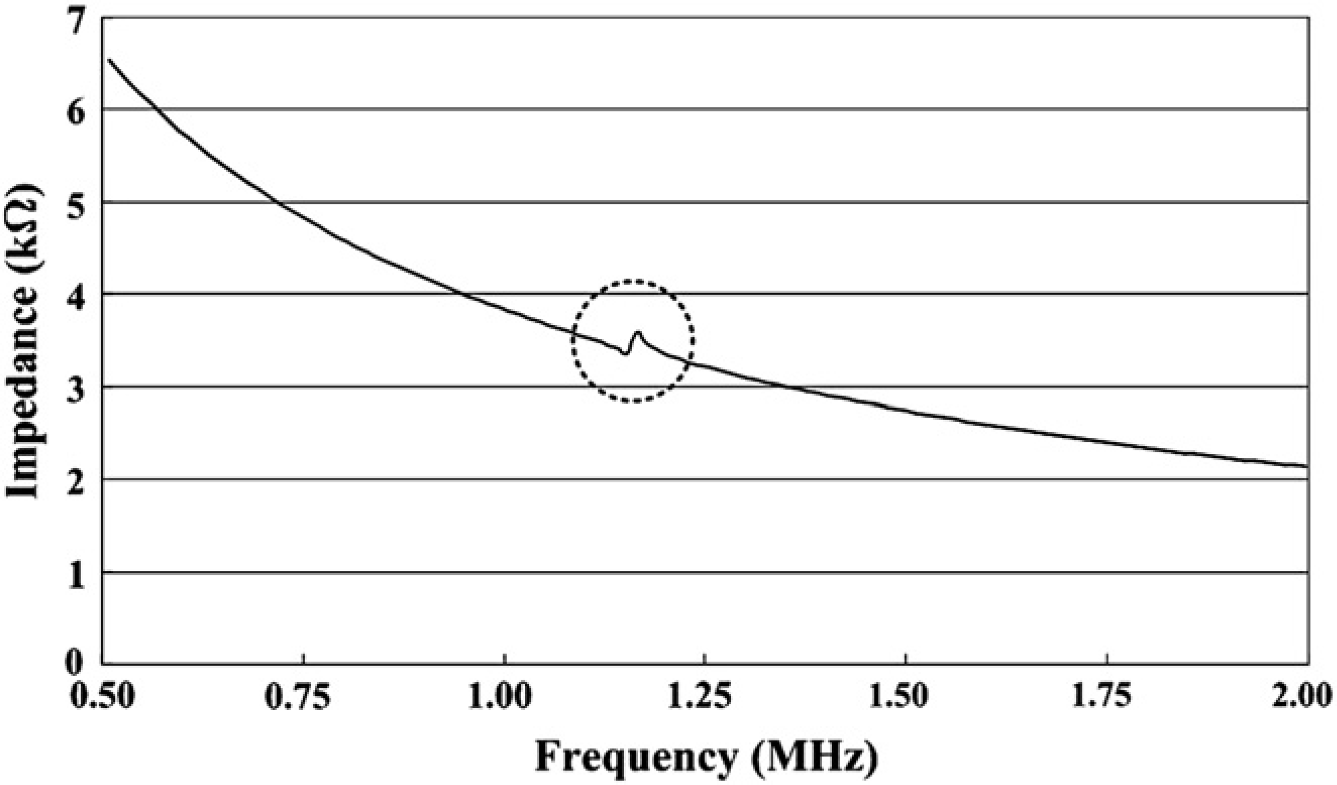
Impedance magnitude of piezoelectric micro cantilever.

Experimental schematic of rectangular pulse test.
Detecting CEA
The piezoelectric micro cantilever sensor detected CEA by the principle of mass micro balancing. As mentioned in the Introduction, CEA is a biomarker of various tumors: cholangiocarcinoma, and cancers of the rectum, breast, prostate gland, cervix, stomach, pancreas, large intestine, and lung. Because CEA is considered to indicate the presence of a tumor when its concentration is 30 pM (ng/mL), the micro cantilever sensor must be sensitive at this level.
Detection Method and Procedures
Change of the fundamental mode frequency of the sensor was measured by sweeping frequency using the LDV and LabVIEW system. The experimental setup is shown in Figure 5. The LabVIEW system swept the input frequency to the cantilever with 1 Hz resolution. Then, the LDV measured the mechanical vibration of the micro cantilever and transferred it to the data acquisition system of LabVIEW. Then, the first resonance frequency was measured by locating the maximum peak point because the output signal reflects the mechanical displacement of the cantilever induced by the swept input signal.

Scheme of detection experiment using LabVIEW and laser Doppler vibrometer.
The detection procedure had three steps as shown in Figure 6. In the first step, the porous silica sol–gel glass containing the CEA antibody was coated on the end of the micro cantilever by dipping the piezoelectric micro cantilever into the sol–gel. After coating the silica sol–gel glass for 1 min, it was dried in air at atmospheric pressure for 1 min to keep the size of the pores in which the antibody is located uniform. This coating was repeated five times. The coated micro cantilever was dried for 8 h to allow the sol to gel. In the second step, the end of the cantilever was dipped into a buffer solution for 5 min so that the sol was no longer gelated. Then, the first resonance frequency of the cantilever was measured. The third step was binding CEA and the antibody. The antigen and antibody were bound for 20 min. For more effective binding, the piezoelectric micro cantilever was excited at 1 Hz by a function generator. To remove any nonproper bindings, the end of the micro cantilever was rinsed in the buffer solution as in the second step while vibrating at 1 Hz. After rinsing, the fundamental mode frequency was measured again. Lastly, the measured resonance frequencies from the second and third step were compared.
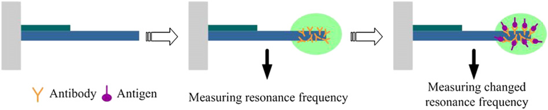
Detection procedures of carcinoembryonic antigen by antigen–antibody bind.
Chemical Conditions and Control Tests
Detection using the porous silica sol–gel glass requires keeping the pore size in the silica sol–gel glass uniform so that the antibody is bound properly with the CEA in the pore. The chemical conditions controlling the pore size are pH and the weight percent (wt%) of polyethylene glycol (PEG) in the silica sol–gel glass. The optimal conditions were found by the following process. Although keeping the concentration of CEA and antibody constant, the change of the resonance frequency was measured as described in Detection Method and Procedures at varying pH values and then at varying PEG concentrations. The detailed processes were as follows. First, the concentration of the PEG was fixed at 20wt%, and the change of the fundamental mode frequencies was measured at pH values of 2.3, 3.5, 4.3, and 5.5. The PEG concentration was fixed at 20wt% because the change of the vibrant mode frequency was the largest when using a quartz crystal in the same pre-experiments. Because the maximum frequency change occurred when the pH was 3.5, the pH was chosen as 3.5. Then, the first resonance was measured at the PEG concentrations of 0, 10, 20, and 30wt% at the pH of 3.5. When the PEG concentration was 30wt%, the resonance frequency changed the most, so the PEG concentration was set at 30wt%. Figure 7 shows the optimal conditions of the pH and PEG concentration.
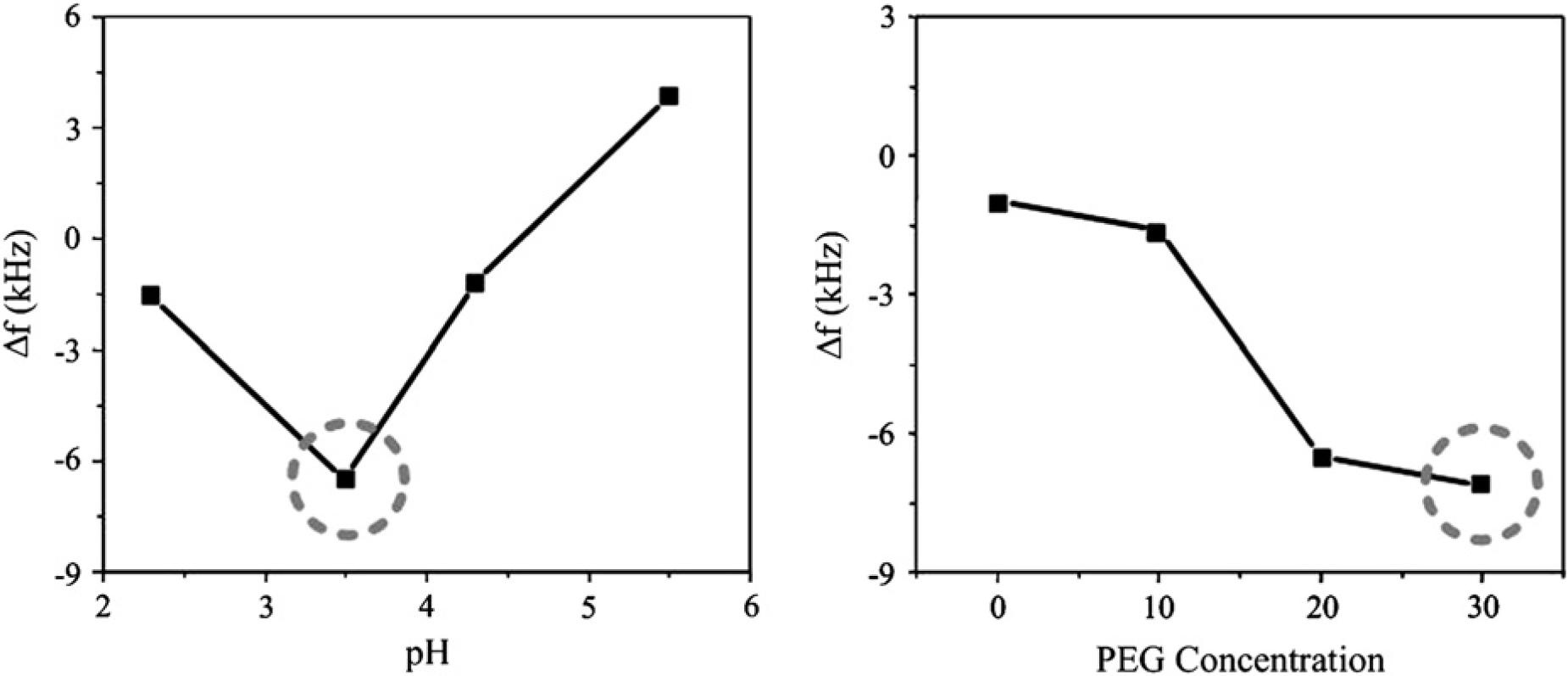
Optimal conditions of pH and polyethylene glycol.
Another important factor in the detection is that the first resonance frequency of the sensor should change only due to the binding of the antigen and antibody. Thus, the CEA must not bind with other biochemical materials except for the antibody, and the first resonance frequency has to remain constant when the antigen–antibody binding does not exist. Therefore, the control tests were examined by the nonspecific binding with bovine serum albumin (BSA) and the binding without the antigen–antibody. As shown in Figure 8, although frequency changes occurred due to the nonspecific binding with BSA and the binding without the antigen–antibody, they were insignificant compared to the frequency change due to the CEA binding (about 6.5 kHz). The tiny change of the frequency from the nonspecific binding is probably caused by other material attaching in various biochemical processes after coating the silica sol–gel glass. This effect might be eliminated by finding more optimal biochemical conditions and rinsing the micro cantilever more in each process. Then, the change of frequency could be regarded as only due to the normal binding of CEA.
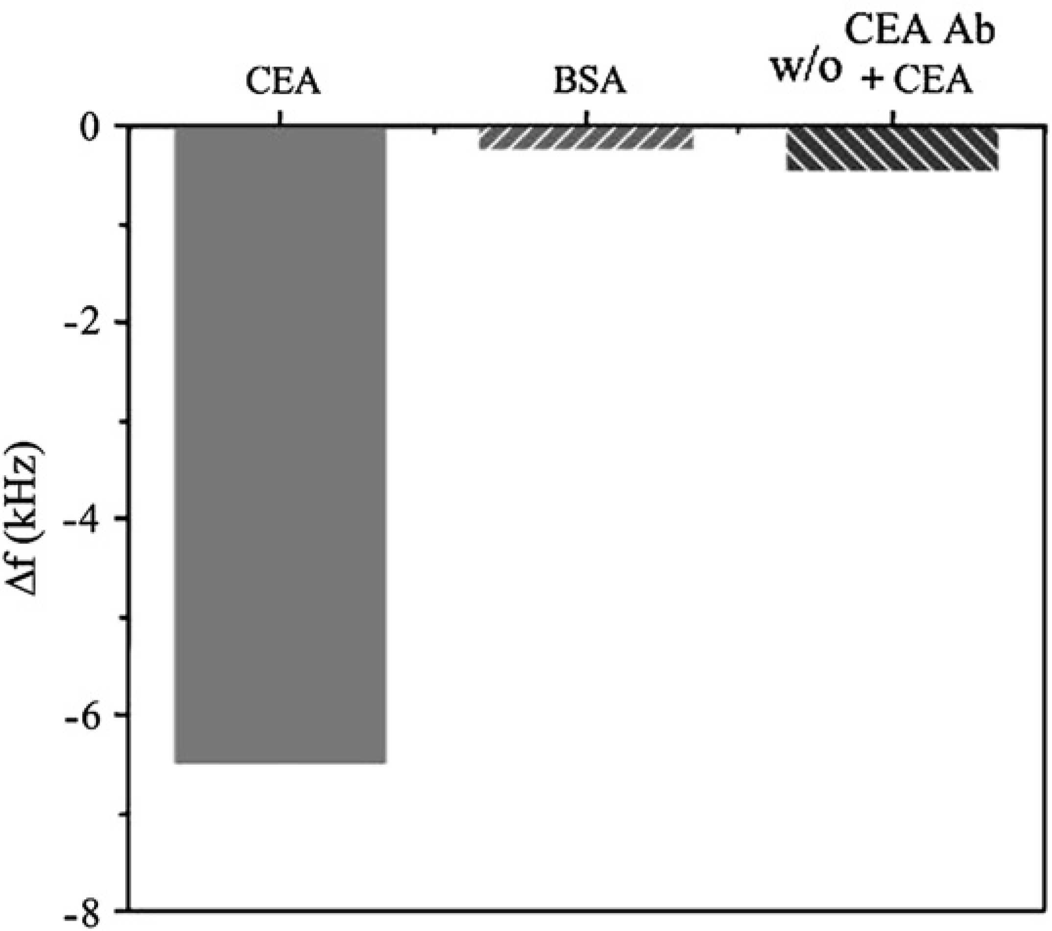
Control tests: detection specificity of sensor.
Results
The chosen conditions of the silica sol–gel glass were used to determine whether CEA can be detected at the criterion concentration, which is 30 pM (5 ng/mL). The concentration of the antibody in the silica sol–gel glass is 5 mg/mL, and as shown in Figure 9, CEA was detected at the concentration of 30 pM. However, the deviation of the frequency change at 30 pM was somewhat large compared with concentrations larger than 30 pM. However, CEA was detected at the criterion concentration, so the existence of the antigen can be checked.
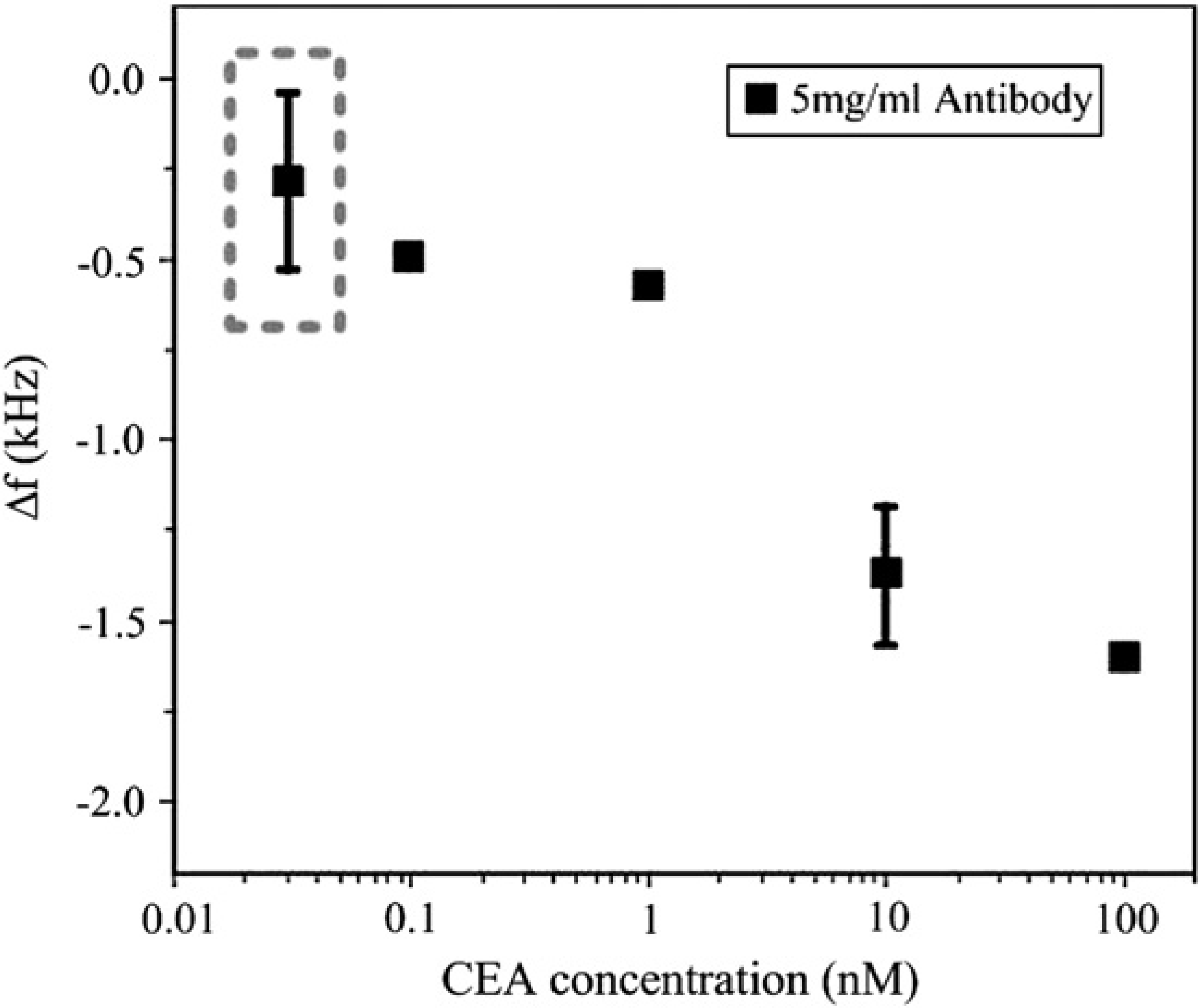
Carcinoembryonic antigen detection with respect to concentration.
Conclusion
A micro cantilever sensor actuated by a PZT film was designed and fabricated for label-free detection of the CEA, which is known to be a biomarker of various cancers. The sensor detected various concentrations of CEA from high concentrations to the critical value of 30 pM (5 ng/mL). These detection results show that the sensor can be used in label-free detection sensors for clinical testing. It can be also applied in diagnostic instruments if more precise experiments and improvements are achieved and if the chemical conditions of silica sol–gel glass, the coating of various biochemical solutions, and the times of coating and binding are optimized.
Acknowledgment
This work was supported by the Development of Next-Generation New Technology project (Development of Intelligent Robot Technologies for Laboratory Medicine by Applying Biotechnology, Project Number: 10024719-2007-13) from the Ministry of Commerce, Industry and Energy of Korea and the Korea Science and Engineering Foundation through the National Research Lab Program funded by the Ministry of Science and Technology (No. R0A-2007-000-20042-0).