
Abstract
We describe three new automated methods for purifying genomic DNA from whole blood. The MagneSil® Blood Genomic, Max Yield System uses MagneSil® paramagnetic particles (PMPs) in a 96-well format to purify the maximal amount of DNA from a 200-μL blood sample. In contrast, the MagneSil® ONE, Fixed Yield Blood Genomic System uses MagneSil® Fixed Yield PMPs to purify a normalized amount of DNA from 60 μL of blood in a 96-well format. These methods are implemented on the Beckman Coulter Biomek® FX automated workstation. The MagneSil® KF Genomic System uses MagneSil® PMPs to purify DNA from 1 to 15 samples of 200-μL blood using the moderate-throughput Thermo Electron KingFisher® mL instrument. The MagneSil® Blood Genomic System typically yields > 4 μg per 200 μL of whole blood, depending on the white blood cell content. The MagneSil® ONE System is best suited where there is a requirement for purification of a narrow concentration range of DNA. This system purifies 1 μg(±50%) of DNA from 60 μL of blood. The MagneSil® KF System purifies 2 to 6 μg of DNA from 200 μL of blood. DNA purified using all of these methods is suitable for PCR, STR, EADIT® SNP genotype analysis, and multiplexed PC analysis.
Introduction
High-throughput, walkaway methods for purifying DNA using MagneSil® paramagnetic particles (PMPs) have been described for a variety of applications, including plasmid purification, 1 PCR cleanup, 1 genomic DNA purification from foodstuffs, 2 and DNA purification from plant seed and leaf samples. 3 Here we describe purification of genomic DNA from human whole blood using MagneSil® PMPs on a variety of automated platforms, including the high-throughput Beckman Coulter Biomek® FX, the Tecan Genesis, and the moderate-throughput Thermo Electron KingFisher® mL workstations.
Many fields of basic and clinical research increasingly require the ability to purify genomic DNA from whole blood in a high-throughput setting. Furthermore, single nucleotide polymorphism (SNP) studies and veterinary applications such as genetic authenticity testing, animal breeding programs, and animal diagnostics for disease susceptibility also benefit from the time saving and precision of automated genomic DNA purification. The development of paramagnetic particle cores enclosed within silica surfaces has enabled the adaptation of well-known, solid-phase extraction methods to flexible formats that are easily scaleable and use magnetic separation rather than centrifugation. Additionally, as a result of the high binding capacity of MagneSil® PMPs, and optimized binding chemistries and washes that limit nonspecific binding, these methods allow more rapid solution kinetics than standard column-based systems.
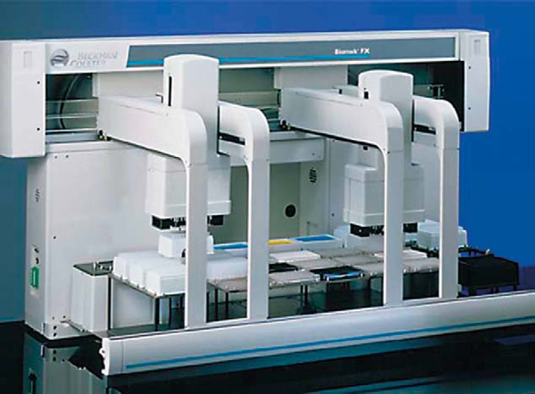
We describe two methods of high-throughput genomic DNA purification from 200-ul whole blood samples. With the MagneSil® Blood Genomic, Max Yield System method, the maximal amount of DNA is purified, and DNA yields vary from sample to sample depending upon the white blood cell content of the sample. However, with the MagneSil® ONE, Fixed Yield Blood Genomic System, a predetermined, normalized range of DNA yield (1 ug ± 50%) is obtained that does not vary with the white blood cell content of the sample. The Beckman Biomek® FX (shown in Fig. 1) and Tecan Genesis robotic platforms can be used in conjunction with 96-well plates for high-throughput automation of DNA purification.
The Beckman Coulter Biomek® FX can be adapted for use with either 96-well or 384-well microtiter plates. The methods described for 200-μL blood purification necessarily are limited to the use of 96-well plates, based on sample volume size.
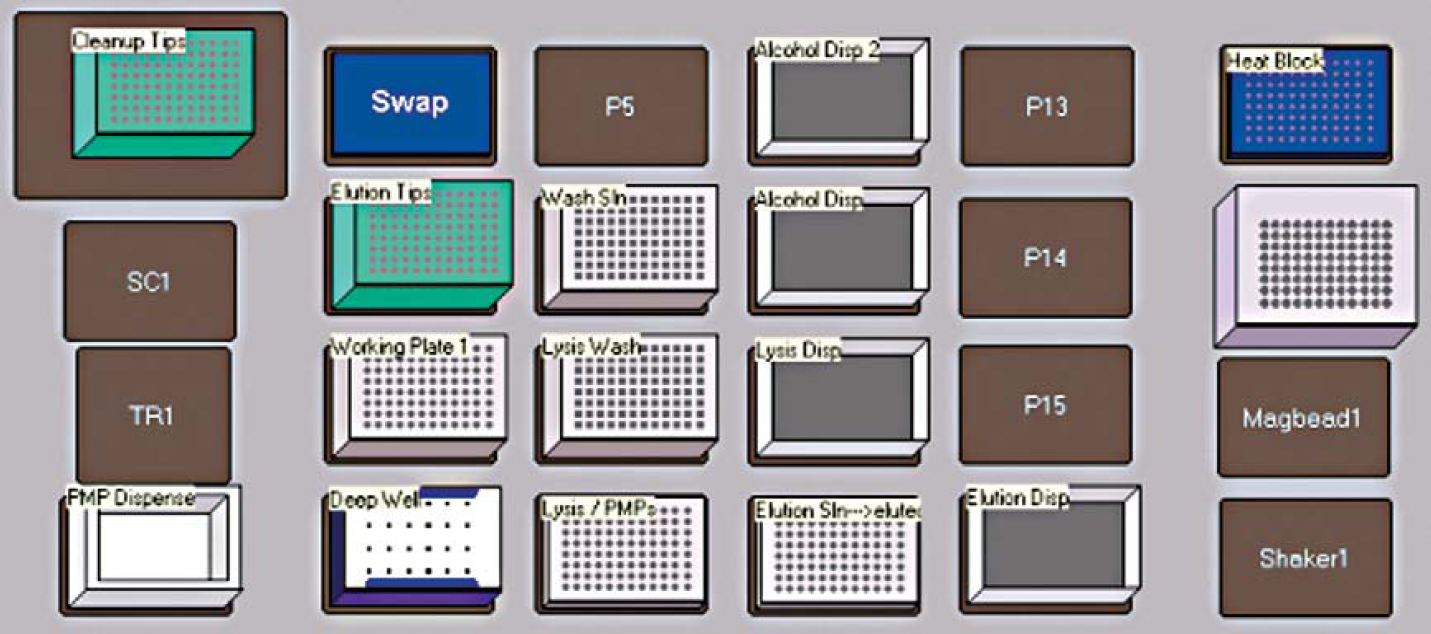
In addition to the high-throughput methods described above, for moderate-throughput purification of genomic DNA from 200 μL of whole blood, we describe the MagneSil® KF, Genomic System that can be used in conjunction with the Thermo Electron KingFisher® mL. The KingFisher® mL is shown in Figure 2. Its chief advantages are lower cost, small footprint, and simplicity of operation, compared to 96-well-plate-based robotic platforms. Figure 3 shows the layout of dispensed reagents used with the MagneSil® KF protocol run on the KingFisher® mL robot.
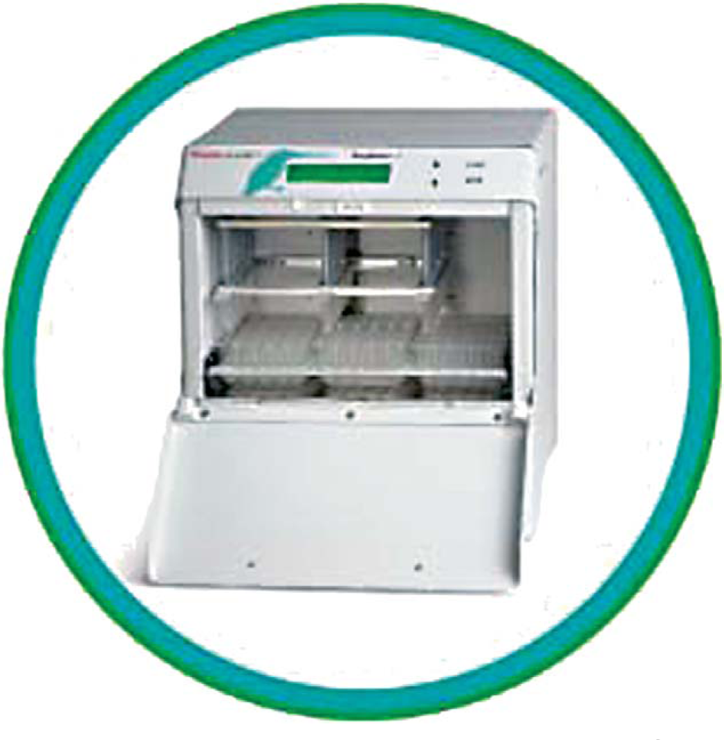
The Thermo Electron Labsystems KingFisher® mL has a small lab bench footprint and can process up to 15 samples per run.
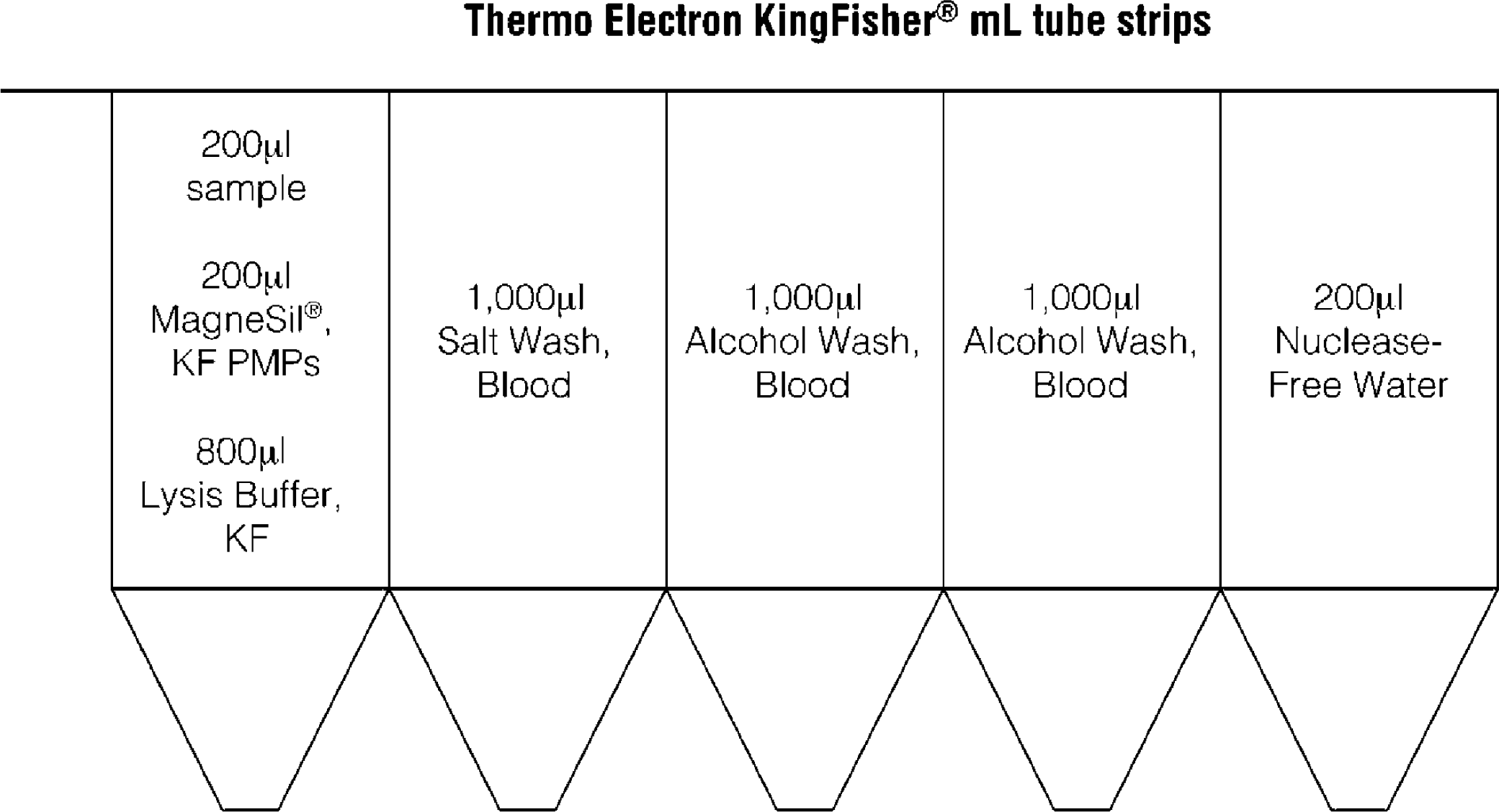
The reagent contents used in the 1 × 5 strip tube used for genomic DNA purification using the MagneSil® KF Genomic System.
Methods
Figure 4 outlines the procedure for purifying genomic DNA from 200-μL whole blood using the MagneSil® Blood Genomic, Max Yield Purification System (Promega Corporation, Madison, WI). This method was developed for the Biomek® FX but could be adapted to other automated systems based on information present in the technical documentation. For single-plate purifications, a 96-well plate containing 200 μL of whole blood per well is placed on the workstation deck, and the required reagents are placed in designated reservoirs. The workstation then dispenses the reagents into 96-well plates and purifies the DNA. After the deck is set up, the process is entirely automated. On a single-POD Biomek® FX instrument, the purification process takes about 1.5 h for one 96-well plate. Using a two-POD Biomek® FX could reduce the overall processing time, as can the simultaneous processing of multiple plates.
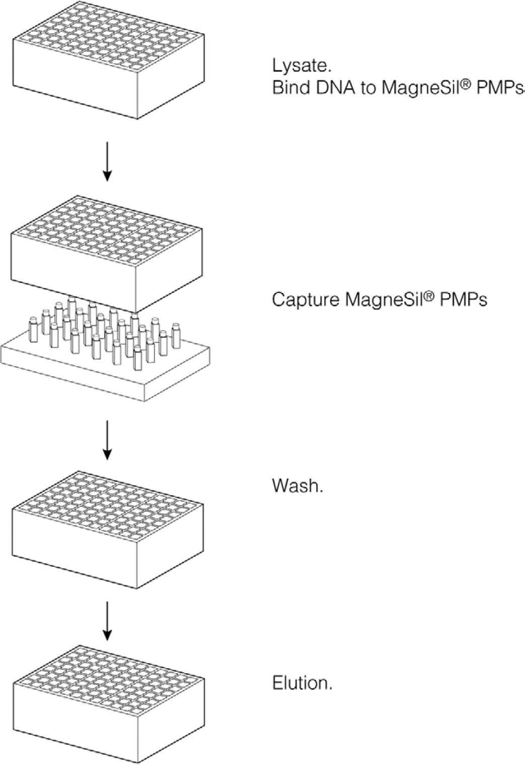
Schematic representation of genomic DNA purification from whole blood in a 96-well format.
When processing a 200-μL blood sample, we recommend using deep-well, 96-well plates such as the Marsh 1.2-mL plate (Marsh cat. # AB-0564/BP). Round-bottom plates that are compatible with the suggested magnetic separation device, the Deep Well MagnaBot® 96 Magnetic Separation Device (Promega Corporation, Madison, WI; Fig. 5), are also required. For larger volumes, deep-well, 96-well plates such as the Marsh 2.2-mL plate (Marsh cat. # AB-0932) can be used for initial lysis, followed by serial transfer to lower capacity, round-bottom plates that are compatible with magnetic separations. A full listing of required robotic instrumentation is available at www.promega.com\automethods (listed by automated instrument and protocol).
The Deep Well MagnaBot® 96 Magnetic Separation Device allows the use of larger volume deep well 96 plates in automated purifications. The magnets are twice the strength of the standard MagnaBot® separation device and efficiently capture paramagnetic particles through the thicker walled, deep well 96 plates.
Results
Purifications typically yield > 4-μg DNA from normal human whole blood that has been stored at 4°C for up to 3 weeks or frozen at −20°C. DNA yield is dependant on the sample's white blood cell content. Figure 6 shows that the isolated DNA is high molecular weight (>20 kb). The size of the DNA results from the shearing forces of the pipetting steps used in the automated protocol. As shown in Figure 7, the total yield begins to decline after 1 week of refrigerated storage. This decline is not observed with frozen, anticoagulated blood. Figure 8 shows that comparable DNA yields are obtained from human whole blood, regardless of whether it is stored in EDTA, ACD, heparin, or citrate anticoagulants.
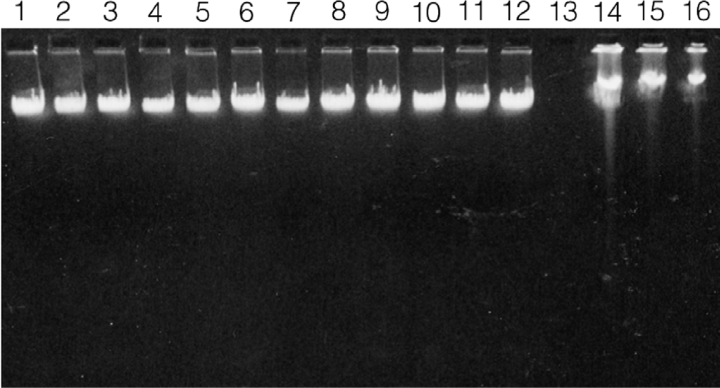
DNA purified from human whole blood using the MagneSil® Blood Genomic Max Yield protocol on the Beckman Coulter Biomek® FX. Lanes 1-12 show 10 μL from a total of 200 μL of purified DNA. Lane 13 is blank, and lanes 14-16 show Promega human genomic DNA (cat. # G3041) at 300, 200, and 100 ng per well.
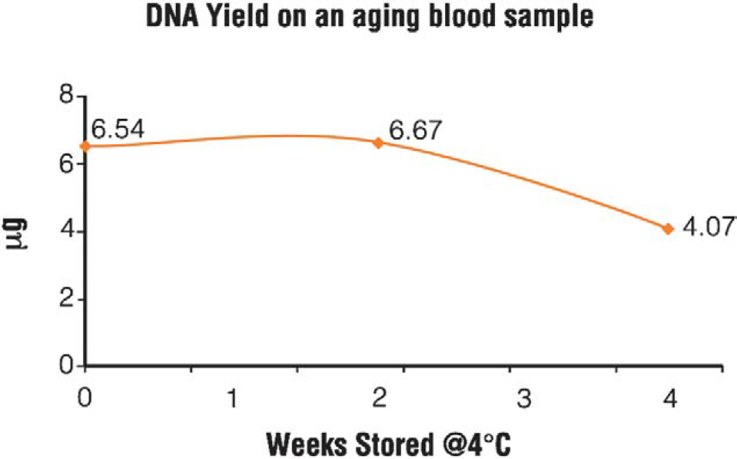
DNA yields determined by PicoGreen® of DNA purified from human whole blood refrigerated at 4 °C for up to 4 weeks using the MagneSil® Blood Genomic Max Yield System on a Beckman Coulter Biomek® FX.
The functionality of the isolated DNA is not dependent on the anticoagulant used. Storage in ACD seems to provide a small benefit over the other three anticoagulants during the first week of storage at 4 °C, and the small boost in DNA yield shown in Figure 8 is typical of this effect. Other than this, we have observed no difference among the four commonly used anticoagulants in terms of DNA yield, purity, or performance in downstream applications. Figure 9 shows an example of a multiplex PCR profile obtained using the Y-Chromosome Deletion Detection System (Promega Corporation, Madison, WI) to analyze normal human male DNA purified from blood using either the MagneSil® Blood Genomic System or the MagneSil® ONE System.
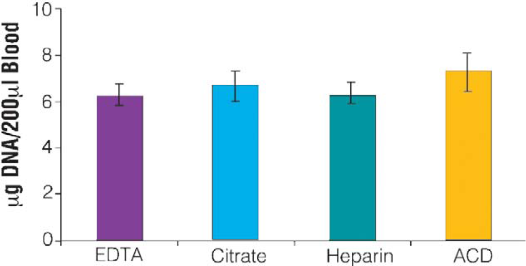
DNA yields determined using PicoGreen® from human whole blood stored at 4 °C for one week using the MagneSil® Blood Genomic Max Yield System on a Beckman Coulter Biomek® FX. Comparable DNA yields were obtained from all four types of anticoagulated blood.
Although refrigerated or frozen anticoagulated blood provides more reliable DNA quality, anticoagulated whole blood is often shipped under ambient conditions (which may vary from freezing to 45 °C, depending on geography and the time of year). Figure 10 shows a comparison of genomic DNA isolated from blood stored in unopened tubes at −20, 4, and 21 ° C. White blood cells stored at elevated temperatures undergo apoptosis, yielding a characteristic apoptotic ladder of purified DNA (Fig. 14). MagneSil® chemistries are able to purify DNA across the broad size range present in the sample. When purifying DNA from blood samples stored under such conditions, reagent chemistries and protocols that facilitate the binding of degraded, lower molecular weight DNA (in addition to the higher molecular weight DNA purified from normal human blood) are required to obtain consistently higher and representative yields. Note that for isolating viral sequences, such as HCV, the blood sample storage conditions can be particularly important. 4
The MagneSil® Blood Genomic System is a scalable system, allowing users to purify DNA from smaller amounts of blood, such as 50 μL of whole blood. This ability to scale down the process to accommodate lower sample volumes saves reagents and enables use of lower volume (and less expensive) 96-well plates. DNA purification can also be scaled up to accommodate sample volumes greater than 200 μL. The chief limitation is the volume restriction of commercial deep-well, 96-well plates, which generally have a maximum capacity per well of 2.2 mL. Larger volumes may be accommodated by using a larger volume plate, such as a square-bottom, 2.2-mL deep-well plate for initial lysis and then transferring smaller volumes to round-bottom, 96-well plates that are compatible with the MagnaBot® Magnetic Separation Device.
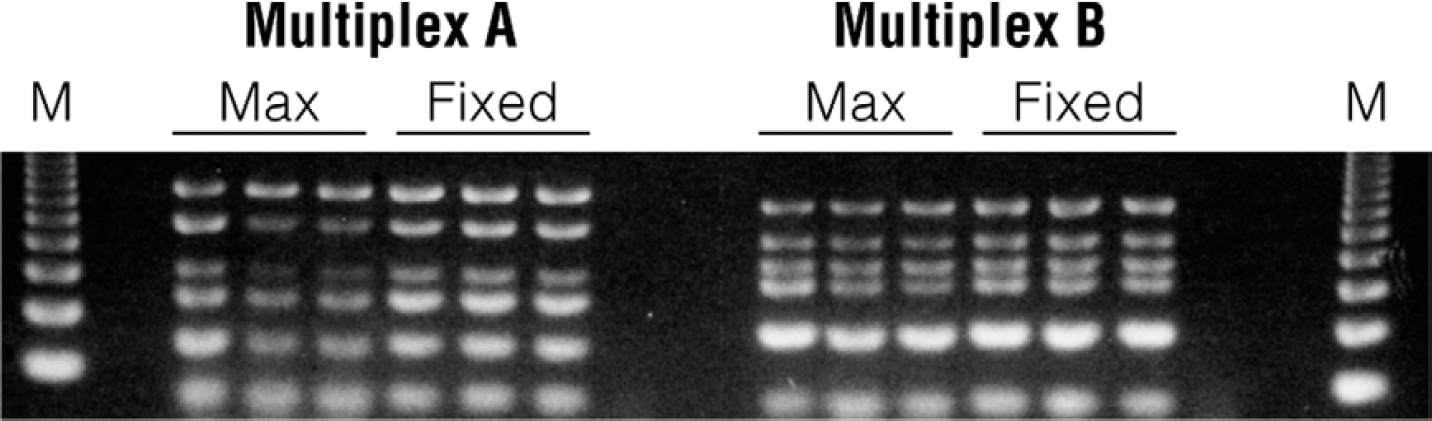
Multiplex PCR results using DNA from a wild-type human male purified using the MagneSil® Blood Genomic Max Yield System (MAX) or the MagneSil® ONE, Blood Genomic System (FIXED) on a Beckman Coulter Biomek® FX.
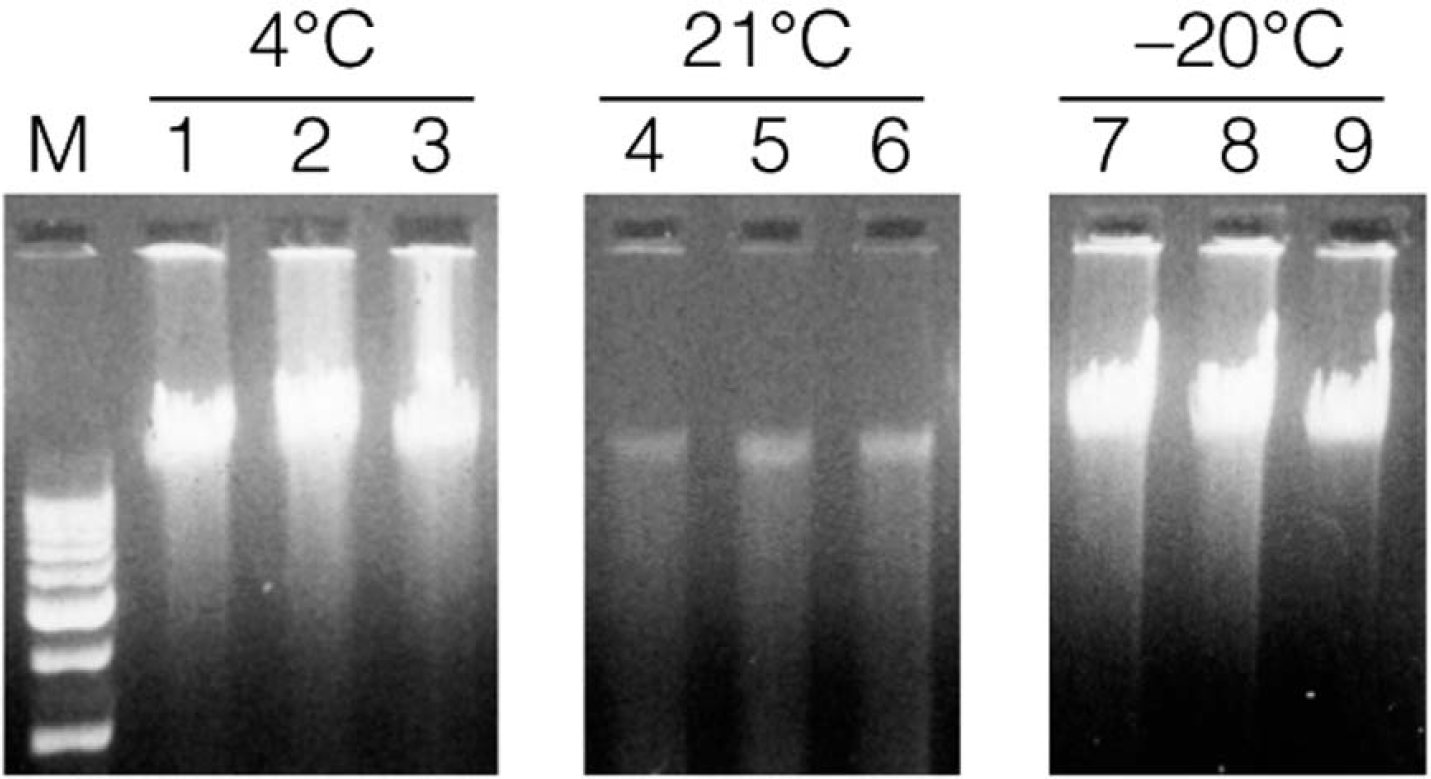
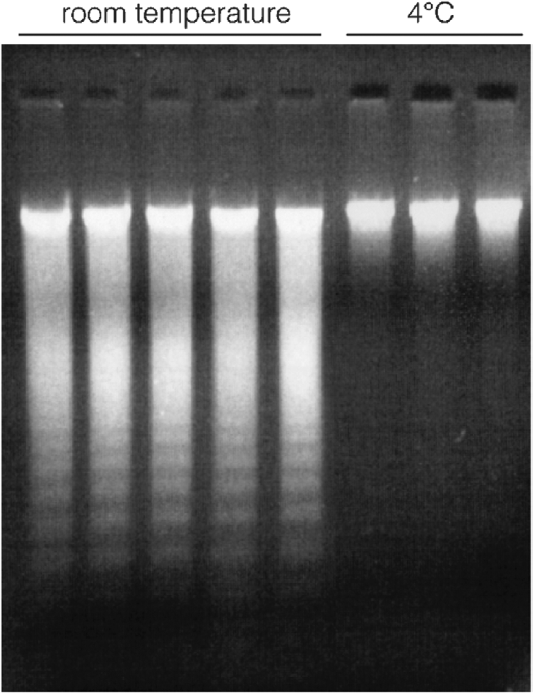
Shows how storage conditions of EDTA anticoagulated blood can affect DNA yield and molecular weight. DNA was purified using the MagneSil® Blood Genomic Max Yield protocol on a Beckman Coulter Biomek® FX. Lane 1 Promega 1-kb ladder, lanes 2-4, shows DNA from blood stored 8 d at 4 °C, lanes 5–7 stored 8 d at +21 °C, and lanes 7–8 show storage at −20 °C for 8 d.
Many applications can be streamlined by using a specific quantity of DNA, rather than the maximum amount of DNA available from a sample. The MagneSil® ONE System allows purification of DNA at a desired yield or concentration range, if the sample with the lowest quantity of DNA lies within the specified range. 5 The MagneSil® ONE System protocol, available for the Biomek® FX workstation (Fig. 11), purifies 1 μg of DNA (±50%) from 60 μL of human whole blood.
The initial deck layout of the MagneSil® ONE Fixed Yield System protocol on a Beckman Coulter Biomek® FX.
By limiting the surface 6 available for DNA binding so that the lowest-yielding sample contains DNA in excess of the available binding surface, users can normalize the samples to generate a narrow range of purified DNA. The primary benefit of this is that quantitation of DNA yield is not required before performing downstream applications. This saves time and money. Additionally, the DNA is of high quality because the reduced surface area presents less opportunity for non-specific binding of contaminants, and there is less surface area to wash.
If a range of blood sample volumes is used because the amount of blood sample is hard to quantify, the samples will generally still fall within the narrow specified yield or concentration range (i.e., using 120 μL of whole blood rather than 60 μL will still generally result in the DNA yields remaining within a 1 μg(±50%) range). However, more or less than 1 (g of DNA can be obtained by altering the protocol, primarily by using more or less MagneSil® PMPs -Fixed Yield. Regardless of the amount of DNA purified, the DNA is suitable for applications such as PCR (see Figs. 12A and 12B below), STR, READIT® SNP Assay analysis, and multiplexed PCR systems (as shown in Fig. 9 above).

Prevention of cross-contamination between samples is a critical issue in most high-throughput purifications. Contamination must be undetectable in the downstream application. For example, in high-throughput SNP screening, a level of cross-contamination of 0.5% might be quite acceptable in terms of allowing complete accuracy in SNP calling, but a level of 1 × 10⁻⁶ would not be acceptable in medical disease diagnostics. We have used three approaches to assess and address these issues.
A commonly practiced method of testing for sample cross-contamination involves the use of empty samples or water blanks. Figure 12A shows that this method will only allow detection of cross-contamination above the level of about 1%. While we have not observed cross-contamination using water blanks, we believe more sensitive methods are generally required to provide a more meaningful semiquantitative evaluation of cross-contamination when using robotic platforms. Cross-contamination below the 1% detection level of water blanks is frequently touted as “no cross-contamination,” rather than the more accurate statement of “cross-contamination less than the 1% detection limit.” To avoid this misleading conclusion, we have used two approaches to better address the issues of cross-sample contamination.
For routine testing of cross-contamination down to levels of 5 × 10⁻³, we have used a DYS214 sequence found on the Y-chromosome 7 [Primers: Left = CCGTGTGTTGCTGG GCTGTC, Right = GGGGTTTATACTGACCTGCC], which is present as five copies in a wild-type male donor and is absent in most female donors. This sequence duplication is a fairly common event on the Y chromosome. 8,9 Some female donors, particularly those who have been pregnant with male children, carry some male cells in their blood. These male cells may persist in the woman's blood for years, 7 and such female donors may generate Y-chromosome DYS214 amplification products in our test. The X-chromosome has a sequence similar to the DYS214 probe sequence, and in the absence of Y-chromosome-specific DNA, this X-chromosome-specific ∼180-bp amplicon will be generated, readily distinguished from the Y-specific ∼140-bp amplicon.
Figure 12B shows a series of dilutions of male DNA into a female DNA sample, followed by amplification using the DYS214 probes. A competitive amplification curve is evident from 1 to 0.05% of male DNA into female DNA. Figure 12B also shows DYS214 amplifications of female blood samples that have been alternated in the 96-well plate with male blood samples so that the female samples have been surrounded by male samples, on four sides but not diagonally, in the 96-well plate. The alternating amplification of the X-chromosome-specific product and the Y-chromosome-specific product is consistent with observing no cross-contamination above the detection limit of 0.05%. Using this test, cross-contamination can be semiquantitatively measured by observing the presence of male DNA cross-contaminating female DNA to a level of about 0.05%.
We have also used a modified TaqMan® (Roche Molecular Systems) qPCR method to measure the presence of the SRY gene sequence (Accession # L08063) that can detect as little as seven copies of a male Y-chromosome target in 100 ng of purified female DNA (about 33,000 copies of a single copy gene). Amplification primers and probes were developed as per Lo et al. 10 Dual-labeled TaqMan® probes were synthesized and HPLC purified at Synthegen (Houston, TX). TaqMan® amplification reactions were at a 50-μl scale, containing 5-μl 10x Buffer A; 300-nM forward and reverse amplification primers; 100-nM TaqMan® probe; 3.5-mM MgCl2; 200-(M each dATP, dCTP, dGTP, and dTTP; and 1.25-U AmpliTaq Gold® DNA Polymerase. DNA extracted from whole blood using a MagneSil® system was quantitated by Picogreen® assay (Molecular Probes, Eugene, OR) and 100 ng (5-10 μL) was added to each reaction. 11 The thermal cycling procedure consisted of a 12-min initial denaturation at 95 °C followed by 40 cycles of 95 °C for 15-s denaturation and 1 min at 60 °C anneal/extend step. Figure 13 shows amplification curves from the TaqMan® system with 100-ng female DNA spiked with 335, 35, 17, and 7 copies of male DNA. As can be seen in Figure 13, cross-contamination down to 1/5000 (0.05%) can be detected with this qPCR method, consistent with the semiquantitative PCR method shown in Figure 12B.
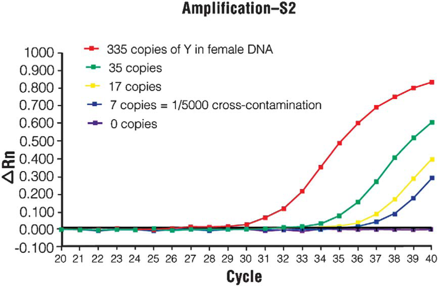
Amplification plot of Taqman® assay after seeding 335, 35, 17, or 7, or 0 copies of human male DNA into 100 ng (∼33,000 copies) of human female DNA. Amplification of 7 copies clearly produced product within 40 cycles.
For more sensitive cross-contamination detection, we have used blood samples spiked with an attenuated poliovirus vaccine strain surrounded by poliovirus negative blood samples. For example, whole blood samples containing 108 of poliovirus surrounded by blood samples that do not contain poliovirus routinely allows the detection of 1 × 10⁻⁶ cross-contamination using a TaqMan® qPCR analysis. For more specific tests, the target molecule being detected can be used for greater test specificity (e.g., for HIV detection, use samples of 109 HIV sequences surrounded by negative samples).
For processing blood samples in a moderate-throughput (up to 15-150 blood sample purifications per day), we have used the Thermo Electron KingFisher® mL workstation, which is shown in Figure 2. The KingFisher® mL processes samples of 200-μL whole blood and uses 5-well purification strip-tubes that can be run in multiples of up to 15 samples per run. Each run is performed in under 24 min. Depending on white blood cell content, purifications typically yield 2- to 6-μg DNA from normal human whole blood that has been stored either at 4 °C for up to 3 weeks or frozen at −20 °C, using EDTA, ACD, heparin, or citrate as anticoagulants. Figure 14 shows that the DNA is of high molecular weight and that blood samples shipped or stored under ambient conditions can also be processed using the KingFisher® mL.
Similar to the DNA shown in Fig. 10, DNA can also be purified using the MagneSil KF Genomic System on the Thermo-Electron KingFisher mL: lanes 1–5 show DNA purified from EDTA anticoagulated blood stored at +21 °C for 7 d (showing an apoptotic ladder), and lanes 6–8 show DNA purified from the same blood stored for 7 d at 4 °C.
The use of EDTA, ACD, heparin, or citrate anticoagulated blood produces similar DNA yields using this protocol, and the DNA produced functions equally well in downstream applications regardless of which anticoagulant is used.
Discussion
The use of MagneSil® paramagnetic particles and MagneSil® PMPs — Fixed Yield resin in walkaway, high-throughput DNA purification on the Beckman Coulter Biomek® FX provides attractive alternatives to multicolumn (and multi-96-well plate) manual genomic DNA purifications from whole blood. Automated purification methods developed for the Biomek® FX can serve as a starting point for adoption to other robotic platforms. In addition to purifying the maximal amount of DNA from 200-μL blood samples, users can also purify a fixed amount of DNA with the MagneSil® ONE System protocol described. Both the MagneSil® Blood Genomic System and MagneSil® ONE System can be scaled to process larger or smaller sample sizes, saving reagents and money and adding flexibility. This flexibility, along with the lower costs associated with MagneSil® purifications relative to many column-based, 96-well methods, provides advantages for automated processing of liquid blood.
In addition, automated purification of up to 15 200-μL blood samples per batch can also be performed on moderate-throughput robotic platforms, such as the Thermo Electron KingFisher® mL. This flexibility of throughput along with the scalability of the MagneSil® DNA purification technology allows users to directly couple automated DNA purification with automated downstream genetic analyses in a very cost-effective analytical system.