
Abstract

Microfluidic systems have become important platforms for diagnostic and therapeutic applications. 1 –6 However, as channel dimensions decrease within these systems, the surface properties of these microchannels become increasingly important. When biological fluids are flowed through such channels, nonspecific protein adsorption can occur due to surface interactions, often resulting in reduced efficiency of the system. When small quantities of biological sample are involved, any loss of sample through the system can be critical. Poly(ethylene glycol) (PEG) films are known to reduce protein adsorption, but the existing technology for coating PEG to surfaces is not always appropriate for the micro/nanoscale features common in microfluidics systems. Therefore, we have developed a vapor deposition technique to modify microchannels with PEG. The vapor deposition technique is effective in coating miniature size features since it forms uniform, conformal and ultrathin films on the surface. These films have been extensively characterized by contact angle measurement, X-ray photoelectron spectroscopy (XPS) and atomic force microscopy (AFM). Surface modification approaches using CVD show promise for a variety of bioMEMS applications.
Silicon is one of the commonly used materials for microfluidic systems since it can be easily microfabricated. Surface modification of silicon seems to be the most appropriate approach to make silicon-based microfluidic systems more compatible with the biological milieu. In recent years people have shown more interest in developing microfluidic systems using glass since it is cheaper and all the silicon microfabrication processes as well as chemistries can be repeated on it. PEG has been shown to form thin films on silicon surfaces in their solution form. 8,9 For PEG, silanes are used as precursors or bridges to connect PEG molecules to a surface. Silane precursors are highly sensitive to moisture. They tend to form aggregates and lumps on the silicon surface in the presence of moisture, which would clog up sites or mask micron-size features on devices. In this study we focus on the vapor deposition of silane and subsequently PEG on silicon surfaces in a moisture free nitrogen atmosphere. 7,10 To deposit PEG on the surface, we start from the basic molecule — ethylene oxide with a gas catalyst. Vapor phase deposition has the following advantages over solution phase coating:
Control of the coating environment (i.e., a moisture free environment)
Uniform coatings on micro and nano size patterns such as microchannels and pores
Solvent free process yields no contamination
Direct injection of reactant into the stream of nitrogen to form film directly on the surface
Silicon wafers <100> (1cm×1cm) were treated with 3ml ammonium hydroxide and 3ml hydrogen peroxide (30%) in 15ml distilled water at 80°C for 15 minutes to clean the surface. This was followed by treating the wafers with 3ml concentrated hydrochloric acid (50%) and 3ml hydrogen peroxide (30%) in 18ml distilled water at 80°C for 15 minutes to make the surface hydrophilic (i.e., attach −OH groups on the surface). The samples were thoroughly washed with distilled water after each step to avoid any impurities and were dried using nitrogen gas. Ethylene oxide in vapor phase was used to grow PEG on the silicon surface. The surface was first silanized with a reactive end group silane like 3-aminotripropyltrimethoxy silane (APTMS). Boron triflouride was used as a gas catalyst with ethylene oxide since it is a weak Lewis acid. Lewis acids are electron pair acceptors, hence will accept a free pair of electrons of −NH2 on APTMS, and make the reaction site available for reactive ethylene oxide molecules to attach and then additional polymerization reactions will form PEG on the surface (Figure 1). PEG composition can be controlled by the concentration of ethylene oxide and polymerization reaction time. The reaction can be terminated by flowing inert gas over the surface. 20 and 40mmol/cm2 of ethylene oxide concentrations were used with BF3 in 2:1 ratio. Formation of PEG on a surface is a polymerization reaction, hence increasing the concentration of ethylene oxide will result in more PEG on the surface. 11 The reaction was allowed to proceed for four hours. APTMS concentration of 4mmol/cm2 was used. Our previous studies show that this concentration of silane is optimum to achieve the desired composition on the surface for further modification. 10
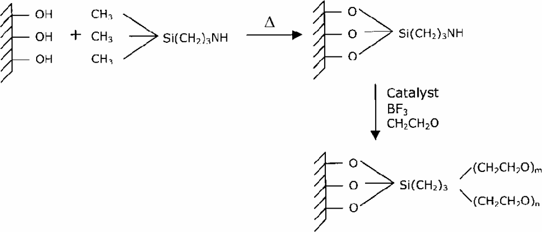
Reaction mechanism on silicon/glass surface for surface modification with PEG.
In order to investigate the physiological behavior of these surfaces, protein adsorption on these surfaces was studied. PEG modified (20 and 40 mmol/cm2, four hours) and clean silicon samples were transferred into wells of standard 24-well plates (14mm across internal diameter) and 1 ml of 0.5 mg/ml fibrinogen solution and 2 mg/ml FITC-labeled BSA solution in PBS was added. Adsorption was allowed to proceed in an incubator (0.5% CO2) for two hours at 37 °C. Upon completion of adsorption, the samples were thoroughly washed three times with deionized water for removal of non-adsorbed proteins and salts from the buffer. The samples were dried with nitrogen and their surface composition was determined using XPS for fibrinogen adsorption and fluorescence images were taken using a CCD camera attached to a microscope for FITC-labeled BSA adsorption. The analysis was conducted on a Perkin-Elmer 5600 XPS/SIMS instrument with a monochromatic Al-Kα-X-ray source (1486.6 eV) with an Omni Focus III small area lens and multichannel detector. A concentric hemispherical analyzer (CHA) was operated in the constant analyzer transmission mode to measure the binding energies of emitted photoelectrons. In order to investigate the surface topography after protein adsorption on these surfaces, atomic force microscopy was used. Atomic force microscopy (AFM) was performed on a commercial Nanoscope III (Digital Instruments, Santa Barbara, CA) using optical beam deflection to monitor the displacement of a microfabricated silicon cantilever having a spring constant of 80 N/m. Since these surfaces will be used for biological applications, we were interested in seeing the topography of the surface which cells will see after protein adsorption. The AFM was performed with silicon nitride probes mounted on cantilevers in tapping mode to avoid surface damage at a scan rate of 1 Hz. The force of interaction was approximately 10-9 N. This method significantly improves lateral resolution on soft surfaces and thin films. The AFM images were obtained under ambient laboratory conditions and all the scans were two microns in size. In order to investigate the efficacy of these films, the same reaction protocol was repeated on glass capillaries of 250μm diameter and two inches long to modify their surface with PEG. 1mg/ml of BSA solution in PBS was flown through unmodified and PEG-modified capillaries with a micro-pump at 10μl/sec for 30 minutes to show improved repeatability of lesser loss of protein due to wall interaction. By using glass capillaries comparable in size and properties to microchannels, the process occurring can be studied in a simple way and our principle can be proved.
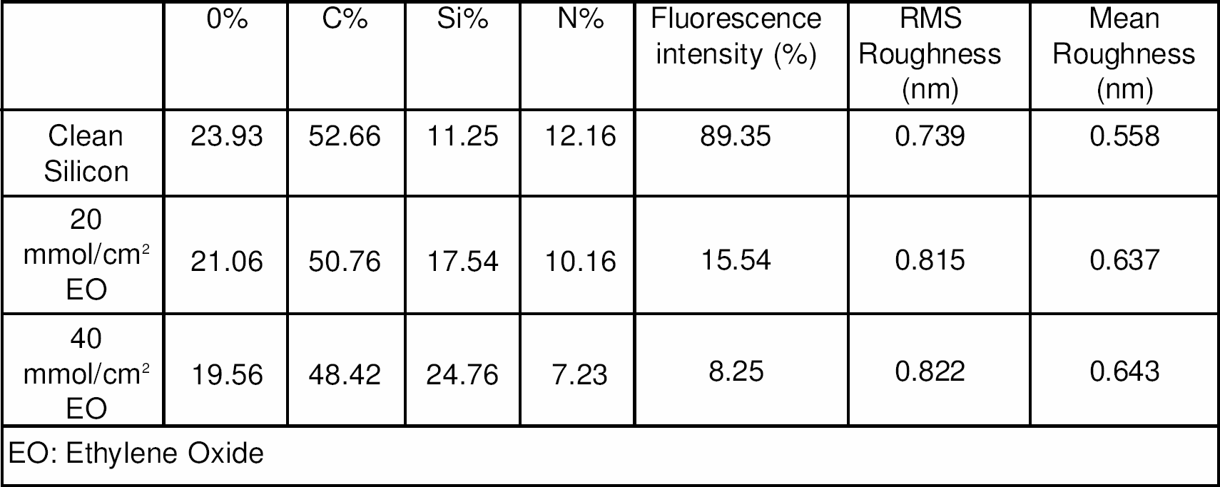
EO: Ethylene Oxide
PEG is known to absorb less protein due to steric stabilization effect, chain mobility and chain density. 12 XPS analysis was used to verify that adsorption of fibrinogen was lower on modified surfaces compared to unmodified surfaces. There is a sharp increase in the Si2p (100eV) peak while a significant decrease in C1s (285eV) and N1s (410eV) peak for protein adsorption on PEG modified silicon (20 and 40mmol/cm2, four hours) compared to clean silicon. Increases in ethylene oxide concentration results in decreases in the amount of protein adsorbed on the surface, which is supported by the elemental composition of the surface (Table 1). FITC-labeled BSA adsorbed surfaces were observed under fluorescence microscopy. In our case we defined fluorescence intensity as the amount of green color on the surface since we are using FITC (green color) labeled protein. 100% fluorescence intensity means the surface is totally green whereas 0% fluorescence intensity means the surface is totally black. This intensity can be directly correlated with the amount of protein adsorbed on the surface. PEG-modified silicon shows lower fluorescence intensity as compared to unmodified silicon (Table 1). Also, a lower fluorescence intensity is observed for 40mmol/cm2 compare to 20mmol/cm2 suggesting lower protein adsorption with greater PEG concentration. The contact angle for PEG modified surface is around 40° compared to that of clean silicon which is around 8°. Contact angle does not depend on the concentration of ethylene oxide and remains the same for both 20 and 40mmol/cm2 concentrations. Figure 2 shows 3D AFM images of protein adsorbed on modified and unmodified silicon obtained using WSxM 1.2 software for scanning probe microscopy. The scale for the images is made standard using this software. The protein is sparsely adsorbed on PEG modified surfaces compare to unmodified surfaces. This can be seen in the 3D images of protein adsorption. The roughness of the modified surfaces with protein adsorption is greater compared to unmodified surfaces indicating lower adsorption with few proteins adsorbed sparsely adsorbed on the surface (Table 1). The ability to control surface roughness will be useful for both microfluidic and biomedical microdevices applications in vitro and in vivo. BSA solution was flown through unmodified and PEG modified glass capillaries to investigate the efficacy of these films on surface (Fig. 3). PEG modified surfaces adsorb less protein compared to unmodified silicon surface under flow conditions. The protein adsorption is slower in PEG modified and takes more time to saturate compared unmodified surface. This is clearly indicated by the slope of the line. After saturation is reached, the concentration reaches back to initial.
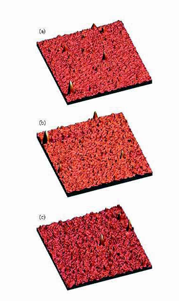
3D AFM Images for fibrinogen adsorption on (a) unmodified Silicon surface, (b) 20 mmol/cm2 (c) 40 mmol/cm2 ethylene oxide concentration.
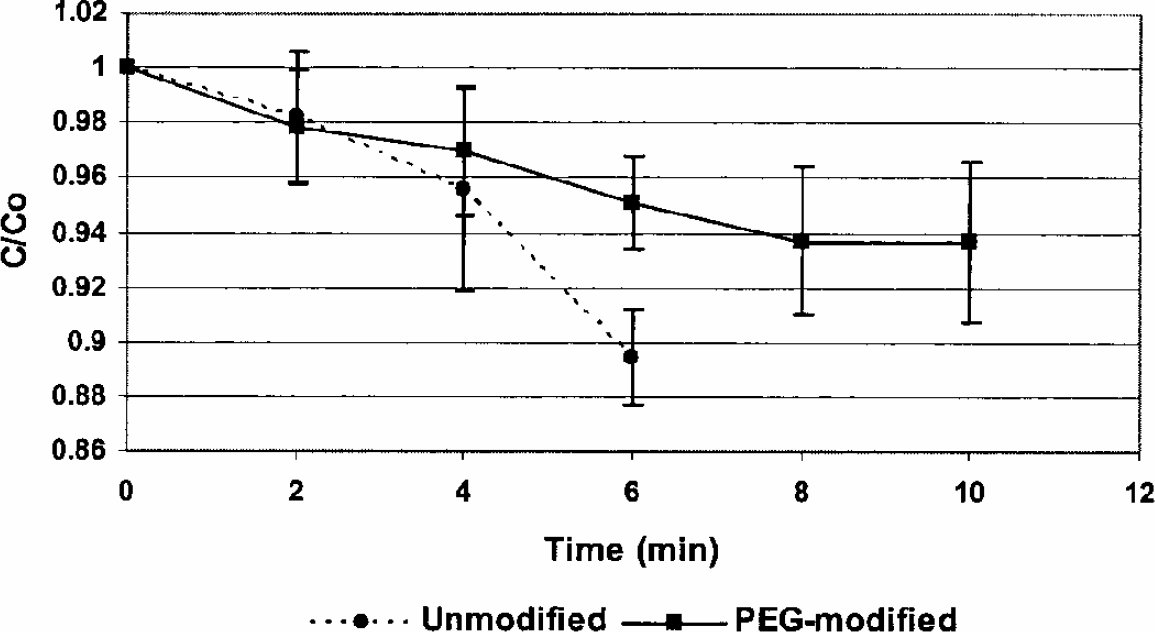
Flow of 1mg/ml BSA solution through 2 inch long glass capillary.
The development of uniform, conformal, ultrathin and stable films on silicon surfaces is an important criterion for the development of useful biomedical micro-/nano-devices and microfluidic systems. This work concentrates on the development and characterization of vapor deposited PEG films on silicon to achieve controlled surface wettability and roughness. Silanes are often terminated by desired end groups. They can be used as a precursor layer for further modification with more biocompatible functional groups such as Poly (ethylene glycol). In our studies, PEG not only modified the surface in terms of wettability and chemical reactivity, but also modified the surface topography and protein adsorption, which is extremely important for subsequent biological interactions with the surface. A simple experiment of flowing protein solution through unmodified and PEG modified glass capillaries shows promising results. Further studies are now directed toward applying this technique to various microfluidic devices.