
Abstract
Micro-capillaries with an engineered hidden surface were produced via the self-rolling of polymer thin films whose surface was photochemically modified so as to coat it with silver nanoparticles. The polymer films consisted of a poly(4-vinyl pyridine) (P4VP) layer top-coated with a polystyrene layer. When immersed in acid water, they underwent self-rolling due to selective swelling of the poly(4-vinyl pyridine) layer. The generation of Ag nanoparticles at the inner structure of the 40 μm-wide capillaries was carried out by a photochemically initiated reduction process. It was confirmed by UV-VIS spectroscopy, transmission electron microscopy (TEM) and energy-dispersive X-ray spectroscopy (EDX) analysis. The possibility of designing non-uniform distributions of silver particles at the inner surface of the tubes was also established. It consists of photopatterning the top of the film - the polystyrene side - prior to the swelling/self-rolling step. This approach provides new opportunities for the design of micro-compartments for micro-biological research and for micro-fluidics applications.
1. Introduction
Tubular micro-structures of different types achieve a great variety of applications in micro-fluidics, catalysis, micro-biological studies, and many other fields. Recently, a novel method was introduced for producing micro-tubes and micro-scrolls from thin semiconductor, polymer and composite thin films via a self-rolling process [1–3]. Scrolling was caused by the internal bending moment which arises due to opposite in-plane strains in the top and bottom layers of the films, due to a discontinuity of any property at the interface between the two layers. For instance, in semiconductor epitaxially grown heterofilms, the strain is provoked by the mismatch of the crystal lattice constants of the top and bottom layers [4–7]. In polymer multi-layer films, curling can be triggered by an unequal swelling ratio of the top and bottom layers in a given solvent [8–12]. For instance, polystyrene/poly(4-vinyl pyridine) bilayer films roll up in acidic water due to preferential swelling of the P4VP layer [8]. The approach has triggered numerous advanced applications, such as micro-needles for intra-cellular surgery [2], microtubular chemical reactors [3], X-ray microscale waveguides [4], two-dimensionally confined cell culture scaffolds [5], the design of self-propelling catalytic microparticles [6], the temperature-controlled capture and release of micro-particles [9], the fabrication of fibrous materials with a complex inner structure for the fibres [11], and many others.
A very attractive feature of the self-rolling approach is the opportunity for the complex engineering of the interior of the tubes or scrolls. Prior to rolling, the films can be exposed to various techniques for surface functionalization, such as plasma chemical activation and hydrophilization, metallization by sputtering, photolithography, and the anchoring of micro- and nanoparticles, etc. [9,11,12]. Functionalization of the interior of the micro-tubes with nanoparticles can be considered in the context of the broad field of the encapsulation and release of micro- and nano-objects, including catalytic nanoparticles and biological agents, such as drugs and living cells. The last two applications are especially valuable for biological and medical research.
During recent years, one of the authors of this article (L.B) made a significant contribution to the photochemically-assisted synthesis and implementation of metal nanoparticles (MNPs). This powerful in situ approach, involving the photoreduction of an appropriated formulation, induces a homogeneous distribution of MNPs in a cross-linked polymer network. As an extension of this work, specific procedures allowing MNPs to be photogenerated onto polymer surfaces were developed using UV or visible light sources [14–16]. Even more importantly, the possibility of patterning the spatial distribution of MNPs, either by using an amplitude mask or an interferential device to shape the actinic beam, was demonstrated.
In the present paper, we take advantage of this knowledge to design advanced polymer microtubes functionalized with silver nanoparticles. The photochemical route to MNPs provides many decisive advantages, since it combines the characteristic features of light (i.e., versatility and convenience of the process, high spatial resolution and reaction controllability - intensity and wavelength) with the simplicity of the colloidal approach [13–17].
The fabrication of polymer microtubes by the self-rolling method is simple and cheap and offers high outputs, but it allows only reduced control over the functionalization of the hidden walls of the tubes. In consideration of the strong potential of photo-assisted processes used to generate MNPs, arbitrary complex functionalization, including complex geometrical patterning of the inner walls, can be designed.
2. Experimental section
The micro-tubes were prepared as follows: a layer of P4VP (60 kDalton, Sigma-Aldrich) was generated on a glass substrate by spin coating a 3% solution of the polymer in chloroform (speed 2500 rpm, ramp 1000 rpm/sec, time 60 sec). The film was cross-linked by UV radiation (λ=254 nm, irradiation dose ca 2 J/cm2) or by quaternization reaction in saturated vapour of diidodobutan at 80°C over 90 min. The cross-linked P4VP films were top-coated with a layer of polystyrene (PS) (3% solution in toluene, 185 kDalton, Aldrich), deposited under the same dip-coating conditions. In order to promote good adhesion of the PS and P4VP layers, a small amount of the biblock-copolymer (5–10 mg per 10 ml, PS-block-P4VP, 35.5/4.4 kDalton, Polymer Source Inc.) was added to the solutions of PS and P4VP. The thickness of the P4VP and PS layers was measured by AFM via a scratch test as 0.6 and 1.2 μm, respectively. The bilayer films were hydrophilized by air plasma (0.075 Torr, 50 W) for 30 seconds.
Silver nanoparticles were generated by reducing AgNO3 with UV light (365 nm) in the presence of Irgacure 819 (bis(2,4,6-trimethylbenzoyl)-phenylphosphine oxide) and 1-propanol. Irgacure 819 was used as a generator of free radicals capable of reducing silver cations. The reduction of Ag+ to Ag(0) and the formation of NPs involve both benzoyl and phosphonyl primary radicals, resulting from the cleavage of the C—P bond [18]. A few drops of the photosensitive formulation (AgNO3 0.5 wt. %, Irgacure 0.5 wt. %) were poured onto the bilayer film. After drying, the sample was irradiated for three minutes at room temperature. It must be emphasized that no MNPs are generated when AgNO3 is irradiated under the same photonic conditions in the absence of Irgacure. This observation excludes the intervention of any photothermal effect in the process generating NPs. Moreover, no capping agent was used to stabilize the nascent nanoparticles.
The photochemical reactions were carried out under UV irradiation (Xe-Hg source LC5 8253 from Hamamatsu). Absorption spectra were taken with a Perkin-Elmer Lambda 750 spectrometer. Transmission electron microscopy (TEM) was conducted using a Philips CM20 instrument with a LaB6 cathode operated at 200 kV. The tubes were broken by applied pressure in order to produce polymer flakes transparent to the electron beam. The sample was placed on the observation grid. Energy dispersive X-ray spectroscopy (EDX) and scanning electron microscopy (SEM) were performed using a FEI-Quanta 400 model.
Optical images were taken using an Olympus BX51 microscope and a digital camera.
After the photochemical treatment, unidirectional scratches were made with a metallic blade. The samples were soaked in an acid solution (pH = 2, HCl in water ca 0.1 %) in which the tubes formed in a few minutes.
3. Results and discussion
The formation of microtubes by the self-rolling process of P4VP/PS polymer bilayer films coated with an Ag nanoparticles layer is shown in Figure 1, while the whole process is described step by step in the schematic of Figure 2. The tube rolls in the direction opposite to the edge of the silicon wafer (Figure 1). When the picture was taken, a narrow region of the bilayer (ca 1 mm broad) had rolled up to form a 41 μm-wide tube. Assuming a perfect circular cross-section of the nanotube and a negligible thickness relative to the diameter of the tube, the tube would consist of an eight-ply arrangement, as estimated from the relationship n ≈ H/(πd), where H is the width of the rolled-up part of the film and d is the diameter of the tube.

Optical microscope image of the formation of a microtube by the self-rolling of the bilayer P4VP/PS film in acidic water. The white arrow shows the direction of rolling.
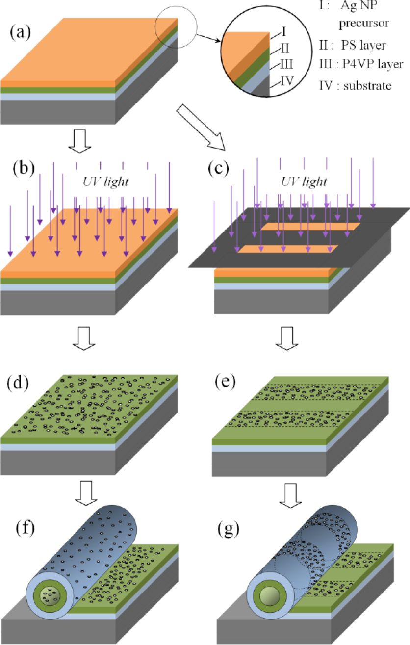
Scheme of fabrication of nanotubes functionalized by silver nanoparticles: (a) initial multi-layer structure; (b) exposition of the sample to UV radiation, or (c) irradiation of the sample through an amplitude mask; (d,e) Ag nanoparticles on the surface of the PS/P4VP film; (f,g) rolled-up microtubes functionalized with nanoparticles.
The diameters of the microtubes were observed to depend upon different fabrication parameters, including but not limited to the thickness of the P4VP and PS films, the irradiation dose and the acid concentration [10].
Most of the tubes were formed via self-rolling not from the wafer edges but from the grooves made in the bilayer film by the sharp blade. If the uniformity of the tube's diameter is not critical for a particular application, the film can be scratched with a metallic brush that allows large areas of the film to be quickly patterned. When the wafer coated with the bilayer polymer film is soaked in the acid solution, the self-rolling and the formation of nanotubes proceeds until the exhaustion of the accessible material. The rolling process can be stopped at any moment by removing the material from the swelling solution and drying it. When the film is patterned by a series of parallel scratches, twin tubes roll from two neighbouring grooves in opposite directions.
Once a polymer film has rolled up, microtubes detached spontaneously or else could be peeled off via gentle shaking of the substrate. In order to obtain concentrated suspensions of microtubes, they were collected with a pipette from several samples and transferred to a storage flask.
Prior to rolling, the upper surface of the bilayer film that corresponds to the hidden walls of the microtubes can be functionalized with silver nanoparticles. The surface of a reference sample (a P4VP/PS film) coated with a liquid formulation containing AgNO3 (0.5 wt. %) and IR 819 (0.5 wt. %) and dried at room temperature for two minutes was exposed to UV radiation (5 mW/cm2) for three minutes. This photo-chemical treatment induced the formation of a layer of silver nanoparticles at the surface of the bilayer film. The UV-vis absorption spectrum of films with and without silver NPs are shown in Figure 3. The UV spectrum of the bilayer film coated with AgNPs exhibits the characteristic surface plasmon band located at 443 nm, while this band is absent in the blank sample. After rolling, the microtubes obtained from silver-coated bilayer films showed a brown colour (Figure 4), which is characteristic of Ag nanoparticles. The UV-Vis spectrum of a suspension of these microtubes exhibits the shape of a slightly diffusive system with a pronounced shoulder in the region that corresponds to the surface plasmon of silver NPs (Figure 3).

Time evolution of absorption spectra of the samples upon irradiation at 365 nm (a) without silver cations, (b) with silver cations, and (c) with silver cations on the rolled-up microtubes. Incident light power 5 mW/cm2. Exposure 180 s.
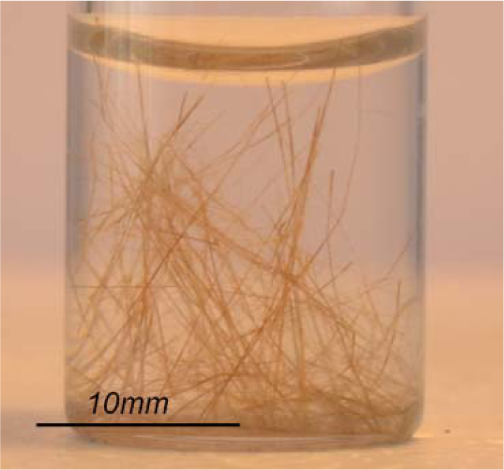
Photograph showing the high aspect ratio tubes in a HCl solution
It can be concluded, therefore, that the (electrostatic) interactions causing the silver NPs to adhere onto the polymer surface are not significantly affected by the rolling up process of the microtubes.
A sample obtained by drying a suspension of nanotubes was analysed by electron microscopy (TEM, SEM) and by energy-dispersive X-ray spectroscopy (EDX). Despite the action of the capillary forces - which tend to flatten the tubes - the dried tubes retained an almost circular cross-section (Figure 5a), due to their tight multi-shell structure. The EDX spectrum (Figure 5b) taken on a small section of the inner surface of a microtube obtained from a silver-modified polymer film (selection on Figure 5a) exhibited an intense signal characteristic of Ag, which confirmed the presence of metal NPs in the chemical elementary composition of the tubes.

Scanning electron microscopy (a) and EDX analysis (b) of the microtubes functionalized with Ag nanoparticles
The presence of AgNPs in the microtubes was also corroborated by TEM. A suspension of flakes produced by the mechanical grinding of dried tubes was dispersed in tetrahydrofuran (THF) and dropped on the copper grid of the instrument. Figure 6a shows the bright field micrograph of this sample. Spherical silver NPs are clearly visible and a statistic analysis carried out on a collection of 100 particles leads to an average diameter of 6.3 ± 0.6 nm (Figure 6b).

TEM image (a) and the corresponding particle size distribution (b) of silver NPs on the polymer micro-tubes. Light power 5 mW/cm2.
One of the main advantages of the photo-chemical approach used to functionalize self-rolled microtubes consists in the possibility of patterning the surface of the bilayer polymer film that will form the inner walls of the microtubes after rolling-up. To exemplify this concept, the PS surface of a P4VP/PS film coated with the photosensitive formulation described above was irradiated through an amplitude mask (Figure 7), positioned perpendicularly to the direction of self-rolled tubes. When an amplitude mask was used to shape the actinic beam, photo-reduction was observed to take place in the bright areas and it was possible to image the incident pattern with silver nanoparticles (Figures 2g and 7).

Optical microscope image of silver patterns onto a polymer sample through an amplitude mask. The white arrow indicates the direction of rolling.
4. Conclusions and perspectives
The innovative photochemical approach described in this work provides an alternative to processes used so far to functionalize microtubes. Silver nanoparticles were photosynthesized and anchored at the surface of thin bilayer polymer films before these films were swelled in acid water to trigger self-rolling. In that way, microtubes with an inner surface covered with unprotected silver nanoparticles can be generated. Functionalizing the interior of polymer micro-tubes with stable and unprotected silver nanoparticles did not affect the integrity of the polymer. This process can be easily implemented; it is very fast, uses only readily available precursors, and does not require any of the conventional S-, N- or P-stabilizing ligands.
The characteristic absorption due to a surface plasmon at ca 443 nm and EDX spectra confirmed the presence of silver nanoparticles with an average diameter of ca. 6 nm, as determined by TEM.
The possibility of controlling the spatial distribution of MNPs at the inner surface of microtubes by shaping the light beam used to photolyze the polymer film precursor was established. In future experiments, more sophisticated and subtle image-wise distributions could be generated with the help of high resolution photo masks.
Micro-capillaries with hidden walls engineered with Ag nanoparticles should generate interest in a great variety of applications in biology, microelectronics, optics and photonics. In the field of micro-biology, such microengineered containers should open up new vistas for the study of the behaviour of micro-organisms (bacteria, living cells) in confined spaces. The spatial distribution of bactericide Ag particles at the inner surface of capillaries - either uniformly covered or else covered only in places - might generate additional controlled stress on cells and bacteria and influence their movements and the life cycle of micro-organisms as well.
Moreover, self-rolled polymer films grafted with AgNPs could be used to explore the catalytic activity of such systems in a microfluidics approach (i.e., with reactors requiring only minuscule amounts of reactants [19]).
Furthermore, such bilayer polymers coated with metal particles could also be used as templates to create new 3D metallic objects, such as microtubes, solenoids and micro-springs.
Prospects along the same lines are concerned with a broader range of NPs, including semiconductors, photochemically generated at the hidden walls of self-scrolled polymer microtubes, and the development of micro-devices using this new process.
Footnotes
5. Acknowledgments
L.B. thanks the Agence Nationale de la Recherche (ANR) for financial support under contract ANR-09-JCJC-0029-01.