
Abstract
This article reports on a study on fluorescence adenosine triphosphate (ATP) detection by InP/ZnS quantum dots (QDs). We present a spectroscopic analysis displaying the effect of enzymatic reactions of glucose oxidase (GOX) and hexokinase (HEX) on the InP/ZnS quantum dots at physiological pH. The InP/ZnS quantum dots act as glucose sensors in the presence of GOX, Glu and ATP, and their luminescence quenches during the release of hydrogen peroxide from the reaction. However, in the presence of adenosine 5′ triphosphate, glucose, and HEX, a significant photobrightening of the InP/ZnS QDs is recorded. This is dependent on the concentration of ATP in the sample. The relationship between the ATP and the emission intensity of InP/ZnS nanocrystals is linear. The present results are the first to report the effect of different by-products released by these enzymatic reactions on the fluorescence of the InP/ZnS QDs.
1. Introduction
Semiconductor Quantum Dots (QDs) have recently replaced organic dyes in life sciences due to their relatively narrow emission line-width and high photostability. They are particularly attractive in the field of biosensors due to their long-term photostability [1, 2–7]. The surfaces of QDs are highly sensitive due to their large surface-to-volume ratio. Therefore, small molecules tend to have a considerable effect on their luminescence. QDs have been used as optical sensors for ions (Cu2+, Mn2+, Hg2+) [8–11], drugs, organic pollutants such as polycyclic aromatic hydrocarbons [12,13], and small biological molecules (glucose, folic acid) [4,14–16].
Adenosine-5-triphosphate (ATP) plays an important role in many biological processes. It is the energy source for biological reactions as well as an extracellular signalling agent [17]. Several techniques have been used for the detection of ATP, including bioluminescence detection [18] and aptamer-based fluorescent detection [19].
Some researchers have employed fluorescence detection for the quantification of ATP. Previously reported detection methods have been based on the use of cadmium-based QDs [14], which are well known for their high cytotoxicity. Therefore, recently many research groups have refrained from using cadmium-containing QDs for cellular imaging and biological applications [20, 21]. Alternatives must therefore be addressed. In this paper, we present a spectroscopic method based on a competitive enzymatic reaction between hexokinase (HEX) and glucose oxidase (GOX) for the detection of ATP. Sun et al. reported an assay to detect ATP via a competitive enzymatic reaction, hexokinase (HEX)/glucose oxidase (GOX) [22–24]. Additionally, various researchers have developed a glucose sensor using quantum dots. The reaction of glucose oxidase (GOX) and glucose (Glu) is an enzymatic reaction that results in the production of hydrogen peroxide (see equation 1 below) [26]. The release of the hydrogen peroxide in the presence of QDs results in the rapid quenching of the luminescence of the QDs [15]. The concept of a glucose sensor using QDs reported previously was based on the quantification of luminescence quenching by hydrogen peroxide to indicate the presence of glucose.

The present study has adopted the idea of competitive enzymatic reactions; InP/ZnS QDs have been used to implement the fluorescence detection of ATP. The spectroscopic study conducted in this paper is the first to report the use of InP/ZnS QDs for the fluorescent detection of ATP via competitive enzymatic reaction. A novel method has been developed to detect the effect of different enzymatic reactions on the photoluminescence of InP/ZnS QDs.
2. Experimental details
2.1 Chemicals
The following chemicals were purchased from Sigma-Aldrich: Adenosine-5′-diphosphate sodium salt at approximately 99% (from bacterial surface), hexokinase and glucose-6-phospahate dehydrogenase from baker's yeast (S.cervisiae). The following were purchased from Fluka: glucose oxidase from Aspergillus niger, D-glucose 6-phosphate sodium salt, and magnesium chloride solution Ultra ∼ 1M in water.
2.2 Synthesis of hydrophilic capped InP/ZnS QDs
The hydrophobic InP/ZnS QDs were prepared using a previously published procedure [25]. 500 μL of InP/ZnS QDs was dispersed in 1-octadecene, and then washed with toluene followed by centrifugation for 10 minutes. The supernatant was then precipitated with ethanol at a ratio of 1:3. InP/ZnS QDs were re-dispersed in 500 μL dimethylsulfoxide (DMSO) followed by the addition of an excess of the ligand 2-mercaptoethanol. The samples were subsequently heated to 60°C and left to stir overnight. A few drops of acetone together with 750 μL of diethylether were added in order to precipitate the water dispersible InP/ZnS QDs. The QDs were centrifuged for 15 minutes at 4000 rpm and the InP/ZnS/ME QDs were re-dispersed in tris-glycine buffer.
2.3 Fluorescence titration of ATP
A solution of the following materials was prepared: InP/ZnS QDs, 4 mM glucose, 20 mg/ml glucose oxidase, 1 KU hexokinase/glucose-6-phsphate dehydrogenase, NADP and 5 mM MgCl2. The ATP was added later during the UV-VIS measurements. The UV/VIS spectra of InP/ZnS QDs were recorded before and after the addition of ATP. The following concentrations of ATP were mixed with InP/ZnS QDs on a microplate: 2.4 mM, 1.2 mM, 0.5 mM, 0.2 mM, 0.1 mM, and 0.01 mM. The fluorescence intensity was recorded using a microplate reader (Floustar Omega microplate reader, from BMG Labtech). The emission wavelength used was 380 nm, with a band-pass filter of 460 nm (BP). The fluorescence intensity was recorded at different time intervals. The data are representative of experiments performed more than three times independently.
2.4 UV-VIS photoluminescence spectrometry
Photoluminescence spectra were recorded on a HORIBA JOBINYVON FL3-11 spectrometer using 370 nm excitation wavelength. Absorbance measurements were carried out on a Cary 5000 Lamda 25 spectrometer.
3. Results and discussion
3.1 Glucose oxidase and InP/ZnS QDs in the presence of ATP
The main criterion of a competitive ATP assay is that the luminescence of the InP/ZnS QDs is quenched by hydrogen peroxide in a similar way. Glucose oxidase was mixed in solution with glucose, ATP and the InP/ZnS QDs. When the reaction between GOX and Glu takes place, the QDs gradually lose their fluorescence. Figure 1 shows the decrease in the luminescence intensity as glucose is being oxidised. The photoluminescence spectra show a maximum at approximately 600 nm and a shoulder at about 525 nm. Although the origin of the shoulder is not apparent at this wavelength, a clear quenching of the QD photoluminescence is observed. It is also apparent that the presence of ATP does not affect this glucose assay.
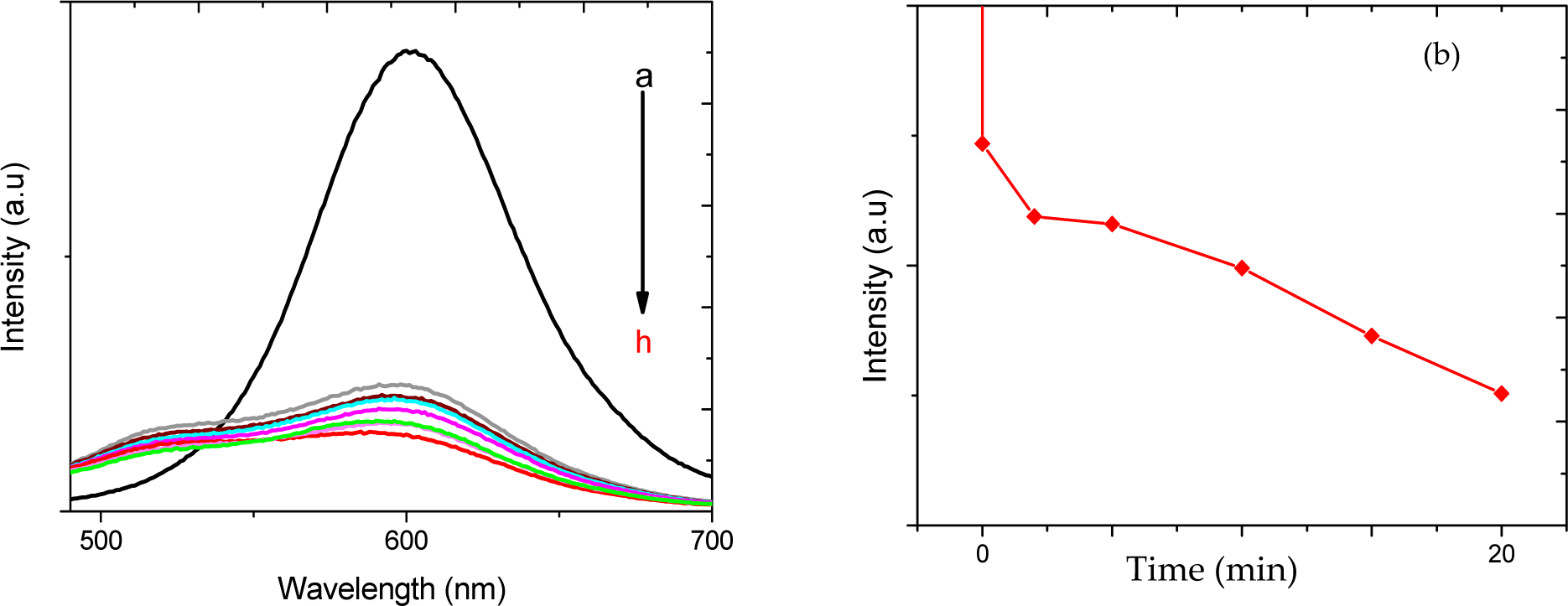
(a) Photoluminescence spectra of InP/ZnS QDs in the presence of glucose oxidase, ATP a) before the addition of glucose b) upon the addition of 4 mM glucose c) after 2 minutes d) after 5 minutes e) after 10 minutes f) after 15 minutes g) after 20 minutes; (b) the decrease in fluorescence intensity of InP/ZnS QDs with time in a solution containing glucose, ATP and glucose oxidase
Furthermore, InP/ZnS QDs behave similarly to previously used cadmium-containing QDs as their photoluminescence intensity decreases in the presence of hydrogen peroxide.
The enzymatic reaction of the glucose oxidase and glucose is time-dependent; one unit of GOX oxidises 1 μmol of glucose/minute at pH 7. The concentration of hydrogen peroxide in the solution increases with time, until all of the glucose has been consumed. Our solution contained 4 mM glucose and GOX was added in excess (20 mg/ml). Following the principle that one unit of GOX oxidises 1 μmol of glucose/minute at pH 7, a 4 mM glucose solution would be oxidised by eight units of GOX in eight minutes. Figure 1 (a), shows the decrease in fluorescence intensity of the InP/ZnS QDs with time. The particles continue to quench even after the enzymatic reaction has been completed. This demonstrates that the quenching kinetics is the rate-determining step in this reaction.
Glucose oxidase is an enzyme that requires the presence of glucose as its main substrate, and it has been reported that GOX has a quenching effect on many types of cadmium-containing quantum dots [15]; however, its effect on InP/ZnS QDs has only been reported in the present study.
3.2 InP/ZnS QDs in the presence of hexokinase, glucose oxidase and ATP
The enzyme hexokinase (HEX) is introduced to the system at this stage. Hexokinase is an enzyme that phosphorylates glucose in the presence of two main substrates, glucose and adenosine triphosphate (ATP), and the presence of Mg2+ ions as a cofactor [23]. Figure 2 shows the photoluminescence spectra of InP/ZnS QDs in the presence of the two enzymes (hexokinase and glucose oxidase) (Table 1). In the presence of ATP, an unsteady change in the intensity of the InP/ZnS QDs is observed (Figure 3). For instance, five minutes after the reaction commenced, the luminescence intensity of the InP/ZnSQDs increased, as shown by the red spectrum.
Summary of the main components of the two-enzyme ATP sensor and their roles in the system

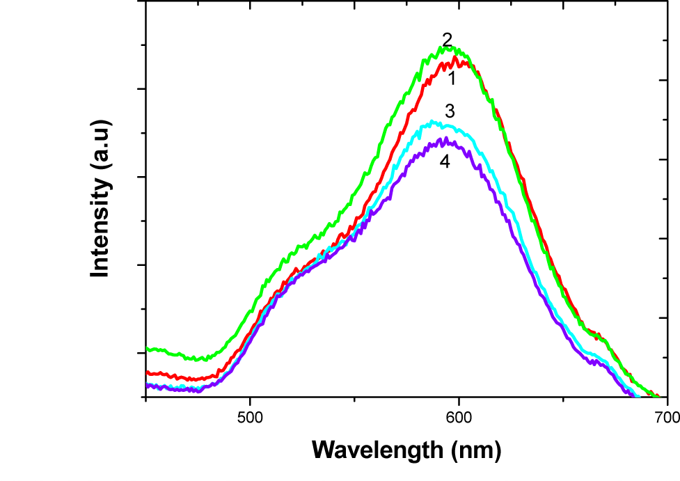
Emission spectra of InP/ZnS QDs+ hexokinase, glucose oxidase, glucose, and ATP at various time intervals during the reaction: (1) at the start of the reaction, (2) time at five minutes, (3) time at 10 minutes, (4) time at 20 minutes

(a) PL spectra of InP/ZnS QDs solution; excitation wavelength 400 nm, emission slit 1 nm, a) before the addition of ATP, b) upon the addition of 5mM ATP c) after 2 minutes, d) after 5 minutes, e) after 10 minutes, f) after 15 minutes, g) after 20 minutes h) after 25 minutes; (b) the effect of the hexokinase enzyme on the luminescence of the InP/ZnS QDs
However, 10 minutes after the reaction time, the luminescence intensity of the InP/ZnS QDs reverted and decreased.
The results shown in Figure 2 of debatable validity due to the unsteady change in the fluorescence intensity. Upon review of the literature we found minimal research on the effect of hexokinase on the luminescence of QDs. Therefore, in order to investigate further, solely the hexokinase enzyme was analysed with the InP/ZnS QDs in the presence of Glu and ATP. Hexokinase was added in excess to a solution containing QDs, Glc, GOD, ATP and NADP. In principle, an additional amount of hexokinase consumes more glucose and ATP, and thus its effect on the InP/ZnS QDs should be more evident.
3.3 InP/ZnS QDs in the presence of adenosine triphosphate and HEX/G-6-P-D
The InP/ZnS QDs were mixed in a solution containing HEX/G-6-P-D, NADP+ and glucose. The fluorescence of the QDs in the solution containing the enzyme was measured before and after the addition of ATP. Figure 3 illustrates the fluorescence spectra of the InP/ZnS QDs. Spectrum ‘a’ (blue) represents the fluorescence intensity of the QDs prior to the addition of ATP, while spectrum ‘b’ (red) corresponds to the fluorescence of the InP/ZnS QDs with the addition of ATP. All the remaining spectra demonstrate the increase in intensity of the InP/ZnS QDs with time for a period of 45 minutes at 5-min intervals; The QDs became brighter when the HEX/G-6-P-Dokinase enzyme started to be active (in the presence of ATP).
The enzymatic reaction is a time-dependent reaction. HEX/G-6-P-Dokinase consumes both glucose and ATP with time. HEX/G-6-P-Dokinase phosphorylates 1 μmol glucose per minute; consequently, phosphorylation of 4 mM glucose will take eight minutes. Figure 3 (a) shows the increase in intensity of the InP/ZnS QDs with time. As the enzymatic reaction takes place, the nanoparticles immediately exhibit photobrightening. This photoenhancement is mostly visible when the reaction gives its maximal effect at around eight minutes. Previous reports have shown the effect of ATP on CdTe QDs [14]. Wang et al. have observed quenching of the CdTe QDs, rather than the photo-enhancement that has been observed in the case of the InP/ZnS QDs.
The InP/ZnS QDs act as a fluorescent sensor of ATP, in the presence of the hexokinase enzyme. The sensitivity of this sensor was tested in order to determine the minimal amount of ATP that could be detected by the InP/ZnS QDs. The corresponding results shown in Figure 4 were performed in PBS media, at physiological pH (7.4). Different concentrations of ATP in solutions were mixed with InP/ZnS QDs+HEX/G-6-P DEH, and an ATP-free sample was tested as a control.

Fluorescence intensities of InP/ZnS QDs as a function of time; in the presence of (a) 0.0 mM ATP, (b) 0.01 mM ATP, (c) 0.1 mM ATP, (d) 0.2 mM ATP, (e) 0.5 mM ATP, (f) 1.2 mM ATP, and (g) 2.4 mM ATP. The excitation wavelength used is 380 nm, and the emission is detected beyond 460 nm
Figure 4 displays the results obtained by the fluorescence intensity detected at a wavelength of 380 nm. The QDs-Enzyme solution was added to the differently concentrated solutions of ATP. It was observed that the fluorescence intensity of the InP/ZnS QDs increases as the concentration of ATP increases. This instant increase of luminescence intensity is due to the start of the enzymatic reaction of hexokinase. Hexokinase phosphorylates glucose in the presence of ATP; the more ATP present, the more the enzyme consumes ATP as a phosphate source.
The results show that the differences in fluorescence intensities of InP/ZnS QDs can determine the difference in concentrations instantly. In Figure 5 it is inevitable to notice that at time 0, as the reaction starts, the fluorescence intensities of the InP/ZnS QDs differ according to the concentration of ATP in each sample. For instance, the sample containing 0.01 mM ATP has higher fluorescence intensity than the initial solution, and the sample containing 2.4 mM ATP has the highest fluorescence intensity when compared with the other samples.

PL intensity of InP/ZnS QDs in terms of ATP concentration at different times: (a) at the time of the reaction, (b) after five minutes, (c) after 20 minutes, (d) after 50 minutes, (e) after 100 minutes. Excitation wavelength 380 nm
Additionally, the fluorescence detection of ATP is a time-dependent process, because it depends on the activity of the hexokinase enzyme. Figure 4 shows the time-dependent fluorescence intensities of InP/ZnS QDs with differently concentrated QDs. This graph confirms the results displayed in Figure 5, which illustrates that the relationship between the concentration and the intensity displays good linearity. It can noticed from Figure 5 that there is an increase in intensity as a function of ATP concentration; however, at high concentrations of ATP (1.2 mM and 2.4 mM) the differences in fluorescence intensities become less visible due to equilibrium being reached. The photobrightening of the InP/ZnS QDs was clearly observed with the enzymatic reaction of the hexokinase. The mechanism of this interaction has not been discussed in the literature yet.
4. Mechanism
The photoluminescence of the QDs is caused by the recombination of an electron hole pair. The efficiency of this recombination can be affected by the presence of molecules or ions and their interaction with the surface atoms of the QDs [28,29]. We have observed a photoluminescence enhancement of QDs in the presence of the enzymatic reaction of hexokinase. In the literature, it has been discussed that phosphate groups have a brightening effect on QDs, unlike other functional groups such as thiols, which tend to quench the PL of the QDs [30–32].
Since the enzymatic reaction of hexokinase causes this photobrightening, we have examined the products of this enzymatic reaction individually to determine which of the products causes the surface passivation of the InP/ZnS QDs. Table 2 lists the molecules that are produced by the enzymatic reaction.
The effect of the molecules in the system on the luminescence of the InP/ZnS QDs

Each molecule was added to a solution of InP/ZnS/ME QDs. None of the molecules had any effect on the PL of the InP/ZnS QDs, with the exception of ADP. The PL spectra of the InP/ZnS QDs, in the presence of ATP alone. The spectra show that the presence of ATP does not affect the PL intensity of the InP/ZnS QDs significantly. This result confirms that ATP is not able to change the luminescence of the QDs. Therefore, since the starting material does not affect the luminescence of the QDs, one of the products of the enzymatic reaction must be responsible for the photobrightening of the QDs.
The InP/ZnS QDs were mixed with ADP molecules. The PL spectra were recorded at the time of addition of ATP, and then every five minutes over a period of 45 minutes. The spectra show that the InP/ZnS QDs increase their luminescence in the presence of ADP with time. The results shown in Figure 2 display a spectra profile similar to the one seen previously, when the hexokinase enzyme was active. When comparing the spectra obtained in the presence of ADP with the spectra obtained with hexokinase-ATP, it is noticeable that ADP affects the QDs in the same manner seen before with the enzyme. From this it can be concluded that ADP is the molecule responsible for the photoenhancement of the InP/ZnS QDs.
The photoluminescence of the InP/ZnS QDs in the presence of ADP increases gradually. The same result was observed when the ATP sensing was taking place in the presence of InP/ZnS QDs-HEX. These results can be explained by the fact that phosphates tend to passivate the surface of QDs, as has been reported elsewhere [33].
PL enhancement of the InP/ZnS QDs is caused by the passivation of trap states on the surface of the QDs. The phosphate groups of ADP can adsorb onto the surface of the QDs and decrease the surface defects, eventually increasing the PL of the QDs. When the concentration of the ADP in the system increases, the PL of the QDs increases. However, at the point where saturation is achieved, or in other words when all of the ATP is converted to ADP, an equilibrium is achieved and the PL of the QDs stabilises. This mechanism is supported by the results of the fluorescence titration of ATP.
5. Conclusions
We have shown that InP/ZnS/ME QDs are suitable alternatives to Cd-containing QDs for glucose sensing. The InP/ZnS QDs lost their luminescence completely in the presence of GOX and Glu. The InP/ZnS QDs were used to design an ATP sensor. Firstly, the ATP sensor was based on the presence of QDs and two enzymes, glucose oxidase and hexokinase. These two types of enzymes compete with glucose, but hexokinase is only active in the presence of ATP. The two enzyme systems did not give the results expected; therefore a single enzyme was used to detect ATP.
The effect of the enzymatic reaction of hexokinase on the InP/ZnS QDs was investigated. The results obtained showed that when the hexokinase is active, the fluorescence of the InP/ZnS QDs exhibit photoenhancement. This is the first report to reveal the effect of ATP, ADP and HEX on InP/ZnS QDs. The InP/ZnS QDs became brighter when the hexokinase is active. The by-products of the enzymatic reaction were studied separately to learn about their effect on the InP/ZnS QDs. It was shown that the ADP released by the enzymatic reaction was responsible for the increase in the luminescence.
In conclusion, the photoenhancement of InP/ZnS QDs is dependent on the concentration of ATP. It has been shown that the ATP sensor designed was successful at detecting ATP at different concentrations, and this fluorescence detection was perceived as an increase in the luminescence of the InP/ZnS QDs.
Footnotes
6. Acknowledgements
The authors thank Dr Shu Xu for the preparation of InP/ZnS quantum dots. Also, they would like to thank the Engineering and Physical Sciences Research Council (EPSRC) for the funding of this project.