
Abstract
Silver (SN) nanoparticles were re-dispersed in aqueous solution of Pluronic F127 and complexed with the antioxidant, alpha- Lipoic acid (LA). The nanoparticle - LA complex (SN-LA) was characterized using SEM. SN-LA exhibited DPPH radical scavenging activity in vitro and anti- inflammatory activity against acute and chronic paw models of edema in mice. SN-LA protected mice from whole body gamma radiation induced body weight losses and mortality revealing its radioprotecting capacity. Meanwhile, SN-LA administration to tumour-bearing mice prior to whole body gamma radiation exposure, aided in better tumour growth delay. The results suggest the feasibility in using SN-LA as a therapeutic adjuvant during cancer radiotherapy.
1. Introduction
Ionizing radiation causes damage to living tissues through a series of molecular events, such as Photoelectric, Compton and Auger effects, depending on the radiation energy. Because human tissues contain 80% water, the major radiation damage is due to the aqueous free radicals, generated by the action of radiation on water. These free radicals react with cellular macromolecules, such as DNA, RNA, proteins, membrane, etc, and cause cell dysfunction and mortality. These reactions take place in tumor as well as normal cells when exposed to radiation [1]. Hence, development of novel and effective approaches using nontoxic radioprotectors is of considerable interest for defense (nuclear wars), nuclear industries, radiation accidents, space flight, etc, besides playing important role in the protection of normal tissues during radiotherapy of tumors [2].
Nanoparticles have particle size in the range between 1 and 100 nm and exhibit unique electronic, optical and catalytic properties [3]. Recently, nanoparticles are gaining interest in the field of radioprotection as cerium oxide nanoparticles, yttrium oxide nanoparticles, carbon nanoparticles, etc. were found to possess antioxidant properties and several works have shown the ability of these nanoparticles to offer protection against radiation damages [4–10].
Silver nanoparticles have been known to posses excellent free radical scavenging, antimicrobial and anti-inflammatory activities [11–17]. The results of several studies suggest that nanocrystalline silver specifically may play a role in altering or compressing the inflammatory events in wounds and facilitating the early phases of wound healing. These benefits are associated with reduced local matrix metalloproteinase levels and enhanced cellular apoptosis [18, 19]. Silver nanoparticles have also gained increasing interest in the field of nanomedicine due to their unique properties and obvious therapeutic potential in treating a variety of diseases, including retinal neovascularization [20, 21] and acquired immunodeficiency syndrome due to human immunodeficiency virus (HIV) [22, 23]. They inhibit vascular endothelial growth factor (VEGF)-induced angiogenesis in bovine retinal endothelial cells [21]. Similar studies have proven their inhibitory effect on vascular permeability induced by VEGF, interleukin (IL)-1β [24] and advanced glycation end product [25] in retinal endothelial cells. Silver oxide nanoparticles exhibit antitumor properties in transplanted Pliss lymphosarcoma tumor models when administered by intravenous injection in the form of aqueous dispersions [26].
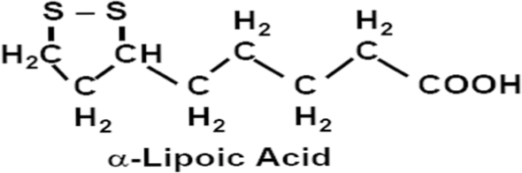
Alpha- Lipoic acid (LA), a disulphide derivative of octanoic acid, has been known to be a crucial prosthetic group of various cellular enzymatic complexes and characterized as an efficient antioxidant, for decades. It is a potential therapeutic agent in the treatment or prevention of different pathologies that may be related to an imbalance of the oxido-reductive cellular status [27, 28]. Lipoic acid or its reduced form, dihydrolipoic acid, quenches a number of oxygen-free radical species in both lipid and aqueous phase, and chelates transition metals. Alpha- Lipoic acid participates in the recycling of Vitamin- C and Vitamin-E, increases cellular levels of glutathione, and suppresses non-enzymatic glycation [29]. Anti-inflammatory and anticancer activities of LA are well established [30–32]. The relatively good scavenging activity of lipoic acid (Scheme 1) is due to the strained conformation of the 5-membered ring in the intramolecular disulfide [33] which confer it with antioxidant and radioprotecting activities [34–37].

SEM Micrograph of SN-LA at (A) 5000X and (B) 10,000X magnifications revealing aggregates of still smaller nanoparticles < 500nm (encircled).
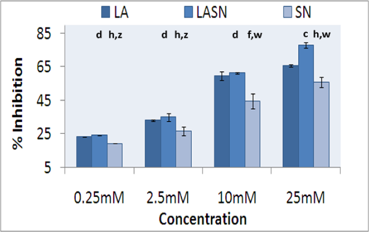
DPPH radical scavenging activity of SN-LA. Values expressed as Mean ± S.D. (c- p<0.05 and d- p>0.05 when compared to respective LA treated group. f- p< 0.01 and h- p>0.05 when compared to respective LA treated group. w- p<0.001 and z- p>0.05 when compared to respective SN-LA treated group)
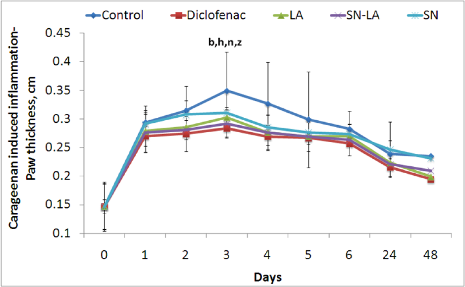
Effect of SN-LA on carrageenan induced paw oedema. Values expressed as Mean ± S.D. (b - p<0.01, h – p>0.05, n- p<0.05, z- p>0.05 for diclofenac, LA, SN-LA and SN treated groups respectively when compared to the control group at 3rd hour of study)

Effect of SN-LA on dextran induced paw oedema. Values expressed as Mean ± S.D. (a - p<0.001, f – p<0.01, l- <0.001, x- p<0.05 for diclofenac, LA, SN-LA and SN treated groups respectively when compared to the control group at 3rd hour of study)
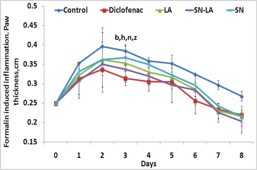
Effect of SN-LA on formalin induced paw oedema. Values expressed as Mean ± S.D. (b - p<0.01, h – p>0.05, n- p<0.05, z- p>0.05 for diclofenac, LA, SN-LA and SN treated groups respectively when compared to the control group on day 3 of study)

Effect of SN-LA on 10 Gy gamma-radiation-induced body weight loss (n=10, the experiment was repeated twice). Statistics of irradiated groups is provided as an inset. (a - p<0.001, b - p<0.01, c- <0.001, d- p<0.05 when compared to the control irradiated group; x - p<0.001, y - p<0.01, e- p<0.05 when compared to LA treated irradiated group).

Effect of SN-LA on 10 Gy gamma-radiation-induced mortality (n=10, the experiment was repeated twice). Statistics of irradiated groups is provided as an inset. (a - p<0.001, b - p<0.01, c- <0.001, d- p<0.05 when compared to the control irradiated group; x - p<0.001, y - p<0.01, e- p<0.05 when compared to LA treated irradiated group) (All 0 Gy points overlap).

Effect of SN-LA on radiation induced tumor growth delay in mice. Values expressed as Mean ± S.D. (statistical analysis data is presented as insert- a - p<0.001, c - p<0.05 when compared to 0 Gy control group. e - p<0.001, f - p<0.01, g - p<0.05, h - p>0.05 when compared to the 4 Gy control group)
Previous studies from our lab have explored the radioprotecting properties of silver nanoparticle complexes of compounds such as glyzyrrhizic acid [38], gallic acid [39] and 6-palmitoyl ascorbic acid 2-glucoside [40].
In the present study, silver nanoparticles (SN) having a particle size less than 50 nm [41], were allowed to form complex with LA by means of a surface stabilizing agent, Pluronic F 127. Efforts were made to explore the free radical scavenging and anti-inflammatory activities of SN-LA. SN-LA was analysed for its radioprotecting properties in terms of its effect on providing protection against body weight losses and mortality in mice exposed to lethal dose of whole body gamma radiation. The impact of the complex on tumor growth delay in whole body irradiated solid tumor bearing mice was also determined in order to predict the feasibility in using SN-LA as a therapeutic adjuvant during cancer radiotherapy.
2. Materials and methods
2.1 Chemicals
Alpha- Lipoic acid was obtained from Sigma-Aldrich Chemie, Steinheim, Germany. Silver nanoparticles prepared using tri-sodium citrate as initial surfactant cum reducing agent and sodium formaldehyde sulfoxylate as secondary reducing agent having a particle size less than 50 nm [41], were provided by Dr. P. K. Khanna, Center for Materials for Electronic Technology (CMET), Pune, India. All other chemicals were of analytical grade procured from reputed Indian manufacturers.
2.2 Animals
Male Swiss albino mice, 8-10 weeks old and weighing 22-25 g were obtained from the Small Animal Breeding Section, Thrissur, Kerala, India. They were kept under standard conditions of temperature and humidity in the Centre's Animal House Facility and provided with standard mouse chow (Sai Durga Feeds and Foods, Bangalore, India) and water ad libitum. All animal experiments in this study were carried out with the prior approval of the Institutional Animal Ethics Committee, strictly adhering to the guidelines of Committee for the purpose of Control and Supervision of Experiments on Animals constituted by the Animal Welfare Division of Government of India.
2.3 Preparation, characterization and the mode of administration SN- LA
Colloidal silver was prepared via re-dispersion of silver nanoparticles (SN) (0.1%) in 1% Pluronic F-127 under ultrasonication to have a clear solution with distinct yellow colour characteristic of silver nanoparticles. The ultrasonication was carried out at a frequency of 20KHz. 250W. The solutions were centrifuged to remove residues. To the supernatant, lipoic acid (LA) was added to a final concentration of 100 mM and subjected to sonication to obtain the SN-LA complex (Scheme 2). Scanning Electron Microscopic (SEM) analysis of SN-LA was done using JEOL Model JSM - 6390LV at Sophisticated Test And Instrumentation Centre, CUSAT, Kerala, India.

The animals were administered with SN-LA by means of oral gavage and dosage was such that the animals received 100 mg equivalents of LA /kg body weight. LA alone and SN alone treated groups were also included as control groups. For all the in vivo experiments, animals were divided into 4 groups comprising of 6 animals as follows: Group 1 – distilled water; Group 2 – SN-LA; Group 3 – LA; Group 4 – SN.
2.4 Determination of in vitro free radical scavenging activity
The free radical scavenging capacity of SN-LA was determined by using DPPH assay [42]. DPPH (1,1diphenyl-2-picryl-hydrazyl) is a stable free radical and has been used as a model free radical compound to evaluate the effectiveness of antioxidants. Freshly prepared methanolic solution of DPPH (6.34 μM) was incubated at ambient temperature with different concentrations of SN-LA / LA/SN and absorbance at 515nm was measured using a spectrophotometer. The percentage of inhibition of DPPH reduction (decolourization) was calculated according to the formula,

where, A0= Absorbance at 0 minute and A30= Absorbance at 30 minutes'.
2.5 Determination of anti-inflammatory activity of SN-LA in acute and chronic paw edema models
2.5.1. Carrageenan and Dextran induced acute paw edema models
Acute inflammation was induced in all groups of animals by sub plantar injection of 0.02 ml freshly prepared 1% suspension of carrageenan/ dextran in normal saline in the right hind paw of mice. Animals of Group 1 received carrageenan/ dextran injection alone and served as negative control. An additional group, Group 5 was also included which were administered with Diclofenac (10 mg/kg body weight) and served as positive control. Animals were administered with single doses of SN-LA/ LA/ SN/ diclofenac / distilled water by means of oral gavage 30 minutes prior to the sub plantar injection of carrageenan/ dextran. The paw thickness was measured using vernier callipers at one hour intervals up to 7th hour followed by 24th hour and 48th hour.
2.5.2 Formalin induced chronic paw edema model
Animals were divided into groups as done for carrageenan/ dextran induced acute paw edema models. In all groups chronic inflammation was induced by sub plantar injection of 0.02 ml freshly prepared 1% formalin in normal saline in the right hind paw of mice. Animals which received formalin injection alone served as negative control. Diclofenac served as the standard reference drug. The administration of SN-LA/ LA/ SN/diclofenac / distilled water was done as mentioned before. The paw thickness was measured everyday for 7 consecutive days following formalin injection.
2.6 Determination of the effect of SN-LA on radiation-induced body weight alterations and survival loss
Animals were administered with SN-LA/ LA/ SN/ to mice at doses mentioned earlier. Thirty minutes after administration, the animals were exposed to a lethal dose of 10 Gy whole-body gamma radiation. Administration of SN-LA/ LA/ SN was continued for 5 more days post radiation exposure. The animals were checked on a daily basis to record the body weight changes and mortalities, if any.
2.7 Determination of the effect of SN-LA on tumor growth delay in Daltons Lymphoma Ascites solid tumor bearing mice exposed to whole body gamma radiation
i) Induction of solid tumor in mice: Solid tumor was induced by injecting DLA (Daltons Lymphoma Ascites) cells (1×10⁵ cells/animal) in to the right hind legs of mice. On the 10th day after tumor induction (when the tumor reached a size of 1.0 cm3), SN-LA/LA/ SN/ were administered to tumor bearing animals at doses mentioned earlier and the dosages were continued for 5 more days post radiation exposure.
ii) Exposure to γ- radiation: Half of the animals from each of the treated groups were subjected to 4 Gy whole body gamma radiation. Irradiation was carried out using a 60Co- Theratron Phoenix teletherapy unit (Atomic energy ltd, Ottawa, Canada) at a dose rate of 1.88 Gy per minute.
iii) Measurement of tumor volume: The hind leg thicknesses were measured using a vernier caliper once in three days from the day of radiation exposure. The tumor volume was calculated as follows:
Tumor thickness = Thickness of tumor induced leg - Thickness of normal leg
Tumor volume = 4/3 πr3, where r is the tumor radius.
2.7 Statistical analysis
The results are presented as mean ± SD of the studied groups. Statistical analyses of the results were performed using ANOVA with Tukey-Kramer multiple comparisons test.
3. Results
SEM analysis confirmed SN-LA as aggregates of still smaller nanoparticles well below 500nm. (Figure 1)
3.1 DPPH free radical scavenging activity of SN-LA
The reduction of DPPH results in a decrease in absorbance at 515nm. Presence of SN-LA/LA/SN at different concentrations caused dose dependent inhibition of DPPH free radical formation (Figure 2). At 25mM concentration, SN-LA, LA and SN exhibited 78.08%, 65.84% and 55.90% inhibition of DPPH radicals.
3.2 Anti-inflammatory activity of SN-LA
The sub plantar injection of carrageenan/ dextran/ formalin in mice produced local inflammatory responses.
In carrageenan model, it reached a maximum intensity of oedema at hour 3 after the application of the phlogistic agent and was quantified by measuring changes in thickness of footpad. The paw oedema was significantly reduced by 9.28% in LA, 12.62% in SN-LA, 6.72% in SN and 14.96% diclofenac administered groups respectively at 3rd hour as shown in fig.3.a.
Dextran induced maximal inflammatory response at hour 3 after the application of the phlogistic agent. The paw oedema was significantly reduced by 16.99% in LA, 26.27% in SN-LA, 17.08% in SN and 25.99% diclofenac administered groups respectively at 3rd hour as evident from fig.3.b.
Formalin induced inflammatory response reached a maximum intensity of oedema on day 3. The paw oedema was significantly reduced by 8.26% in LA, 12.44% in SN-LA, 4.56% in SN and 18.41% diclofenac administered groups respectively at 3rd hour as evident from fig.3.c.
3.3 Effect of SN-LA on radiation-induced body weight alterations and survival loss
From the data presented in Figure 4.a., it can be observed that upon exposure to a lethal dose of 10 Gy gamma radiation, there was a profound loss in the body weight of the survivors. SN-LA treatment delayed this loss in body weight to a much greater extent than other control treatment groups.
The data on survival of the animals exposed to the lethal dose of 10 Gy whole body gamma radiation is presented in fig.4.b. Mortality of the irradiated animals commenced on the 3rd day post radiation in the control irradiated, on 10th day in SN-LA, on 7th day in LA and on 4th day in SN treated groups. The loss of survival continued at a much slower rate in SN-LA group compared to all the control groups. SN-LA group showed a survival of 80% on 10th day which dropped to 50% on 12th day, 40% on 13th, 20% on 14th, 10% on 19th and finally 0% on 20th day post radiation. As all the control irradiated animals died on 9th day and complete mortality of SN-LA groups were observed only on 19th day post radiation, it could be surmised that SN-LA administration bestowed survival advantage to the mice exposed to lethal dose of 10 Gy gamma radiation.
3.4 Effect of SN-LA on radiation induced tumor growth delay in mice
The results of the study on the combined effect of gamma radiation (4 Gy) and SN-LA administration is presented in Fig. 5. The growth of the tumour was found to be delayed or suppressed in animals exposed to 4 Gy gamma radiation compared to un- irradiated animals. The administration of SN-LA/ LA/ SN itself reduced tumour growth to some extent. The tumour growth was found substantially suppressed in the group of animals administered with SN-LA and exposed to 4 Gy gamma radiation than all other treatment groups.
4. Discussion
Radiation is known to produce oxygen free radicals which are implicated in the process of DNA damage, cell killing, mutagenesis, and carcinogenesis; hence, it is reasonable to assume that agents capable of scavenging free radicals would play a significant role in modulating these processes. The radioprotection of normal cells by a number of synthetic and natural compounds are reported to be mediated through free radical scavenging activity [43]. Anti-lipid peroxidation assay using goat liver homogenate and DPPH scavenging test has already established the anti-oxidant potency of the silver nanoparticles [44]. In the present study, the silver nanoparticle combined with lipoic acid to form the complex, SN-LA showed improved DPPH free radical scavenging activity.
Inflammation is a physiological process in response to tissue damage resulting from microbial pathogen infection, chemical irritation, and/or wounding. The relation between inflammation and atherosclerosis, diabetes, cancer, arthritis and Alzheimer's disease has been well substantiated [45–48]. Ionizing radiation has been shown to exaggerate the inflammatory responses and to enhance the release of inflammatory mediators in experimental animals [49–52] and humans [53, 54], a process which probably involves the local vascular system, the immune system, and various cells within the injured tissue. The SN-LA complex alleviated the extent of acute and chronic inflammation in different paw edema models in mice. This may be due to the down regulation of different cytokines by LA that are involved in sustaining the inflammatory response along with the combined radical scavenging and anti-inflammatory activities of SN.
SN-LA protected whole body radiation exposed mice from radiation induced body weight loss and mortality suggesting its usefulness as a radioprotector during different radiation exposure scenarios. Radiotherapy is frequently used as part of cancer treatment to achieve tumor control. Apart from inducing anti-proliferative and cell-killing effects in tumor tissue, radiotherapy also provokes normal tissue damage. For a drug to be used for normal-tissue protection in cancer therapy, it is highly desirable that the drug should give significant protection to normal tissues with no or minimal protection to tumours. Hence, the effectiveness of SN-LA on tumor growth delay and its feasibility as an adjuvant in radiotherapy was evaluated. SN-LA administration aided in significant tumor growth delay and this anti-tumor effect was more prominent in conjunction with gamma radiation treatment (4 Gy). The anti-inflammatory, antioxidant and radioprotecting properties of SN-LA must have aided in a faster recovery of normal tissues in irradiated animals without interfering with the delaying effects of radiation on tumor growth.
5. Conclusion
Thus present results suggest the possibility of using SN-LA as an adjuvant in cancer radiotherapy to protect normal tissues from radiation damages and also to enhance the anti-tumour activity of gamma radiation.
Footnotes
6. Acknowledgements
The authors thank Dr. P. K. Khanna, CMET, Pune for providing the silver nanoparticles. CKKN expresses his gratitude to BRNS, Department of Atomic Energy, Government of India for the financial support as a Research grant awarded to him.