
Abstract
We used temperature-sensitive poly(N-isopropylacrylamide) hydrogels as drug delivery systems, so changes in body temperature induced by pathogens could act like external stimuli to activate controlled release of the drugs incorporated in the hydrogel. In the distilled water combined release studies, we chose two model drugs: aminophylline and triamterene. The amount of drug released was measured by UV-Vis spectroscopy following the evolution of the absorption peaks of aminophylline (271 nm) and triamterene (365 nm). The maximum release time was greater for triamterene than for aminophylline at 37 °C, so these time-release profiles enabled the active ingredients to work over different periods of time. By increasing molar mass or solubility of the drug, we observed that the diffusion coefficient decreased. On the contrary, increasing hydrophobicity of the drug leads to a diffusion coefficient increase. The evolution of pore size distribution of hydrogels during loading and releasing was measured by quasi-elastic light scattering and by environmental electronic scanning microscope. When loading and releasing the drugs, the pore size of the hydrogel decreased and increased again without reaching the initial pore size of the hydrogel, respectively. We observed that the greater the concentration of drug loaded into the hydrogel, the greater the reduction in pore size.
1. Introduction
Recently, combined therapy with drugs of different therapeutic effects shows an effective way to treat diseases and cause tissue to be regenerate. In order to optimize their effects, different drugs should be used at their optimal dose and at different periods in the treatment. One of the main challenges of combined therapy is to control the release behaviour of each drug independently [1]. However, single drug delivery systems cannot fulfil the needs of such therapy. Therefore, developing dual drug delivery systems (DDDS), which can control the release behaviour of each drug, is needed. However, so far there has been limited research on dual drug delivery systems [2–9].
The incorporation of two different kinds of drugs in a single vehicle is a challenging, as well as important, aspect for smart drug delivery. The body's defence mechanisms are very complex and once triggered, they follow several paths in cascade. Thus, it is difficult to target the entire cascade using single drug molecules.
Hydrogels (hydrophilic three-dimensional cross-linked polymer networks which contain a large amount of water [10]) are highly permeable to various drugs and the entrapped molecules can be released through their web-like structures. As compared to conventional administration vehicles, drugs can prolong their duration time through the hydrogel drug delivery system (DDS). Mechanical properties, as well as the swelling and shrinking behaviour of the hydrogel, change in response to physical or chemical stimuli, such as temperature, pH, ionic strength, solvent composition and electric fields. Hence, the hydrogels behave as intelligent materials in drug delivery [1, 11].
Poly(N-isopropylacrylamide) (PNIPA) is one of the most widely studied hydrogels because of its unique properties and it is attracting more and more interest in biomedical applications because it exhibits a phase transition behaviour at a well-defined lower critical solution temperature (LCST) of 32 °C [12–18], which is close to the human body temperature. So, PNIPA hydrogel swells when cooled below LCST and collapses at body temperature. PNIPA thermo-sensitive hydrogel is a good candidate for a drug delivery system [19, 20].
In this work we prepare a dual drug delivery system based on PNIPA hydrogels. Two different kinds of drugs, triamterene and aminophylline, were loaded in this system. Triamterene presents a wide clinical usage sparing potassium diuretic, alone or in combination with other high ceiling diuretics. This drug displays a particular in vivo behaviour, with very irregular oral absorption (because of its low solubility) presenting, even inside the same formulation, wide inter-individual variations. The multi-ring structure (Figure 1) and low molecular weight (Table 1) makes it a typical hydrophobic drug. Triamterene is especially convenient as a model drug in release studies due to its intrinsic fluorescence, which even allows the imaging by fluorescence of the drug intercalated in our hydrogel structure. Finally, triamterene is weak basic, which explains why most of the attempts to form classical salts of this drug have failed. Aminophylline is a bronchodilator used successfully in the treatment of asthma in combination with other drugs. The controlled release profiles were followed by UV-Vis spectroscopy and the mechanisms of single and dual drug release by diffusion were modelled. The morphology changes of hydrogels during loading and releasing were studied by quasi-elastic light scattering (QELS) and observed by environmental electronic scanning microscope (ESEM).

Chemical structure of aminophylline (A) and triamterene (B).
Chemical structures, molar masses, solubilities and hydrophobicites of aminophylline and triamterene.

2. Experimental Part
2.1 Materials
N-isopropylacrylamide (NIPAM), C6H11NO, was used as a monomer (M = 113.16 g·mol−1) and purchased from Acros Organics (purity > 97%). N,N'-Methylenebisdiacrylamide, C7H10N2O2, was employed as a cross-linking agent (M = 154.17 g·mol−1) and sodium disulphide, Na2S2O2, was used as an initiator (M = 190.11 g·mol−1). Both were purchased from Merck. Triamterene, C12H11N7, and aminophylline, C14H16N8O4 · C2H8N2 ·xH2O, from Sigma, were used as model drugs in the release experiments and their structures are shown in Figure 1. Water used in all the experiments was double distilled and deionized.
2.2 Preparation of PNIPA hydrogels
The polymerization of conventional PNIPA hydrogels was carried out in deionized water at room temperature (22 °C) for 6 h using N,N'-Methylenebisdiacrylamide as a cross-linker and sodium disulphite as an initiator. In order to eliminate any unreaccted monomer, oligomer and non-cross-linked polymer chains, the dispersion was dialyzed opposite to water for approximately 3 days by using a dialysis membrane by and changing water daily.
2.3 Drug load experiments
A pre-weighed dried copolymer disk (Wd) was immersed in a drug solution (50 mg drug/mL water) at 22 °C for 1 week to attain swelling equilibrium in a dark environment (in order to avoid degradation). Drug-loaded disks were dried under a vacuum at 37 °C for several days to remove residual water. No presence of residual solution was assumed, because no weight change was observed in the vacuum after this drying period. The weight of the drug-loaded dry disks was defined as Wc. Therefore, Lc (g drug loading/g dry polymer disk):
2.4 Drug release experiments
For drug release experiments, 100 mg disks of drug-loaded xerogels were placed in a vessel containing 100 mL of water at a constant temperature (37.0 ± 0.1 °C), kept with an oil bath, and under magnetic stirring (300 rpm), using a glass support to keep the disks in place. We use a peristaltic pump operating at 30 rpm to maintain a constant flow between the release vessel and the spectrophotometer cell. In this way we did not change the volume in the test vessel. We found that the delay between the measured changes in drug concentration in the cell and in the vessel was 5 ± 2 seconds. All release studies were carried out at least in triplicate and at sink conditions (drug concentrations less than 10% of drug solubility).
2.5 UV-Vis spectroscopy
The amount of released drug was determined by UV-Vis spectroscopy (Cintra 308, GBC Scientific Equipment Pty Ltd., Dandenong, Australia) with a quartz cuvette at an absorbance wavelength of 365 nm for triamterene and 271 nm for aminophylline. As it can be seen in Figure 2, the UV spectra were not overlapped to any extent, so accurate drug released concentrations were determined through this method. The amount of drugs at any selected time was calculated from the triamterene and aminophylline calibration curves obtained fitting the maximum absorbance at different drug concentrations. Using a fluorescence spectrometer, Zhang et al. [33] had problems accurately measuring the drug concentrations using standard curves, due to the interference in their maximal excitation and emission wavelength. So we also considered, in addition to physico-chemical and therapeutic properties, the difference in the maximum wavelength of the absorption band by UV-Vis spectroscopy (Δλ = 92.6 nm for aminophylline and triamterene) when we chose a pair of drugs for the dual release test.

Concentration dependence of the absorption peaks of aminophylline and triamterene.
2.6 Quasi-elastic light scattering
Pore size distributions were evaluated by QELS using a BI-200SM goniometer (Brookhaven Instruments Co., NY, USA) at a scattering angle of 90°, equipped with an Ar-Ion laser source operating at 17 mW and delivering a vertically polarized light of λ = 514 nm. The autocorrelation functions were analysed with the Non-Negative Least-Squares (NNLS) algorithm. All solutions were filtered through a Millipore filter with a pore size of 0.8 μm to remove any dust impurities. All measurements were made at 37 °C.
2.7 Environmental scanning electronic microscopy
The internal morphology of PNIPA hydrogels were observed by ESEM (EVO LS 15, Carl Zeiss Inc., Oberkochen, Germany), operating at an acceleration voltage of 15 kV. The environmental capability allowed for examination of the pore structure of the hydrated hydrogels in a saturated water vapour environment which enabled imaging with minimal drying. Samples were observed at a working distance of 10 mm. To retain the internal nano-structure, the PNIPA hydrogels were equilibrated in pure water at 37 °C for 2 hours and then were frozen quickly to -10 °C by Peltier effect, which allows us to control the temperature from -30 to 50 °C in a few minutes. We operate the ESEM in the variable pressure mode allowing us vacuum pressure levels control between 10 Pa and - 400 Pa at constant temperature, in order to maintain the relative humidity (95% in general, slightly lower sometimes), in order to emphasize hydrogel structure.
3. Results and discussion
The three-dimensional network nano-structure and the matrix morphology of the resulting hydrogels were studied by ESEM. Pictures of the freeze-dried, swollen hydrogel samples are shown in Figure 3. They show a clear dependence of the morphology on the loading and releasing processes of the hydrogel with both drugs. From ESEM pictures, we can see how the morphology of these hydrogels retained the porous nano-structure of the swollen state due to the freeze-drying method employed in the sample preparation. Morphology became more irregular after the release.

ESEM micro-graphs showing the change in pore size in swollen and frozen hydrogels when the drug was loaded (B) and released (C). For comparison a micro-graph of the pore size of the hydrogel without drug loaded was included (A). Scale bar is 50 μm for left hand pictures and 20 μm for right hand ones.
We can follow the evolution of pore size during the load and release of the drugs also by QELS. At body temperature (37 °C), PNIPA hydrogel is collapsed and the drug release generally involves simultaneous absorption of water and desorption of the drug. In Figure 4 we can observe the pore size distributions. An increase in pore size is found when the drug is loaded (from 192 nm to 281 nm), but the pore size already gets smaller when the drug is released (from 281 nm to 173 nm) without increasing the pore size of the unloaded hydrogel (192 nm) due to the enhancement of the hydrogen bonds between the hydrophilic segments of the PNIPA network in the absence of the drug. If we increased the amount of drug loaded and released, we found a reduction (from 173 nm to 149 nm) in the final pore size of the hydrogel (Figure not shown).

Pore size distribution changes when both drugs ([aminophylline] = 10 μg·mL−1 and [triamterene] = 5 μg·mL−1) were loaded (B) and released (C) from the PNIPA hydrogels at 37 °C. For comparison the pore size distribution of the hydrogel swollen without any drug loaded is included (A).
3.1 In vitro single drug release
The two model drugs were first separately incorporated in the PNIPA hydrogels to demonstrate that various drug/hydrogel interactions could be exploited to control the kinetics of drug release. Figure 5 and 6 show the cumulative amounts of drugs released from the hydrogels at 37 °C. Hydrogels showed an initial burst release of aminophylline already reported and considered to be due to the localization of this drug near the hydrogel surface [21–24]. At the initial stages of the release, there was a high concentration gradient between the hydrogel surface and the release medium. Considering that it is the driving force for drug diffusion, initial high release rates were expected for aminophylline. The initial burst release of aminophylline from the hydrogel can also be explained by the good water-solubility of aminophylline (See Table 1). Concretely, the aqueous solubility of aminophylline at 20 °C is 20.0 mg·mL−1, which is much higher than that of triamterene (0.048 μg·mL−1). Swollen PNIPA hydrogel with a network structure contains a large amount of water, thus the water-soluble aminophylline can be dissolved directly in the water phase of the hydrogel [33]. So when the drug releases, aminophylline will diffuse directly from the hydrogel allowing the observed fast release behaviour. After the burst period, the hydrogel serves as a diffusion barrier and the drugs are mainly released by the diffusion mechanism. It was found that the triamterene release rate is slower than the aminophylline one. The maximum release time for triamterene is several days, greater than the many hours (h) for aminophylline. In fact, aminophylline was quantitatively released after 2.5 h of incubation at 37 °C; instead triamterene needs over 3.5 days. The mutual interactions between the drugs and the PNIPA hydrogel influence the differences in the release profiles for the drugs. In fact, interactions between amine groups in the drugs and in the hydrogel matrix have been proven to entrap and greatly restrict the remaining drug from releasing [21]. The initial release rate of aminophylline was 40% in half an hour and 58% of the loaded drug was released in 150 minutes. This rapid release is expected for a drug with a minimal hydrogel interaction. However, only 20% of triamterene loaded in the hydrogel was released after 150 minutes of incubation at 37 °C. This quick initial release was followed by two slow steps, the first with a rate of 6% per hour between 2 and 5 hours of incubation and the second of 2% of release per hour until the 75% of drug was released in 25 h.
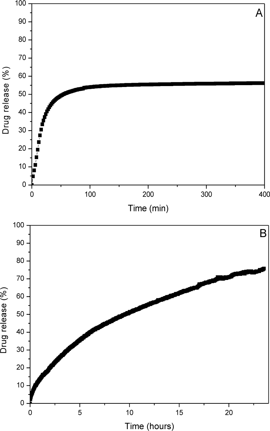
Time dependent fractional aminophylline (A), and triamterene (B) drug releasing behaviour of PNIPA hydrogel in distilled water at 37 °C.
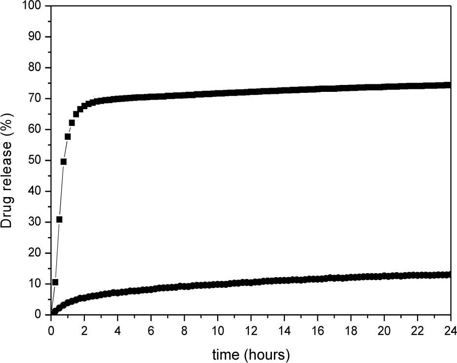
Time dependent fractional aminophylline (squares) and triamterene (circles) combined drug releasing behaviour of PNIPA hydrogel in distilled water at 37 °C.
3.2 In vitro dual drug release
The two drugs chosen to be incorporated in the PNIPA hydrogels (aminophylline and triamterene) have very different chemical structures (Figure 1), hydrophobicity, solubility and molar masses (Table 1). When they were included in the hydrogel using the Korsmeyer and Peppas method [25–28], the yield of the process was 73% for aminophylline and 51% for triamterene. The encapsulation efficiency, until proven different, can be considered high for both drugs.
To test the utility of PNIPA hydrogels in delivering combinations of drugs, xerogels were loaded with the two drugs simultaneously and their release monitored spectrophotometrically. Figure 6 shows time profiles of triamterene and aminophylline release from PNIPA hydrogels incorporating both drugs. Approximately 5% of triamterene and 65% of aminophylline was released for the initial 2 h. Triamterene was released slowly at an initial rate of 5% per hour followed by a continuous and also slow release of another 5% in 13 h. On the other hand, aminophylline showed a quick release of 25% per hour during the first 2 h followed by a very slow release of 3% in 13 h.
In comparison, we obtained a bigger amount of aminophylline release when both drugs were present and a smaller quantity of triamterene. The aminophylline release rate was almost the same, in contrast with triamterene, which had a slower release rate in dual release experiments. Zhang et al. [33] also found a decrease of the release rates for the dual release of 5-fluoracil, a pyrimidine analogue, and one adjuvant leucovorin calcium, a water-soluble salt of levofolinic acid, from hyper-branched poly(amine-ester) based hydrogels in comparison with single release studies.
3.3 Single drug release kinetics and diffusion model
To obtain a more quantitative understanding of the transport kinetics in the hydrogel, the drug release data were analysed as a function of the time for 0 < (Mt/M∞) < 0.6. The drug release data from a polymeric disk in glassy state is mainly modelled with the following empirical kinetic power equation to estimate the release kinetic parameters [25–28]:


According to Equation (2), the classification of drug diffusion through elastomer polymers in thermodynamic equilibrium with the solvent matrix is as follows: (a) Fickian diffusion (n = 0.45) occurs when the rate of diffusion is slower than the relaxation one, so we have a diffusion-controlled drug release; (b) Relaxation- or swelling-controlled transport (n = 0.89) occurs when diffusion is very rapid compared with the relaxation process; (c) Non-Fickian or anomalous transport (0.45 < n < 0.89) occurs when the diffusion and relaxation rates are comparable. In this case, the drug release behaviour can be regarded as the superposition of both phenomena [1].
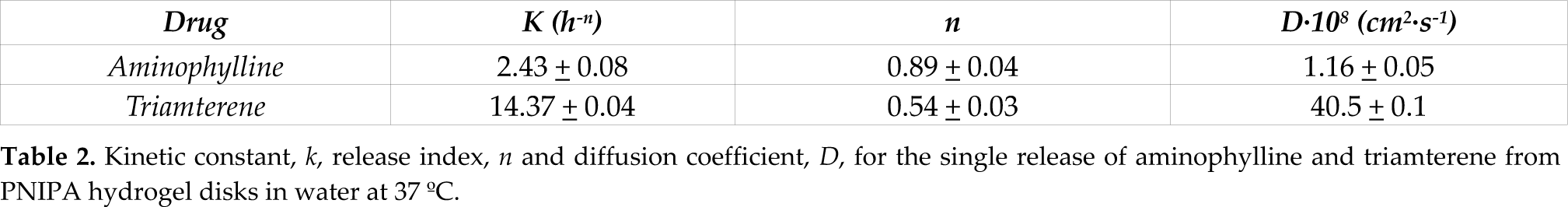
The corresponding values for k and n of the single release experiments of both drugs from the PNIPA hydrogel are listed in Table 2. The diffusion exponents, n, found for the two drugs at 37 °C are 0.89 and 0.54, respectively. So the drug release mechanism from the hydrogel is a relaxation- or swelling-controlled transport for aminophylline and close to Fickian diffusion for triamterene. Chen et al. obtained a non-Fickian diffusion mechanism for aminophylline from poly(N,N-diethylacrylamide-co-N-hydroxymethyl acrylamide) hydrogel, reporting a value for the diffusion exponent of 0.63 [22]. Brazel and Peppas also found Fickian release behaviour for triamterene from poly(2-hydroxyethyl methacrylate-co-methyl methacrylate) with a value for n of 0.45 [29]. Therefore, the drug release kinetics can be determined partially through the swollen polymer network and partially through the water-filled pores in the network structure.
Kinetic constant, k, release index, n and diffusion coefficient, D, for the single release of aminophylline and triamterene from PNIPA hydrogel disks in water at 37 °C.
At an early stage, (Mt/M∞) < 0.6, according to Fick's second law, the diffusion process in hydrogels may be expressed using the Higuchi kinetic equation as [30,31]:

Where D is the effective diffusion coefficient and 2L is the sample thickness. Using Equation (4), the diffusion coefficients of aminophylline and triamterene from the hydrogels, D, are reported in Table 2. To the authors' knowledge, there are no reports of aminophylline and triamterene release from PNIPA hydrogels with which to compare these results. However, PNIPA hydrogels polymerized with other monomers have been employed in the release of these drugs. For instance, Katime et al. [23,32] studied the kinetics of aminophylline release from PNIPA copolymerized with itaconic acid (IA). They reported a diffusion coefficient value of (0.89 ± 0.05)·107 cm2·s−1 for a PNIPA hydrogel with an IA percentage of 0%, which is very close to the one reported in our study. Brazel and Peppas estimated values for the diffusion coefficient of triamterene from (22.2 ± 4.0) to (96.2 ± 50.2)·108 cm2·s−1 in glutaraldehyde cross-linked poly(vinyl alcohol) hydrogels with different degrees of hydrolysis and cross-linking ratios.[28]
3.4 Dual drug release kinetics and diffusion model
We have incorporated the two drugs simultaneously into the cross-linked PNIPA hydrogels and also have observed two different release profiles. The first model applied to the study of the dual drug release from hydrogels was the same as in the case of single drug release. Wei et al. [1] have also applied this model to the study of a dual drug delivery system composed of hydrogels and polypeptide micelles with success. The different release behaviours of the two drugs in DDDS achieved by them, aspirin showing a short-term release while doxorubicin showed a sustained long-term release, are well fitted by this classical empirical power law.
We plotted log (Mt/M∞) against log t of the experimental data according to equation 3. A typical plot of log (Mt/M∞) versus log t for aminophylline and triamterene at 37 °C showed linearity, indicating that the Peppas equation is applicable to the present system. As a first approach, release exponents, n, rate constants, log k and diffusion coefficients, D, from the dual drug delivery system were obtained by these plots and are listed in Table 3. The value of n for aminophylline is 1.41, greater than the value obtained for the single release (0.89), indicating that the release of aminophylline is an anomalous transport instead of a swelling-controlled transport. For the triamterene release, the n value is very close to 0.80, which corresponds to a relaxation- or swelling-controlled transport instead of the Fickian diffusion that we found in the single release. The different release mechanisms of aminophylline and triamterene suggest that the hydrogel has a marked influence on their release behaviour when combination delivery is studied. As can be seen in Table 3, the value of k for aminophylline is 77.5, greater than at single release, while this parameter for triamterene decreases (3.70) when both drugs are present. This indicates that both release rates of aminophylline and triamterene have been influenced by the presence of the other drug in the hydrogel.
Kinetic constant, k, release index, n and diffusion coefficient, D, for the dual release of aminophylline and triamterene from PNIPA hydrogel disks in water at 37 °C.

4. Conclusion
The amount of drug released was measured by UV-Vis spectroscopy following the evolution of the absorption peaks of aminophylline (271 nm) and triamterene (365 nm). The maximum release time is greater for triamterene than for aminophylline at 37 °C, so these time-release profiles enabled the active ingredients to work over different periods of time. By increasing molar mass or solubility of the drug, we observed that the diffusion coefficient decreased. On the contrary, the diffusion coefficients increased when the hydrophobicity of the drug was higher. The evolution of pore size distribution of hydrogels during loading and releasing was measured by QELS, using the algorithm NNLS, and by ESEM. When loading and releasing the drugs, pore size of the hydrogel decreased and increased, respectively, without reaching the initial pore size of the hydrogel. We observed that the greater the concentration of drug loaded into the hydrogel, the greater the reduction in pore size. The data in this paper demonstrate how a single and well understood PNIPA hydrogel may be used to deliver multiple drugs either simultaneously or sequentially. This may simplify multi-drug delivery and material development and regulatory approval for biomedical uses.
Footnotes
5. Acknowledgments
The authors thank Ministerio de Ciencia e Innovación (MICINN) for projects MAT2010-21509-C03-02, ACI2008–0789 and Gobierno Vasco (Grupo Consolidado). E.C. acknowledges the Angeles Alvariño (Xunta de Galicia, Spain, and FSE, European Union) programme.
References
 -CD complex from thermo-sensitive poly(N-isopropylacrylamide) hydrogels
 -cyclodextrin moieties