
Abstract
The risk of cardiovascular complications is increased in patients with obstructive sleep apnea (OSA). Continuous positive airway pressure (CPAP) is the most effective way to treat clinically significant OSA. We hypothesized that the concentrations of the cardiac risk markers N-terminal brain natriuretic peptide (NT-proBNP) and high-sensitive troponin T (hs-TropT) correlate with the effectiveness of CPAP therapy in patients with OSA and coexisting coronary artery disease (CAD). Twenty-one patients with severe OSA and coexisting CAD (group 1) and 20 control patients with severe OSA alone (group 2) were treated with CPAP and monitored by laboratory-based polysomnography. NT-proBNP and hs-TropT levels were measured before and after CPAP. Apnea-hypopnea index (AHI) and oxygen desaturation were similar in both groups. In group 1, hs-TropT levels correlated with AHI and oxygen desaturation upon CPAP. Elevated NT-proBNP levels in group 1 were significantly reduced by CPAP. NT-proBNP levels correlated with AHI and showed negative correlation with ST-segment depression. No such correlations were found in group 2. CPAP has the potential to normalize elevated NT-proBNP serum levels in patients with severe OSA and coexisting CAD. Levels of NT-proBNP and hs-TropT correlated with AHI and oxygen desaturation.
Introduction
Obstructive sleep apnea (OSA) is linked to known cardiovascular risk factors, including obesity, insulin resistance, and dyslipidemia, and shows an association with adverse long-term outcome. 1 3 OSA is characterized by repetitive interruption of ventilation during sleep with a fall in oxygen saturation. OSA is an independent risk factor for hypertension and has also been implicated in the pathogenesis of congestive cardiac failure. 4 Repetitive apneas leading to intermittent nocturnal hypoxemia and sympathetic hyperactivity may result in sub-clinical myocardial injury. 5
High-sensitive troponin T (hs-TropT) is a sensitive marker of myocardial damage and a clinical outcome predictor. 6 8 Ventricular myocytes are also the major source of synthesis and secretion of cardiac N-terminal brain natriuretic peptide (NT-proBNP). In response to increased myocardial wall stress due to volume- or pressure-overload states, NT-proBNP levels are elevated in cardiac failure.9,10 Natriuretic peptide production is also upregulated in the area adjacent to ischemic myocardium. 11
Other studies have shown higher hs-TropT and NT-proBNP serum concentrations associated with frequent apneas or hypoxemia in sleep apnea patients.5,12–14 Nasal continuous positive airway pressure (CPAP) is the gold standard therapy for clinically significant OSA.15,16
We hypothesized that the serum concentrations of the cardiac risk markers NT-proBNP and hs-TropT correlate with the effectiveness of CPAP therapy in OSA patients with coexisting coronary artery disease (CAD).
Materials and Methods
The study obtained ethics approval from the ethics committee of the State Medical Association (FF 6/2012). All participants provided written informed consent. All aspects of the study were performed according to the principles of the Declaration of Helsinki.
Study participants
Initially, 80 patients were screened for clinically significant OSA by polygraphy. After a subsequent laboratory-based polysomnography, 39 participants did not satisfy the study criteria (Fig. 1). Twenty-one patients with severe OSA and coexisting CAD proven by coronary angiography (group 1) were recruited for this study. Twenty patients with OSA alone served as a control group (group 2).
Study flow diagram.
Inclusion criteria were age ≥18 years, apnea-hypopnea index (AHI) >15/h, oxygen saturation ≤80% during apnea, and CAD (group 1). Exclusion criteria were heart failure [Left ventricular ejection fraction (LVEF) <40% quantified by echocardiography] and renal failure (glomerular filtration rate <50 mL/min).
Polysomnography and CPAP therapy
Nocturnal, laboratory-based polysomnography (Alice 5; Philips Respironics) was performed in order to confirm the diagnosis and to grade the severity of OSA. Polysomnographic registration and scoring were performed according to the recommendations of the American Academy of Sleep Medicine. 17 Bedtime was 11 pm to 6 am. A cessation of airflow for ≥10 seconds was specified as apnea. Hypopnea was characterized as a decrease in airflow lasting ≥10 seconds with a ≥30% reduction in airflow and with at least a 4% oxygen desaturation from baseline. The AHI is the number of apneas or hypopneas recorded during the study per hour of sleep. Based on the AHI, the severity of OSA was classified as follows: mild (AHI 5-14/h), moderate (AHI 15-29/h), and severe (AHI ≥30/h). Optimal CPAP was calibrated in the sleep laboratory during a CPAP titration study in the following night. CPAP was increased until obstructive respiratory events were reduced or eliminated. 18 After titration, all patients were monitored under CPAP therapy.
Measurement of cardiac risk markers
The methods of hs-TropT and NT-proBNP measurement and ST-segment analysis have been previously described in detail. 19
Briefly, venous blood samples (5 mL) were collected at different time points: before (9 pm) and after (7 am) CPAP. The measurements were performed immediately after the blood samples were drawn. An immunoassay was used for the measurement of hs-TropT (Cobas e 411; Roche Diagnostics Inc.). Within the normal healthy population, 99% of people will have an hs-TropT <14 ng/L. NT-proBNP levels were measured using an immunoassay for the in vitro quantitative determination of NT-proBNP in heparinized venous blood (Cobas Roche Cardiac proBNP+; Roche Diagnostics). The measuring range is 60-9000 pg/mL, with a mean of the variation coefficients below 15% in the range of 60-1200 pg/mL and below 20% in the range of 1200-9000 pg/mL. The reference values of NT-proBNP depend on age and gender. A value of 125 pg/mL has been suggested as a cut point to rule out left ventricular systolic dysfunction. NT-proBNP has a half-life of 120 minutes.
Electrocardiogram analysis
A continuous electrocardiogram (ECG) recording during sleep was screened for nocturnal ST-segment depression. The ECG sign of subendocardial ischemia is ST-segment depression. A horizontal or down-sloping depression of 1 mm (100 μV) or more or up-sloping depression of the same magnitude 80 ms beyond the J point was considered positive signs of ischemia. After CPAP titration, ST-segment depression at the time of lowest oxygen saturation was analyzed and averaged in 10 cardiac cycles.
Statistics
Continuous variables were expressed as mean and standard deviation. For the descriptive statistics, the first, second, and third quartiles from a set of numerical data were calculated. The Mann-Whitney Utest was used to compare differences between two independent groups. The Wilcoxon matched pairs test was performed to compare matched subjects. Linear regression analysis was used to investigate the relationship between two parameters. Analysis was done using statistical software (BiAS for Windows 10.12). Pvalues less than 0.05 were reported as statistically significant.
Results
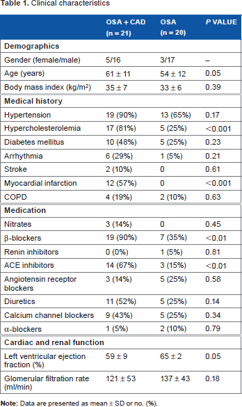
Clinical characteristics
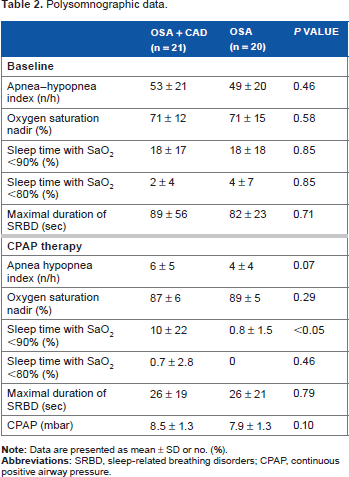
Polysomnographic data.
Clinical heart failure symptoms and signs were absent in all patients. Echocardiography showed normal systolic LV function (LVEF ≥50%) in 19 patients and moderately reduced LV function (LVEF 40%) in 2 patients in group 1. All patients in group 2 had normal LV function. Natriuretic peptide concentrations were higher before and after CPAP therapy in group 1 compared to group 2, but the difference between groups was not significant (Fig. 2). Mean NT-proBNP before and after CPAP treatment measured 475 ± 654 and 352 ± 573 pg/mL in group 1 and 105 ± 59 and 93 ± 58 pg/mL in group 2, respectively. In group 1, CPAP significantly reduced NT-proBNP. A moderate but significant correlation was noted between NT-proBNP levels and AHI in group 1 (Fig. 3), while we found no such correlation in group 2.
NT-proBNP levels pre and post CPAP therapy. Middle horizontal line inside box indicates median. Bottom and top of the box are 25th and 75th percentiles, and the error bars outside the box represent maximum and minimum values, respectively. Correlation between NT-proBNP levels and AHI under CPAP therapy.

Troponin T levels were not different between the two groups. CPAP had no significant effect on hs-TropT levels. After CPAP therapy, hs-TropT was detectable (≥3 ng/L) in 17 (81% of group 1) and 19 (95% of group 2) patients, respectively. Mean hs-TropT levels before and after CPAP were 9 ± 8 and 10 ± 8 ng/L in group 1 and 7 ± 3 and 7 ± 3 ng/L in group 2. We found a moderate but significant correlation between hs-TropT levels and AHI and a weak negative correlation between hs-TropT levels and oxygen saturation nadir in group 1 (Fig. 4). In group 2, these correlations were not significant.
Correlation between high-sensitive troponin T levels and AHI and oxygen saturation under CPAP therapy.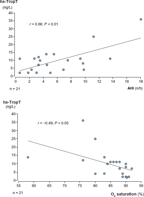
In both groups, ST-segment analysis showed no ST depression ≥100 μV at the time point of lowest oxygen saturation under CPAP treatment. Mean ST depression was -9 ± 47 μV in group 1 and -4 ± 32 μV in group 2. The difference was not significant. In group 1 patients, a moderate negative correlation of ST depression with NT-proBNP concentration was noted (Fig. 5). No such correlation was found in group 2.
Correlation between NT-proBNP levels and ST-segment depression.
Discussion
The prevalence of cardiovascular disease is increased in patients with OSA. 20 In patients with OSA and CAD, repetitive hypoxia due to sleep-induced apnea and/or hypopnea adversely affects the interaction between myocardial oxygen demand and supply, resulting in nocturnal myocardial ischemia. 21 In some studies, insufficient oxygen supply in patients with CAD has been assumed as the main cause for myocardial ischemia, while others postulate a rise in oxygen consumption due to an accelerated heart rate and a sympathetic stimulation following an apnea episode.13,21–24 CPAP therapy is the leading treatment for clinical relevant OSA. CPAP is effective in eliminating hypopnea/apnea and in improving apnea sequelae. 15
There is only limited information about the effect of CPAP therapy on the concentrations of hs-TropT and NT-proBNP in individuals with OSA and coexisting CAD. In the current study, we found only slightly elevated hs-TropT (>13 ng/L) in a few patients. CPAP had no effect on hs-TropT levels. Hs-TropT levels were moderately correlated with AHI under CPAP and negatively correlated with oxygen saturation nadir. The results of other studies that investigated the association between OSA and myocardial injury quantified by troponin measurement are not unequivocal. Also the effect of CPAP on troponin levels remains unclear. Several studies found higher troponin concentrations independently associated with AHI in untreated OSA patients free of CAD.5,12,25 Geovanini et al. 26 showed that OSA severity was independently associated with hs-TropT levels in patients with refractory angina. In contrast, no significant difference in troponin levels between moderate/severe and mild/no OSA was reported by Maeder et al. 27 In this study, short-term CPAP also had no impact on troponin concentrations before and after sleep. Colish et al. 28 also showed no significant change of hs-TropT levels from normal baseline values following 12 months of CPAP therapy. Sharafkhaneh et al. 29 found that an increase in hs-TropT in unselected patients with OSA was associated with severity of hypoxia but not AHI. 29 The effect of CPAP was greatest to those with pronounced hypoxia as assessed by nadir oxygen saturation at baseline. Barceló et al studied patients with OSA without coexistent CAD and assessed the effect of CPAP on hs-TropT levels. In this study, the amount of participants with detectable hs-TropT was higher in patients with OSA than in controls. 30 Interestingly, a significant increase in hs-TropT levels was observed after an effective treatment with CPAP. The authors supposed that to a certain extent, CPAP therapy might cause cardiac stress, resulting in higher hs-TropT levels.
We found significantly higher NT-proBNP levels in group 1 compared to group 2. Interestingly, CPAP significantly reduced NT-proBNP serum levels in group 1. NT-proBNP levels were moderately correlated with AHI under CPAP therapy. In line with our findings, a number of studies described elevated NT-proBNP concentrations linked to OSA but the study results are not consistent.5,14,31–34 NT-proBNP is also associated with the extent of CAD in patients with unimpaired LV function. 35 N-terminal B-type natriuretic peptide predicts ischemia in patients with stable angina pectoris.36,37 Increase in upper airways resistance in untreated OSA patients can cause high negative intrathoracic pressures and enhanced venous inflow, resulting in intensified right ventricular diastolic filling and secretion of natriuretic peptides. Stimulated sympathetic activity, repetitive rises in blood pressure, and apnea-induced wall stress may also contribute as a trigger to release N-terminal B-type natriuretic peptide in OSA. CPAP treatment eliminates negative intrathoracic pressure fluctuations, and this effect could be the reason for a decline in natriuretic peptides secretion. 1 In a meta-analysis, therapy with CPAP was associated with improvements in ejection fraction, diastolic blood pressure, and heart rate in patients with sleep-disordered breathing and congestive heart failure. 38 In several other studies, a decrease of elevated natriuretic peptide concentrations was noted as a response to therapy with CPAP.27,32,39–42 A study by Maeder et al. 27 found larger relative overnight reduction in BNP concentration in patients with moderate/severe OSA than in those with mild/no OSA. However, a study by Cifci et al in 33 consecutive patients with OSA did not detect any significant difference between severity of OSA syndrome and serum pro-BNP levels. CPAP had no impact on natriuretic peptide serum concentration after six months of CPAP therapy. 14 In a prospective study of 47 patients with OSA, levels of NT-proBNP did not change significantly from normal baseline values following one year of CPAP therapy, despite improvement in cardiovascular remodeling. 28
In our study, CPAP significantly decreased elevated NT-proBNP levels but had no significant impact on slightly increased hs-TropT levels. The difference might be explained by the fact that NT-proBNP is not only a marker of myocardial ischemia but also relate strongly to changes in the hemodynamic status. CPAP treatment has relevant hemodynamic effects because it eliminates negative intrathoracic pressure fluctuations and therefore causes a reduction in right ventricular preload and wall stress.
In the present study, ECG in group 1 patients revealed only minor ST-segment depression (<100 μV) under CPAP therapy. ST-segment depression showed a significantly negative correlation with NT-proBNP levels. Nocturnal ST-segment depression has been reported in about a third of untreated OSA patients with CAD during apneas with oxygen desaturation.13,21 Treatment with CPAP significantly ameliorated the nocturnal ST depression. Interestingly, Hanley at al also found ST depression during sleep in symptom-free patients with OSA without a history of CAD. 43 The exact explanation for nocturnal ST-segment depression in patients with OSA remains unclear. ST depression during sleep in OSA patients may be caused by the combination of increased myocardial oxygen consumption and decreased oxygen supply due to oxygen desaturation and peak hemodynamic changes during the rebreathing phase of the obstructive apnea. Repeated respiratory efforts against an obstructed pharynx cause negative intrathoracic pressure fluctuations and changes in cardiac preload and afterload. This mechanism may also contribute to myocardial ischemia even without hypoxemia. 44
Study limitations
The study is limited by the fact that the sample size is relatively small. Factors like sympathetic nervous system overactivity in OSA patients causing accelerated heart rate and increased blood pressure and their impact on myocardial ischemia were not evaluated. The majority of patients of group 1 were on medication (β-blockers, angiotensin converting enzyme [ACE] inhibitors), which may have affected our findings. For analysis of ST-segment depression, we selected the ECG recording at the time of minimal oxygen saturation. However, it is possible that the time of maximum ischemia was during the postapnea tachycardia. Most of the patients in group 1 were treated with coronary interventions before, which could have influenced ischemic thresholds.
Conclusion
Hs-TropT serum concentrations in patients with severe OSA and coexisting CAD were within normal range and similar to individuals with OSA alone. Before sleep, NT-proBNP serum concentrations in patients with OSA and CAD were increased compared to patients with OSA alone. CPAP treatment normalized elevated NT-proBNP serum levels in these patients. Levels of NT-proBNP and hs-TropT correlated with AHI and oxygen desaturation.
Author Contributions
Developed the study hypothesis: MV, CT. Acquired data and drafted the manuscript: RS. Conducted the analysis and drafted the manuscript: MV, CT. All authors provided critical revision of the manuscript and have read and approved the final manuscript.
Footnotes
Acknowledgment
The authors thank Simone Rack of Krankenhaus Sachsenhausen Sleep Laboratory for her excellent technical assistance.