
Abstract
Biotransformations of capsaicinoids such as capsaicin and 8-nordihydrocapsaicin and phenylpropanoids such as cinnamic acid, p-coumaric acid, caffeic acid, and ferulic acid have been investigated using cultured plant cells. Capsain and 8-nordihydrocapsaicin were converted into the corresponding glycosides which are three glycosides respectively using the cultured cells of Catharanthus roseus. In a time-course study under sterile conditions, the changes in amounts of their reaction products were determined. Furthermore phenypropanoid, such as cinnamic acid, p-coumaric acid, caffeic acid and ferulic acid have been biotransformed using the cultured cells of the Eucalyptus perriniana, and then cinnamic acid was converted into two glycosides. In addition, p-coumaric acid, caffeic acid and ferulic acid were converted into four, four and three glycosides respectively. Then in time-course study under sterile conditions, the change in amounts of their reaction products were determined. Finally it was found that the cultured plant cells have the ability to glycosylate the phenolic group of capsacinoids and phenylpropanoids regioselectively.
Introduction
The synthesis of the biologically active compounds using living cells by biotranformation of various organic compounds has been investigated using mammalian cells, bacteria, enzymes and plant cells in the biotechnology application. As plant cells have glycosyltransferases in their cells, glycosylation is a characteristic biotransformation reaction using cultured plant cells.
Plants of the Capsicum species have been important sources of food, spices and medicines worldwide for centuries. The chemical constituents in the fruits of Capsicum plants, such as hot chilli pepper fruits, which are responsible for the sensory effects associated with pungency, are a series of branched or straight-chain alkylvanillylamides, capsaicinoids.
Capsaicin is the most pungent principle among naturally occurring capsaicinoids. 1 Capsaicin has also been reported to decrease adipose tissue weight and serum triacylglycerol content in rats by enhancing energy metabolism. 2 It has also shown a wide range of pharmacological properties, such as body heat generation, 3 lipid metabolism,4,5 antioxidant activity,6–8 antibacterial activity, 9 anodyne effect, 10 and perspiration effect. 11 However capsaicinoids possess extensive neurological toxicity and direct irritant effects on skin and mucous membrane. 12
Furthermore, capsaicinoids are almost insoluble in water and poorly absorbed after oral administration. These disadvantages prevent capsaicinoids from being used as food additives and medicines.
Recently, it has been reported that capsaicin was glucosylated by cultured plant cells to its mono-glucoside, capsaicin-β-D-glucopyranoside. Its pungency was approximately 1/100 of that of capsaicin and its water solubility was enhanced. 13
We report here the biotransformations of capsaicin and 8-nordihydrocapsaicin, both of which are naturally occurring capsaicinoids, into the corresponding monoglucosides and disaccharides, β-primeverosides which are more soluble in water. The Catharanthus roseus cell cultures were used.
Phenylpropanoids such as cinnamic acid, p-coumaric acid, caffeic acid and ferulic acid are naturally occurring anti-oxidants which act as effective scavengers of free radicals.14–16 It is well known that shikimic acid is metabolized, in plant cells, to cinnamic acid and p-coumaric acid, which is further converted into caffeic acid and ferulic acid.
We report here the biotransformations of cinamic acid, p-coumaric acid, caffeic acid, and ferulic acid into the corresponding monoglucosides and disaccharides, which are more soluble in water, using cultured plant cells of the Eucalyptus perriniana.
Experimental Procedures
Substrates
Capsaicin and 8-nordihydrocapsaicin were purchased from Tokyo Kasei Kogyo Co. Ltd.. Cinnamic acid, p-coumaric acid, caffeic acid, and ferulic acid, which were used as substrates, were purchased from Aldrich Chemical Co.
Cell suspension culture and culture conditions
The cultured cells of C. roseus were prepared as described previously. 17 A cell suspension culture was started by transferring static cultured callus (fr. wt 40 g (0.40 g fr. wt of cells per ml of medium) to 100 ml of Schenk and Hidebrandt (SH) medium (pH 5.7) in a 300-ml conical flask. The cells were incubated for 2 weeks on an illuminated (4000 lx) rotary shaker (120 rpm) at 25 °C.
The cultured cells of E. perriniana were subcultured at 4-week intervals on solid Murashige and Skoog (MS) medium (100 ml in a 300-ml conical flask) containing 3% sucrose, 10 mM 2,4-dichlorophenoxyacetic acid, and 1% agar (adjusted to pH 5.7) at 25 °C in the dark. A cell suspension culture was started by transferring subcultured cells (fr. wt 40 g) to 100 ml of MS liquid medium (pH 5.7) in a 300-ml conical flask. The cells were incubated for 2 weeks in a rotary shaker (120 rpm) at 25 °C in the dark.
Biotransformation and purification of products
Biotransformation experiments of capsaicin and 8-nordihydrocapsaicin were performed by adding 0.1 mmol of substrate to each of eight 300-ml conical flasks containing 70 ml of SH medium and 30 g of cultured cells suspension of C. roseus. The cultures were incubated for a 6 h interval to 48 h on an illuminated (4000 lx) rotary shaker (120 rpm) at 25 °C. After incubation, the products were extracted and purified according to the previously reported method. 18 Concerning cinamic acid, p-coumaric acid, caffeic acid and ferulic acid, time course experiments were performed by adding 0.1 mmol of each substrate to six 300-ml conical flasks containing 70 ml of MS medium and 30 g of cultured cells suspension of E. perriniana. The cultures were incubated for a 12 h interval to 72 h on rotary shaker at 25 °C in the dark. After incubation period, the cells and medium were separated by filtration with suction. Extraction and purification procedures of biotransformation products were same as described above. The yield of the products was determined on the basis of the peak area from HPLC and expressed as a relative percentage to the total amount of the whole reaction products extracted.
Analysis of the products
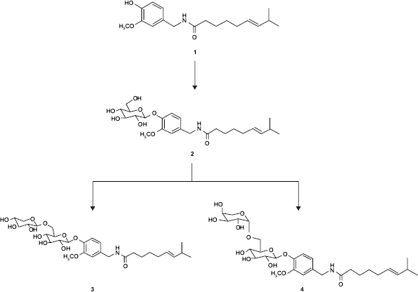
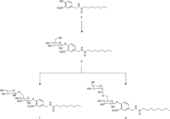
HPLC was carried out with a YMC-Pack R&D ODS column (150 × 30 mm) using MeOH-H2O (9:11, v/v) as the eluent [detection: UV (280 nm); flow rate: 1.0 ml/min]. 1 H and 13 C NMR, H-H COSY, C-H COSY, NOE, and HMBC spectra were measured in CD3OD on a Varian XL-400 spectrometer and the chemical shifts were expressed in δ (ppm) referring to TMS. HRFABMS spectra were taken on a JEOL MStation JMS-700 spectrometer. The structures of the products were determined by HRFABMS, ESIMS, 1 H and 13 C NMR, H-H COSY, C-H COSY, NOE, and HMBC spectra. As regarding capsaicin and 8-nordihydrocapsaicin, the absolute configuration of the sugar moieties in disaccharide products was determined as follows. Each disaccharides, 3, 4, 7, and 8 was added to a vial containing 100 μl of 4.0 M HCl, heated to 80 °C for 2 h, and then cooled to room temperature. The solvent was removed in a stream of N2 and each hydrolysate was converted into the corresponding pentafluoropropionate with pentafluoropropionic anhydride (400 μl) in 400 μl CH2Cl2 in a sealed tube at 120 °C for 2 h. Excess reagents were removed under a stream of N2 and the derivatives were analyzed by chiral GLC (Shimadzu GC-17A with FID) on CP cyclodextrin β236M-19 (Chrompack, column temp: 100 °C, injector temp: 200 °C, detector temp: 200 °C, split ratio: 50, carrier gas: N2, 100 kPa). The peaks of the derivatives from primeverosides 3 and 7 were assigned to those of D-xylose and D-glucose, and the peaks of derivatives from vicianosides 4 and 8 were assigned to those of L-arabinose and D-glucose.
Result and Discussion
Glycosylation of capsaicin
A time-course experiment was carried out to investigate the ability of the cultured cells of C. roseus to biotransform capsaicin. Three glycoside compounds (named products 2–4) were detected in this experiment. The product 2 was identified as capsaicin 4-O-β-D-glucopyranoside by comparison of its 1 H and 13 C NMR spectroscopic data with previously reported data.13,19,20
Structures of products 3 and 4 were determined on the basis of their HRFABMS, 1 H and 13 C NMR spectra, and chiral-GLC as capsaicin 4-O-(6-O-β-D-xylopyranosyl)-β-D-glucopyranoside and capsaicin 4-O-(6-O-α-L-arabinopyranosyl)-β-D-glucopyranoside. The 1 H and 13 C NMR spectra of sugar moiety of 3 and 4 indicated that they were β-primeverose and β-vicianose, respectively.21,22
Capsaicin 4-O-(6-O-β-D-xylopyranosyl)-β-D-glucopyranoside: HRFABMAS: m/z 622.2850 [M+Na]+ (calcd for C29H45NO12Na, 622.2839), 1 H NMR (400 MHz, CD3OD): δ 0.95 (6H, d, J = 6.8 Hz, H-16, 17), 1.37 (2H, q, J = 7.6 Hz, H-11), 1.60 (2H, q, J = 7.6 Hz, H-10), 1.98 (2H, m, H-12), 2.20 (3H, m, H-9,15), 3.15 (1H, dd, J = 11.6, 9.6 Hz, H-5a″), 3.28–3.60 (7H, m, H-2′, 2″, 3′, 3″, 4′, 4″, 5′), 3.75–3.81 (2H, m, H-5b″, 6a′), 3.83 (3H, s, OCH3), 4.05 (1H, dd, J = 11.6, 1.6 Hz, H-6b′), 4.28 (2H, s, H-7), 4,32 (1H, d, J = 7.6 Hz, H-1″), 4.91 (1H, d, J = 7.2 Hz, H-1′), 5.30–5.40 (2H, m, H-13, 14), 6.80 (1H, dd, J = 8.4, 1.6 Hz, H-6), 6.92 (1H, d, J = 1.6 Hz, H-2), 7.10 (1H, d, J = 8.4 Hz, H-5); 13 C NMR (100 MHz, CD3OD): 8 23.1 (C-16, 17), 26.5 (C-10), 30.3 (C-11), 32.3 (C-15), 33.2 (C-12), 36.8 (C-9), 43.7 (C-7), 56.6 (OCH3), 67.1 (C-5″), 69.4 (C-6′), 71.4 (C-4″), 71.6 (C-4′), 74.7 (C-2″), 75.2 (C-2′), 77.8 (C-3″), 77.9 (C-3′, 5′), 102.5 (C-1′), 105.0 (C-1″), 113 (C-2), 118.0 (C-5), 121.2 (C-6), 127.9 (C-13), 135 (C-1), 139.0 (C-14), 147.1 (C-4), 150.7 (C-3), 175.9 (C-8).
Capsaicin 4-O-(6-O-α-L-arabinopyranosyl)-β-D-glucopyranoside (4): HRFABMAS: m/z 622.2841 [M+Na]+(calcd for C29H45 NO12 Na, 622.2839), 1 HNMR (400 MHz, CD3OD): δ 0.95 (6H, d, J = 6.8 Hz, H-16, 17), 1.36 (2H, q, J = 7.6 Hz, H-11), 1.60 (2H, q, J = 7.6 Hz, H-10), 1.98 (2H, m, H-12), 2.20 (3H, m, H-9, 15), 3.28 (1H, dd, J = 12.2, 1.8 Hz, H-5a″), 3.30–3.60 (7H, m, H-2′, 2″, 3′, 3″, 4′, 4″, 5′), 3.73–3.81 (2H, m, H-5b″, 6a′), 3.83 (3H, s, OCH3), 4.05 (1H, dd, J = 11.6, 1.6 Hz, H-6b′), 4.28 (2H, s, H-7), 4.30 (1H, d, J = 6.4 Hz, H-1″), 4.91 (1H, d, J = 7.2 Hz, H-1′), 5.30–5.39 (2H, m, H-13, 14), 6.80 (1H, dd, J = 8.4, 1.6 Hz, H-6), 6.92 (1H, d, J = 1.6 Hz, H-2), 7.10 (1H, d, J = 8.4 Hz, H-5); 13 C NMR (100 MHz, CD3OD): δ 23.1 (C-16, 17), 26.5 (C-10), 30.3 (C-11), 32.2 (C-15), 33.2 (C-12), 36.9 (C-9), 43.7 (C-7), 56.6 (OCH3), 67.0 (C-5″), 69.3 (C-6′), 70.0 (C-4″), 71.8 (C-4′), 72.2 (C-2″), 74.0 (C-3″), 75.2 (C-2′), 78.0 (C-3′, 5′), 102.6 (C-1′), 104.6 (C-1″), 113 (C-2), 117.9 (C-5), 121.2 (C-6), 127.9 (C-13), 134.9 (C-1), 139.0 (C-14), 147.0 (C-4), 150.8 (C-3), 175.9 (C-8).
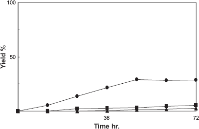
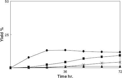
Figure 1 showed that capsaicin was converted into product 2 at early stage incubation and products 3 and 4 were accumulated after 12 h of incubation. At 48 h reaction, the amounts of products 2, 3, and 4 were 36.2%, 18.8%, and 15.0%, respectively, relative to the total amount of reaction products on a molar basis. This result indicated that product 1 was first converted to monoglucoside 2 and further product 2 was converted to disaccharides 3 and 4, as shown in Scheme 1.
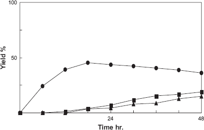
Time-course of the biotransformation of capsaicin by cultured cells of C. roseus. Yield is expressed as a percentage relative to the total amount of reaction products on a molar basis. Yields of 2(•), 3(■), and 4(▲) are plotted.
Glycosylation of capsaicin by cultured cells of C. roseus.
Glycosylation of 8-nordihydrocapsaicin
In a similar way to capsaicin, a time-course experiment was carried out biotransformation of 8-nordihydrocapsaicin. Three glycoside compounds (named products 6–8) were produced by this experiment. The product 6 was identified as 8-nordihydrocapsaicin 4-O-β-D-glucopyranoside by comparison of its 1 H and 13 C NMR spectroscopic data with previously reported data. 19
Structures of products 7 and 8 were identified on the basis of their HRFABMS, 1 H and 13 C NMR spectra, and chiral-GLC as 8-nordihydrpcapsaicin 4-O-(6-O-β-D-xylopyranosyl)-β-D-glucopyranoside and 8-nordihydrocapsaicin 4-O-(6-O-α-L-arabino pyranosyl)-β-D-glucopyranoside. The 1 H and 13 C NMR spectra of sugar moiety of 3 and 4 were shown that they were β-primeverose and β-vicianose, respectively.21,22
8-nordihydrpcapsaicin 4-O-(6-O-β-D-xylopyrano-syl)-β-D-glucopyranoside: HRFABMAS: m/z 610.2833 [M+Na]+ (calcd for C28H45NO12Na, 610.2839), 1 H NMR (400 MHz, CD3OD): δ 0.90 (3H, t, J = 6.8 Hz, H-16), 1.19–1.26 (10H, m, H-11, 12, 13, 14, 15), 1.62 (2H, q, J = 7.6 Hz, H-10), 2.22 (2H, t, J = 7.6 Hz, H-19), 3.16 (1H, dd, J = 11.6, 9.6 Hz, H-5a″), 3.32–3.60 (7H, m, H-2′, 2″, 3′, 3″, 4′, 4″, 5′), 3.75–3.81 (2H, m, H-5b″, 6a′), 3.85 (3H, s, OCH3), 4.01 (1H, dd, J = 11.6, 1.6 Hz, H-6b′), 4.30 (2H, m, H-7), 4.33 (1H, d, J = 7.6 Hz, H-1″), 4.91 (1H, d, J = 7.2 Hz, H-1′), 6.85 (1H, dd, J = 8.4, 1.6 Hz, H-6), 6.94 (1H, d, J = 1.6 Hz, H-2), 7.15 (1H, d, J = 8.4 Hz, H-5); 13 C NMR (100 MHz, CD3OD): δ 14.5 (C-16), 23.7 (C-15), 27.1 (C-10), 30.5 (C-11, 12, 13), 33.0 (C-14), 37.2 (C-9), 43.8 (C-7), 67.2 (C-5″), 69.7 (C-6′), 71.4 (C-4″), 71.6 (C-4′), 74.7 (C-2″), 75.1 (C-2′), 78.0 (C-3′), 77.9 (C-3″, 5′), 102.6 (C-1′), 105.1 (C-1″), 113.2 (C-2), 118.4 (C-5), 121.5 (C-6), 135.2 (C-1), 146.9 (C-4), 151.0 (C-3), 176.2 (C-8).
8-nordihydrocapsaicin 4-O-(6-O-α-L-arabinopy-ranosyl)-β-D-glucopyranoside: HRFABMAS: m/z 610.2838 [M+Na]+ (calcd for C28H45NO12Na, 610.2839), 1 H NMR (400 MHz, CD3OD): δ 0.89 (3H, t, J = 6.8 Hz, H-16), 1.18–1.32 (10H, m, H-11, 12, 13, 14, 15), 1.63 (2H, q, J = 7.6 Hz, H-10), 2.24 (2H, t, J = 7.6 Hz, H-9), 3.29 (1H, dd, J = 12.0, 2.0 Hz, H-5a″), 3.32–3.61 (7H, m, H-2′, 2″, 3′, 3″, 4′, 4″, 5′), 3,74–3.82 (2H, m, H-5b″, 6a′), 3.85 (3H, s, OCH3), 4.05 (1H, dd, J = 11.6, 1.6 Hz, H-6b′), 4.30 (2H, m, H-7), 4.31 (1H, d, J = 6.4 Hz, H-1″), 4.92 (1H, d, J = 7.6 Hz, H-1′), 6.85 (1H, dd, J = 8.4, 1.6 Hz, H-6), 6.94 (1H, d, J = 1.6 Hz, H-2), 7.15 (1H, d, J = 8.4 Hz, H-5); 13 C NMR (100 MHz, CD3OD): δ 14.4 (C-16), 23.7 (C-15), 27.1 (C-10), 30.3 (C-11, 12, 13), 32.9 (C-14), 37.1 (C-9), 43.8 (C-7), 67.1 (C-5″), 69.5 (C-6′), 70.0 (C-4″), 71.7 (C-4′), 72.2 (C-2″), 74.1 (C-3″), 75.3 (C-2′), 78.0 (C-3′, 5′), 102.6 (C-1′), 104.6 (C-1″), 113.1 (C-2), 118.3 (C-5), 121.4 (C-6), 135.1 (C-1), 146.7 (C-4), 150.8 (C-3), 176.0 (C-8).
Figure 2 showed that 8-nordihydrocapsaicin was converted into product 6 at early stage of incubation and products 7 and 8 were accumulated after 12 h of incubation. At 48 h reaction, the amounts of products 6, 7, and 8 were 45.5%, 27.9%, and 19.4%, respectively, relative to the total amount of reaction products on a molar basis. The same as capsaicin, 8-nordihydrocapsaicin was first converted to monoglucoside 6 and further product 6 was converted to disaccharides 7 and 8, as shown in Scheme 2.
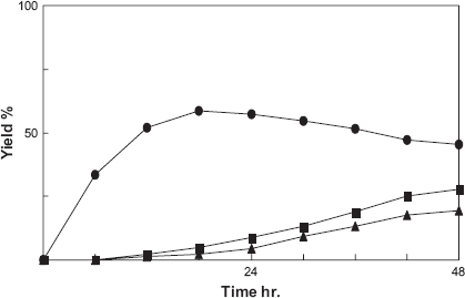
Time-course of the biotransformation of 8-nordihydrocapsaicin by cultured cells of C. roseus. Yield is expressed as a percentage relative to the total amount of reaction products on a molar basis. Yields of 6(•), 7(■), and 8(▲) are plotted.
Glycosylation of 8-nordihydrocapsaicin by cultured cells of C. roseus.
Glycosylation of cinnamic acid
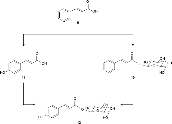
In order to examine the ability of the cultured cells of E. perriniana to biotransform cinnamic acid (9), the time course in the conversion of 9 was followed. Three compounds (named products 10–12) were obtained and the structure of the product 11 was identified as p-coumaric acid by HPLC spectra. Structures of products 10 and 12 were determined on the basis of their ESIMS, 1 H and 13 C NMR spectra as cinnamoyl-O-β-D-glucoside, and p-coumaroyl-O-β-D-glucoside, respectively.
Cinnamoyl-O-β-D-glucoside (10): ESIMS: m/z 333 [M+Na]+, 1 H NMR (400 MHz, CD3OD): δ 5.61 (1H, d, J = 7.6 Hz, H-1′), 6.57 (1H, d, J = 16.0 Hz, H-8), 7.85 (1H, d, J = 16.0 Hz, H-7); 13 C NMR (100 MHz, CD3OD): δ 62.2 (C-6′), 70.9–78.7 (C-2′, 3′, 4′, 5′), 95.7 (C-1′), 118.0 (C-8), 129.1 (C-2, 6), 129.8 (C-3, 5), 131.6 (C-4), 135.3 (C-1), 147.4 (C-7), 166.9 (C-9).
p-Coumaroyl-O-β-D-glucoside (12): ESIMS: m/z 349 [M+Na]+, 1 H NMR (400 MHz, CD3OD): δ 5.57 (1H, d, J = 8.0 Hz, H-1′), 6.37 (1H, d, J = 16.0 Hz, H-8), 6.81 (2H, d, J = 8.8 Hz, H-3, 5), 7.48 (2H, d, J = 8.4 Hz, H-2, 6), 7.72 (1H, d, J = 16.0 Hz, H-7); 13 C NMR (100 MHz, CD3OD): δ 62.2(C-6′), 71.1–78.7 (C-2′, 3′, 4′, 5′), 95.7 (C-1′), 114.3 (C-8), 116.8 (C-3, 5), 126.9 (C-1), 131.2 (C-2, 6), 147.8 (C-7), 161.4 (C-4), 167.5 (C-9).
Figure 3 showed that cinnamic acid (9) was converted into 10 at early stage of incubation and the products 11 and 12 were produced at a low level. At 72 h reaction, the amounts of products 10, 11, and 12 were 28.8%, 5.2%, and 2.6%, respectively, relative to the total amount of reaction products on a molar basis. This result indicated that glucosylation at carboxyl group of cinnamic acid is the predominant modification compared to hydroxylation at 4-position of cinnamic acid to p-coumaric acid. Then, it was suggested that hydroxylation at 4-position of cinnamoyl-O-β-D-glucoside, or glucosylation at carboxyl group of p-coumaric acid was occurred. Biotransformation pathway of cinnamic acid is shown in Scheme 3.
Time-course of the biotransformation of cinnamic acid by cultured cells of E. perriniana. Yield is expressed as a percentage relative to the total amount of reaction products on a molar basis. Yields of 10(•), 11(■), and 12(▲) are plotted.
Biotransformation of cinnamic acid by cultured cells of E. perriniana.
Glycosylation of p-coumaric acid (11)
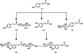
Five compounds (named products 12–16) were produced by incubation of the cultured cells of E. perriniana with p-coumaric acid (11). The structure of the product 14 was identified as caffeic acid by HPLC spectra. Structures of products 13, 15, and 16 were determined on the basis of their ESI, 1 H and 13 C NMR spectra as 4-O-β-D-glucosyl coumarate, 3-O-β-D-glucosyl caffeate, and (3-O-β-D-glucosyl)-caffeoyl-O-β-D-glucoside, respectively.
4-O-β-D-Glucosyl coumarate (13): ESIMS: m/z 349 [M+Na]+, 1 H NMR (400 MHz, CD3OD): δ 4.96 (1H, d, J = 7.6 Hz, H-1′), 6.38 (1H, d, J = 16.4 Hz, H-8), 7.11 (2H, d, J = 8.8 Hz, H-3, 5); 13 C NMR (100 MHz, CD3OD): δ 62.4 (C-6′), 71.2–78.1 (C-2′, 3′, 4′, 5′), 101.7 (C-1′), 117.8 (C-3, 5, 8), 130.3 (C-1, 2, 6), 146.5 (C-7), 160.3 (C-4), 170.8 (C-9).
3-O-β-D-Glucosyl caffeate (15): ESIMS: m/z 365 [M+Na]+, 1 H NMR (400 MHz, CD3OD): δ 4.82 (1H, d, J = 7.2 Hz, H-1′), 6.32 (1H, d, J = 15.6 Hz, H-8), 6.86 (1H, d, J = 8.8 Hz, H-5), 7.17 (1H, dd, J = 8.0, 2.0 Hz, H-6); 13 C NMR (100 MHz, CD3OD): δ 62.4 (C-6′), 71.4–78.4 (C-2′, 3′, 4′, 5′), 104.2 (C-1′), 117.1 (C-8), 117.3 (C-2), 117.9 (C-5), 125.7 (C-6), 128.1 (C-1), 145.7 (C-3), 146.9 (C-7), 150.7 (C-4), 171.2 (C-9).
(3-O-β-D-Glucosyl)-caffeoyl-O-β-D-glucoside (16): ESIMS: m/z 527 [M+Na]+, 1 H NMR (400 MHz, CD3OD): δ 4.82 (1H, d, J = 7.2 Hz, H-1″), 5.56 (1H, d, J = 8.4 Hz, H-1′), 6.41 (1H, d, J = 16.0 Hz, H-8), 6.87 (1H, d, J = 8.4 Hz, H-5), 7.20 (1H, dd, J = 8.0, 2.0 Hz, H-6), 7.56 (1H, d, J = 1.6 Hz, H-2), 7.61 (1H, d, J = 16.0 Hz, H-7); 13 C NMR (100 MHz, CD3OD): δ 62.3 (C-6′), 62.5 (C-6″), 71.1–78.5 (C-2″-5″), 71.4–78.7 (C-2′, 3′, 4′, 5′), 95.7 (C-1′), 104.2 (C-1″), 115.3 (C-8), 117.4 (C-2), 118.1 (C-5), 126.3 (C-6), 127.7 (C-1), 146.9 (C-3), 147.6 (C-4), 151.2 (C-7), 167.4 (C-9).
Figure 4 showed that p-coumaric acid (11) was converted into 13 at early stage of incubation, and products 12 and 15 were accumulated after 12 h of incubation, and products 14 and 16 were slightly produced. At 72 h reaction, the amounts of products 12–16 were 11.0%, 12.8%, 1.2%, 9.5%, and 2.3%, respectively, relative to the total amount of reaction products on a molar basis. This result indicated that glucosylation at phenolic hydroxyl group and carboxyl group of p-coumaric acid preferentially occurred. And the cultured cells of E. perriniana regioselectively hydroxylated at 3-position of p-coumaric acid (11) to produce caffeic acid (14). Subsequently, produced caffeic acid (14) was predominantly glucosylated at 3-position of phenolic hydroxyl group. It was considered that disaccharide product 16 was subsequently biotransformed from p-coumaroyl-O-β-D-glucoside (12) and. 3-O-β-D-glucosyl caffeate (15) Biotransformation pathway of p-coumaric acid is shown in Scheme 4.
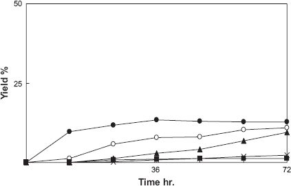
Time-course of the biotransformation of p-coumaric acid by cultured cells of E. perriniana. Yield is expressed as a percentage relative to the total amount of reaction products on a molar basis. Yields of 12(○), 13(•), 14(■), 15(▲), and 16(×) are plotted.
Biotransformation of p-coumaric acid by cultured cells of E. perriniana.
Glycosylation of caffeic acid (14)
Four compounds 15–18 were produced by incubation of the cultured cells of E. perriniana with caffeic acid (14). Structures of products 17 and 18 were determined on the basis of their ESIMS, 1 H and 13 C NMR spectra as 4-O-β-D-glucosyl ferulate and (4-O-β-D-glucosyl)-feruloyl-O-β-D-glucoside, respectively.
4-O-β-D-Glucosyl ferulate (17): ESIMS: m/z 379 [M+Na]+, 1 H NMR (400 MHz, CD3OD): δ 3.89(3H, s, OCH3), 4.98 (1H, d, J = 7.2 Hz, H-1′), 6.39 (1H, dd, J = 16.2, 1.2 Hz, H-6), 7.61 (1H, d, J = 15.6 Hz, H-7); 13 C NMR (100 MHz, CD3OD): δ 56.6 (OCH3), 62.4 (C-6′), 71.1–78.1 (C-2′, 3′, 4′, 5′), 102.0 (C-1′), 112.1 (C-2), 117.1 (C-5), 117.6 (C-8), 123.3 (C-6), 130.4 (C-1), 145.9 (C-7), 149.8 (C-3), 150.7 (C-4), 170.4 (C-9).
(4-O-β-D-Glucosyl)-feruloyl-O-β-D-glucoside (18): ESIMS: m/z 541 [M+Na]+, 1 H NMR (400 MHz, CD3OD): δ 3.89 (3H, s, OCH3), 4.98 (1H, d, J = 7.2 Hz, H-1″), 5.69 (1H, d, J = 8.0 Hz, H-1′), 6.48 (1H, d, J = 16.4 Hz, H-8), 7.73 (1H, d, J = 16.0 Hz, H-7); 13 C NMR (100 MHz, CD3OD): δ 56.7 (OCH3), 62.4 (C-6″), 62.3 (C-6′), 71.0–78.7 (C-2′, 3′, 4′, 5′), 71.2–78.2 (C-2″, 3″, 4″, 5″), 95.8 (C-1′), 102.0 (C-1″), 112.4 (C-2), 116.5 (C-8), 117.2 (C-5), 123.6 (C-6), 130.1 (C-1), 147.3 (C-7), 150.1 (C-3), 150.8 (C-4), 167.2 (C-9).
Time course experiment showed that caffeic acid (14) was converted into 15 at early stage of incubation, and subsequently product 16 was accumulated after 12 h of incubation (Figure 5). It was confirmed that products 17 and 18 were slightly produced. At 72 h reaction, the amounts of products 15–18 were 11.6%, 9.3%, 1.2% and 4.2%, respectively, relative to the total amount of reaction products on a molar basis. This result indicated that glucosylation at phenolic hydroxyl group of caffeic acid (14) occurred. It showed that no glycosylation at carboxyl group of caffeic acid (14) occurred. And the cultured cells of E. perriniana regioselectively methylated at 3-position of caffeic acid (14). However, in this experiment ferulic acid which is methylated at 3-position of caffeic acid was not confirmed, probable due to low yield of it. Subsequently, produced 3-O-β-D-glucosyl caffeate (15) was preferentially glucosylated at 3-position of phenolic hydroxyl group. Biotransformation pathway of caffeic acid is shown in Scheme 5.
Time-course of the biotransformation of caffeic acid by cultured cells of E. perriniana. Yield is expressed as a percentage relative to the total amount of reaction products on a molar basis. Yields of 15(•), 16(■), 17(▲), and 18(×) are plotted.
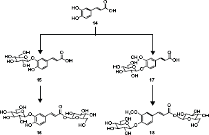
Biotransformation of caffeic acid by cultured cells of E. perriniana.
Glycosylation of ferulic acid (19)
Three compounds (named products 17, 18, and 20) were produced by incubation of the cultured cells of E. perriniana with ferulic acid (19). The structure of product 20 was determined on the basis of its ESIMS, 1 H and 13 C NMR spectra as feruloyl-O-β-D-glucoside.
Feruloyl-O-β-D-glucoside (20): ESIMS: m/z 379 [M+Na]+, 1 H NMR (400 MHz, CD3OD): δ 3.89 (3H, s, OCH3), 5.58 (1H, d, J = 7.6 Hz, H-1′), 6.40 (1H, d, J = 15.6 Hz, H-8), 6.81 (1H, d, J = 8.0 Hz, H-5), 7.09 (1H, dd, J = 8.4, 2.0 Hz, H-6), 7.20 (1H, d, J = 2.0 Hz, H-2), 7.72 (1H, d, J = 15.6 Hz, H-7); 13 C NMR (100 MHz, CD3OD): δ 56.4 (OCH3), 62.3 (C-6′), 71.0–78.7 (C-2′, 3′, 4′, 5′), 95.7 (C-1′), 111.6 (C-2), 114.6 (C-8), 116.4 (C-5), 124.2 (C-6), 127.4 (C-1), 148.1 (C-7), 149.2 (C-3), 150.7 (C-4), 167.5 (C-9).
Time course experiment showed that ferulic acid (19) was converted into 17 in short time incubation, and 18 was accumulated following to the formations of 17 and 20 (Figure 6). At 72 h reaction, the amounts of products 17, 18, and 20 were 20.3%, 3.4% and 2.5%, respectively, relative to the total amount of reaction products on a molar basis. This result indicated that glucosylation of ferulic acid predominantly occurred at 4-position of phenolic hydroxyl group rather than at carboxyl group. Biotransformation pathway of ferulic acid is shown in Scheme 6.
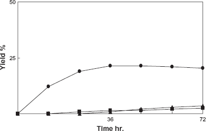
Time-course of the biotransformation of ferulic acid by cultured cells of E. perriniana. Yield is expressed as a percentage relative to the total amount of reaction products on a molar basis. Yields of 17(•), 20(■), and 18(▲) are plotted.
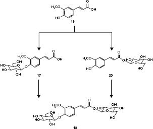
Biotransformation of ferulic acid by cultured cells of E. perriniana.
The result of this study established that plant cultured cells of C. roseus and E. perriniana can glycosylate capsaicinoids such as capsaicin and 8-nordihydrocapsaicin, and phenylpropanoids such as cinnamic acid, p-coumaric acid, caffeic acid, and ferulic acid. The cultured cells of C. roseus preferentially glycosylated 8-nordihydrocapsaicin rather than capsaicin, probably due to the difference in the structure of their side chain. It was found that the cultured cells of C. roseus convert capsaicin and 8-nordihydrocapsaicin into their mono- and disaccharides. From this study, the glycosylation of capsaicinoids by the cultured cells of C. roseus was attempted to reduce their pungency and enhance their solubility in water. It is well known that glycosylated drugs reduce their toxicity and enhance their oral absorption. Therefore, glycosylated capsaicinoids may be useful as drugs, functional foods, and cosmetics. A chemically synthesized capsaicinoid glucoside, vanillylnonamide β-D-glucopyranoside, 23 has been reported to have a remarkable in vivo effect on lipid metabolism in rats. 24 Studies on the physiological activities of glycosylated capsaicin and 8-nordihydrocapsaicin are now in progress.
From this study, it was found that both phenolic hydroxyl group and carboxyl group of phenylpropanoids such as cinnamic acid, p-coumaric acid, caffeic acid, and ferulic acid were glycosylated using the cultured cells of E. perriniana to give the corresponding mono- and disaccharides. Furthermore, the cultured cells of E. perriniana were able to catalyze hydroxylation and methylation as well as glucosylation on cinnamic acid, p-coumaric acid, and caffeic acid. Phenylpropanoids such as caffeic acid and ferulic acid has been reported to have a good effect as topical photoprotective agents. 25 But physiological studies of glucosylated phenylpropanoids have not been reported yet. On the other hand, from this study, the glycosylation of phenylpropanoids using the cultured cells of E. perriniana was attempted to enhance their solubility in water and enlarge effects. Further studies on the physiological properties of glucosylated phenylpropanoids are now in progress. Also, this biotransformation method is of considerable interest in green chemistry. Studies of biotransformation of another foreign substrate using the cultured cells of C. roseus and E. perriniana are now in progress.
Disclosure
This manuscript has been read and approved by all authors. This paper is unique and is not under consideration by any other publication and has not been published elsewhere. The authors and peer reviewers of this paper report no conflicts of interest. The authors confirm that they have permission to reproduce any copyrighted material.