
Abstract
Adenoid cystic carcinomas (ACCs) constitute 0.1–1 % of all malignant breast tumors. They have better prognosis than other breast malignancies. To date, there have been about 933 cases reported as per English literature. To the best of our knowledge, this case may be the second well-documented case of ACC of breast at younger age.
Keywords
Adenoid cystic carcinomas (ACCs) constitute 0.1–1% of all malignant breast tumors. They have better prognosis than other breast malignancies. To date, there have been about 933 cases reported as per English literature.1,2 To the best of our knowledge, this case may be the second well-documented case of ACC of breast at a younger age.
A 19-year-old girl was admitted in YCR Hospital, Latur, with a complaint of lump in right breast, which was gradually increasing in size since past one year. The lump was 2 × 2 cm in size, mobile, and not fixed to deeper tissues and skin. There was no evidence of palpable axillary lymphadenopathy. Mammography was suggestive of benign breast lesion—fibroadenoma. The Fine Needle Aspiration Cytology (FNAC) was planned and performed. Cytology revealed atypical epithelial cells with absence of bare nuclei and reported it as atypical epithelial hyperplasia (Fig. 1). In view of young age, core biopsy was planned and performed and the sample was sent to histopathology department to confirm the diagnosis. The core biopsy revealed prominent solid, microcystic, and focally tubuloglandular patterns of growth with intact myoepithelial layer (Fig. 2). On immunohistochemical evaluation, the tumor cells were immunopositive for CK7 expression (Fig. 3) and myoepithelial cells were positive for p63 (Fig. 4). Thus, the case was finally diagnosed as adenoid cystic carcinoma of breast and treated by breast conservative surgery with adjuvant radiotherapy, and on follow-up, it was found that the patient was doing well until date, since the last 18 months.

FNAC showing loosely cohesive ductal atypical epithelial cells—atypical epithelial hyperplasia.
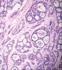
Prominent solid, microcystic, and focally tubuloglandular patterns of growth with intact myoepithelial layer (10×).

Tumor cells showing cytoplasmic positivity for CK7.

The p63 expression by myoepithelial cells.
The histological characteristics of ACC in the breast are similar to those of ACC of the salivary glands. However, the prognosis of ACC of the breast is better than that of other locations with prolonged survival. Breast-conserving treatment including postoperative radiotherapy seems to be equivalent to mastectomy alone with respect to survival. The value of adjuvant systemic therapies is not established. Late relapses can occur, so long-term follow-up is mandatory for these patients. 3 Little has been published to date on its radiological features. 2 The diagnosis is made by careful histological examination, presenting a difficult differential diagnosis with cribriform carcinoma. Therefore, it is necessary to use immunohistochemical techniques. 4
To conclude, it is an interesting incidental core biopsy diagnosis in sharp contrast with the clinical diagnosis. Thus, in every palpable breast lump irrespective of age, core biopsy can be used as an effective diagnostic tool to hit the correct diagnosis.
Author Contributions
Conceived and designed the experiments: SBI. Analyzed the data: SBI. Wrote the first draft of the manuscript: NPJ. Jointly developed the structure and arguments for the paper: BDA. Made critical revisions and approved final version: CRH (I) and SS. All authors reviewed and approved of the final manuscript.