
Abstract
Primary cutaneous adenoid-cystic carcinoma (PCACC) is a rare slow-growing neoplasm of disputed histogenesis characterized by a cribriform pattern at histology and local aggressive behaviour. Up to date about 60 cases of PCACC have been reported in the literature. This tumour is most common in the scalp, affects middle-aged and older individuals (mean age 59) and has predilection for women. We describe an unexpected case of PCACC in a 32-years-old woman referred to our clinic for a subcutaneous nodule in the scalp showing a slow growth and indolent course. The differential diagnosis and the clinical management of this PCACC patient, successfully treated with a wide local excision, are presented and discussed.
Introduction
Primary cutaneous adenoid cystic carcinoma (PCACC) is a rare tumour that affects middle aged and older individuals. It can occur in the scalp (32–41%), chest and abdomen and is characterized by indolent course and local aggressive behaviour.1–7 The average duration of the tumour prior to diagnosis is about 10 years. Patients typically present with slowly expanding skin coloured nodules from 0.5 to 9 cm in size.1–7 If the scalp is involved, alopecia is generally an associated finding. 3 In the past PCACC has been regarded as an eccrine lesion, but the possible origin from apocrine glands has been also proposed. 2
Case Report
A 32 years old woman presented with a subcutaneous nodule of the scalp with associated alopecia (Figure 1). Three years prior to the presentation she noted an indolent, slowly expanding firm nodular lesion clinically resembling a cutaneous cylindroma or trichoblastoma. In her medical history there was no evidence of previous skin lesions or systemic tumour diseases. At physical examination no signs of systemic disease were found and superficial lymph nodes were not palpable. Haematological tests were also in the normal range. The lesion was surgically excised and submitted for histological examination.
The clinical picture of the scalp lesion with associated alopecia.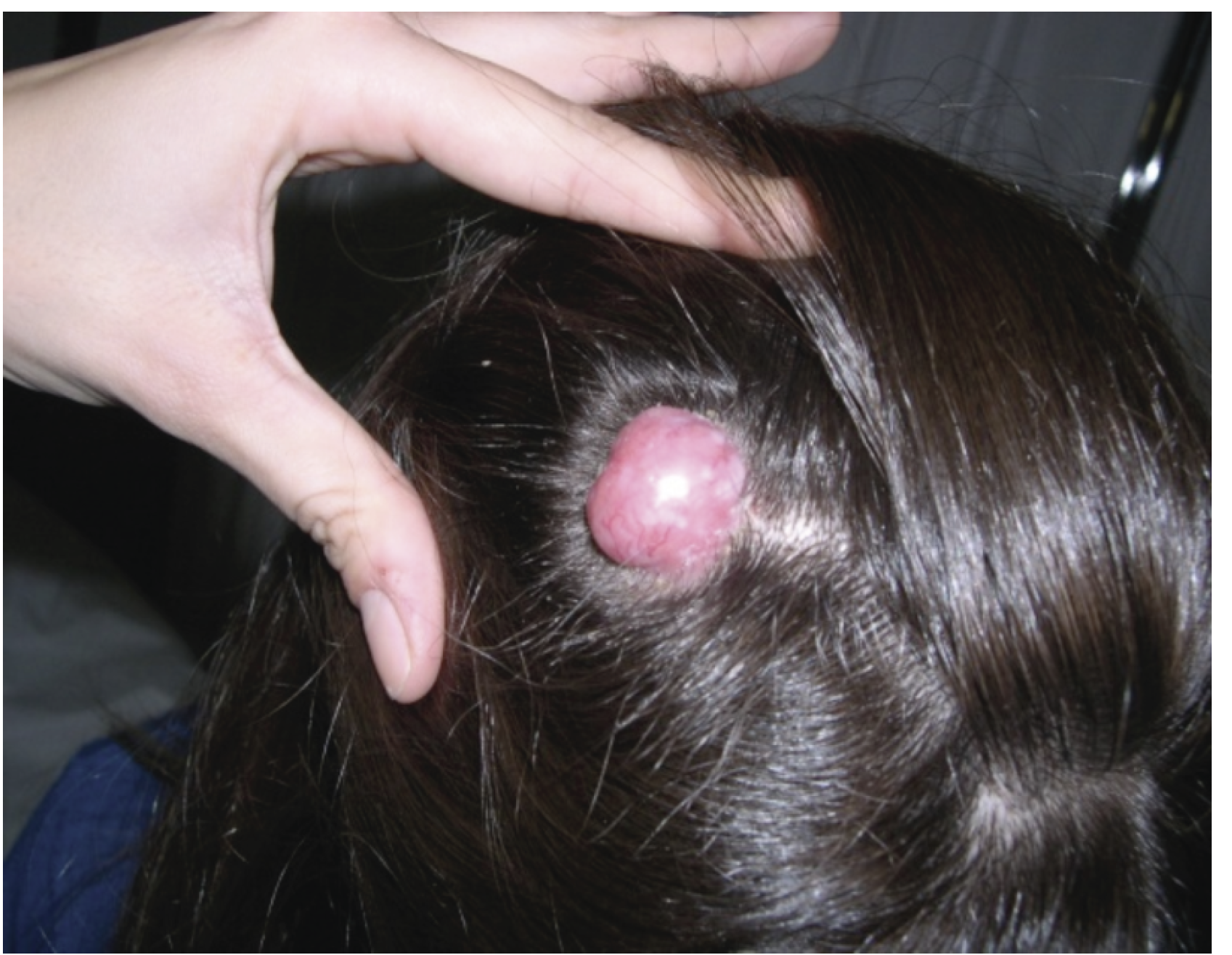
Macroscopically the lesion was a 1.9×1.7×2.3 cm grey-tan, flesh tumour, with poorly circumscribed borders. Microscopically it showed a characteristic basophilic appearance on low power magnification due to the nuclear hyperchromatism. The tumour was poorly circumscribed and consisted of medium-small basaloid cells, with scanty cytoplasm and small and inconspicuous nucleoli, arranged in islands, cords and strands with glandular, cystic, cribriform architecture, embedded in a fibrous stroma (Figure 2A and B). The lesion invaded the mid and deep dermis but not the subcutaneous fat. The basaloid cells were never displaced in a palisades fashion and no connection to the overlying epidermis was observed. Mitotic activity was very low (1 mitosis ×10 high power field), whereas necrosis, perineural invasion, lymphatic and/or blood vessels infiltration were not present.
Histological and immunohistochemical features of a primary cutaneous adenoid cystic carcinoma A) Low power view of the tumour demonstrating a poorly circumscribed neoplasm, which is composed of collection of basophilic cells with cribriform pattern. B) Higher magnification of the lesion showing tumour nests surrounded by an eosinophilic stroma. Nuclear palisading and epidermal contact are absent. C) Immunostaining with mAb to EMA depicts the apical aspect of pseudoglandular spaces. D) The hyaline deposits in small luminal areas among the basophilic cells are immunoreactive with mAb to collagen IV.
At immunohistochemistry, as expected, the tumour expressed low and high molecular weight keratins. A variably expression of S-100 protein and carcino-embryonic antigen (CEA) was also observed. The latter was restricted to the luminal spaces (data not shown). To better analyze the tumour at phenotypic level, selected monoclonal antibodies to type IV collagen and epithelial membrane antigen (EMA) were also considered. The former highlighted the hyaline deposits among the basaloid cells and the latter the apical aspects of pseudoglandular areas (Figure 2). The basophilic intraglandular material, variably detected in the tumour, was also highlighted with alcian blue staining at pH 2.5 and showed immunoreactivity for laminin (data not shown).
Both histological and immuno-phenotypical evaluation suggested the diagnosis of adenoid cystic carcinoma (ACC), but considering the rarity of this entity as a primary skin tumour it was necessary to rule out the possibility of a skin metastasis arising from other malignancies with histological features of ACC.
Adenoid cystic carcinoma, in fact, is most commonly seen as a neoplasm of the salivary glands and seromucinous glands of the upper respiratory tract. It has also been reported to occur in the breast, lung, uterine cervix, prostate, and lacrimal gland.1,8 As a consequence a careful breast and an otolaryngology examination was performed at specialist level, followed by head and neck ultrasonographic scan. A PET-CT scan was also considered to complete the oncological screening. Fortunately, the clinical examination was negative and all the radiological exams resulted in the normal range. All together, the clinical, morphological and immuno-phenotypical features described above, supported the final diagnosis of a primary cutaneous adenoid cystic carcinoma (PCACC). To avoid local recurrences a wide surgical excision with 2 cm disease-free margins was finally performed. After 12 months of follow up the patient is fully recovered without any evidence of disease.
Discussion
Primary cutaneous adenoid cystic carcinoma is a rare skin tumour. It was first described by Boggio in the 1975 3 and, at the best of our knowledge, only 61 cases of PCACC have been reported in the literature.1–7 This lesion frequently arises in the scalp of middle-aged or elderly patient, with a slight predilection for women with an average age of 59 years. 1 In about 76% of the cases perineural invasion may be observed and this feature seems to have a prognostic value, with a double increase of relative-risk of local recurrences when it is detected (46% vs. 22%). 1 In the case reported herein the patient is surprisingly young (32 years), with only two youngest patients (14 years) registered in the literature. 1 The lesion didn't show images of perineural invasion nor lymphatic or blood vessels infiltration (several histological preparations were examined). As aforementioned adenoid cystic carcinoma is relatively frequent in the salivary glands and in this contest it can be an aggressive tumour. ACC has been reported to occur also in the breast, lung, vulva, cervix and prostate.1,8 Considering the age of the patient and the rarity of this tumour in the skin it was imperative to rule out a skin metastasis from an ACC arising in other organs. This fact has important clinical implications because the occurrence of a skin metastasis from a “clinically occult ACC” requires a more complex oncological approach for advanced tumour disease. In our case the negative PET-CT scan and the careful clinical evaluation (in particular breast and salivary glands) allowed the diagnosis of PCACC. As aforementioned PCACC is characterized by a less aggressive behaviour compared to the analogous tumour arising in the salivary gland. The occurrence of lung and lymph nodes metastasis, in fact, is exceptionally rare in PCACC,7–10 but, although infrequently, they can be found in ACCs arising from salivary gland and breast. Moreover perineural invasion, a hallmark feature of the salivary gland ACCs, may be absent in PCACC, as in the case described here. 8 An indolent course is the major feature of PCACC with a high tendency of local recurrence.1,8 For this reason the recommended treatment for PCACC is a wide surgical excision with at least 2 cm tumour free margins.6–11
Differential diagnosis
When a skin metastasis from an ACC arising in other organs is definitively ruled out, the differential diagnosis should include several primary skin tumours: the adenoid-cystic variant of basal cell carcinoma (BCC), mucinous carcinoma (MC) dermal cylindroma (DC) and the primary cutaneous cribriform apocrine carcinoma (PCCAC). All the mentioned lesions closely mimic the PCACC at histology.8,12–13 The lack of basalioid cells disposed in peripheral palisades in an adenoid-cystic lesion without connection to the overlying epidermis help the distinction between a PCACC and the more common BCC adenoid-cystic type. 8 An other helpful histological feature for this differential diagnosis is the presence of artefactual clefts between tumour cells and stroma in BCC. 8 MC can be distinguished from a PCACC by histochemistry; in fact the former differs from PCACC in the produced sialomucins. 8 In the DC the tumour lobules contain nodular deposits of hyaline-pink material. Furthermore the lobules of cylindroma show a mosaic or jigsaw pattern, are surrounded by hyaline material and present peripheral small cells with scanty cytoplasm and dark nuclei. 8 Spiradenoma and spiroadenocylindroma can also have an adenoid cystic like pattern, as reported by Petersson and colleagues.8,13 However, in these tumours this feature is always focal (maximum of 2.5 mm). For this reason a single punch biopsy of such lesions may be not representative for a definitive histological diagnosis. Another findings that lacks in PCACC is the lobular architecture of spiradenoma. 8 Primary cutaneous cribriform apocrine carcinoma (PCCAC) shows a diffuse cribriform pattern in the entire neoplasia, interconnections of the tumour cell aggregates, nuclear polymorphism and authentic elongated nuclei, features that are absent in the PCACC. 12 Opposite to PCCAC, PCACC presents uniform size and shape spaces between tumour cell aggregates and deposits of basement membrane material. 8 Finally, the neurotropism, which may be found in PCACC, has been never observed in PCCAC.1,8
The histogenesis of PCACC is still uncertain and its apocrine or an eccrine derivation is debated. 14 Concluding, the prompt recognition of a PCACC is important. Integration of the clinical and histopathological data is essential for a correct diagnosis. This will avoid both patients distress and unnecessary over treatment.