
Abstract
Hair follicle stem cells (HFSCs) are potentially useful for the treatment of skin injuries and diseases. To achieve clinical application, a prerequisite must be accomplished: harvesting enough HFSCs from limited skin biopsy. The commonly used sorting approach for isolating HFSCs, however, suffers from its intrinsic disadvantages, such as requirement of large-scale skin biopsy. Here, we report an efficient organ culture method to isolate and expand rat HFSCs from limited skin biopsy and these HFSCs could reconstitute the epidermis and the hair follicles (HFs). Seventy-three percent of cultured HFs formed hair follicle stem cell colonies from the bulge, and a single hair follicle provided all the HFSCs used in this research, demonstrating the high efficiency of this method. Quantitative RT-PCR and immunofluorescent staining results revealed that these stem cells obtained from the bulge highly expressed basal layer markers K14 and alpha-6 integrin, epithelial stem cell marker P63, and bulge stem cell marker K15. After long-term culture in vitro, GFP-labeled hair follicle stem cells formed new hair follicles, epidermis, and sebaceous glands following xenotransplantation into the back of nude mice. This study indicated that multipotent hair follicle stem cells could be efficiently harvested through organ culture from limited skin material—even a single hair follicle—and reconstitute hair follicles in vivo after long-term expansion culture, providing the basis for future clinical applications.
Introduction
Most of adult mammalian skin is covered by hair, and hair follicle stem cells (HFSCs) residing in the bulge serve as a reservoir to provide transient amplifying cells that could give rise to various cell types during hair follicle (HF) regeneration (7,12,22,36). These stem cells also repair epidermis during wound healing (2). Based on the successful clinical application of adult stem cell therapies such as bone marrow and cornea transplantation, it has been proposed that similar stem cell therapy is useful for the treatment of severe skin injuries due to burning or scalding (38). Epidermal stem cell transplantation was clinically applied as early as 1991 (13), but the grafted skin did not form hair follicles (1), thus leading researchers to focus on the investigation of HFSCs.
A prerequisite to achieve HFSC therapy is to isolate and expand the stem cells in vitro prior to transplantation (6,29). Mouse HFSCs were isolated by fluorescence-activated cell sorting (FACS) using alpha-6 integrin and CD34 antibodies (35), and cultured mouse HFSCs reconstituted the hair follicles, the epidermis, and the sebaceous glands (3). Using a cocktail of antibodies, Ohyama et al. also isolated human scalp HFSCs following cell sorting (23). These are excellent works, and HFSCs from the skin were successfully isolated by cell sorting. However, some aspects of this isolation approach still need improvement. It requires large amount of skin for single cell suspension preparation, but this is impracticable for patients with severe skin injury (37). In addition, trypsinized cells tend to undergo anoikis during long-term suspension in vitro (17,28,32). Furthermore, mechanical impairment was also inevitable during flow cytometry, leading to low viability and colony-forming efficiency (CFE).
In the present study, we report organ culture as a simple and convenient method to harvest HFSCs from limited skin material. Using this method, HFSCs growing in their innate niche escaped from damages caused by trypsinization and physical impairment encountered in the traditional cell sorting method. These HFSCs also showed high proliferative capacity, allowing the reconstitution of hair follicles after xenotransplantation in nude mice.
Materials and Methods
Animals
Adult and newborn Wistar rats and nude mice were obtained from Vital River Laboratories (Beijing, China). Animals were housed for more than a week before used for experiments in an environment with constant photo-period (12-h light, 12-h dark cycle). They were given food and water ad libitum, in accordance with the Guideline of the Ethical Committee of the Institute of Zoology, Chinese Academy of Sciences.
Organ Culture to Harvest HFSCs
Vibrissa hair follicles were obtained from rats euthanized with chloroform and microdissected as described previously (15). Then each intact HF was cultured in a well of 24-well plate or 10 in every 60-mm dish (Corning, New York, USA) at 37°C/5% CO2 in growth medium of William's E medium (Gibco Laboratories, Carlsbad, CA, USA) containing 15% fetal bovine serum (FBS) (Hyclone, Logan, UT, USA), penicillin-streptomycin, and supplements (35). The growth medium was changed every second day.
HFSC Purification and Passage
In our culture, HFSCs adhered to the plate more tightly than fibroblasts. Taking advantage of this character, HFSCs were easily purified using a two-step trypsinization procedure. After treatment of the culture with 0.1% trypsin-0.008% ethylene diamine tetra-acetic acid (EDTA) at room temperature for 3–5 min, non-HFSCs, which mainly included fibroblasts, were detached from the plate, whereas the HFSC colonies remained adhering to the plate. After rinsing in phosphate-buffered saline (PBS) and trypsin inactivation using FBS, the dissociated fibroblasts were collected and used for future coculture. Supplemented with the digestion solution, remaining HFSC colonies in the plate were further incubated at 37°C for about 5 min until they were entirely dissociated. After trypsin inactivation, single cell suspension of HFSCs was plated, supplied with appropriate number of fibroblasts. The growth medium was changed every second day. This HFSC expansion via organ culture method was repeated for more than three times.
To estimate cell growth, 500 cells were seeded into a 100-mm plate (Corning, New York, USA), collected every third day, and counted. For CFE assay, 1,000 cells were seeded and cultured for 10 days before most of the colonies began to merge. To visualize the keratinocyte colonies, cells were fixed with PBS-buffered 4% paraformaldehyde and stained with 1% rhodamine B (Sigma, St. Louis, MO, USA). For routine passage, low-density seeding was carried out (i.e., 1,000 cells were allowed to grow for 2 weeks in a 100-mm plate until they became confluent).
Karyotype Analysis
Karyotype analysis was conducted using standard murine chromosome analysis protocols (41).
HFSC Identification
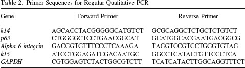
To test the expression of stem cell markers on transcript level, P7 and P12 HFSCs, as well as rat dermal fibroblasts that were used as control cells, were harvested and total RNAs were extracted using TRIzol reagent (Invitrogen, Carlsbad, CA, USA) according to the manufacturer's protocol. Based on absorbance at 260 nm, mRNA samples were adjusted to 1 μg/μl. Reverse transcription was performed with Moloney murine leukemia virus (M-MuLV) reverse transcriptase (New England Biolabs, Beverly, MA, USA). Quantitative RT-PCR was performed in 20-μl reaction volume containing 10 μl 2× GoTaq qPCR master mix (Promega, Madison, WI, USA), 2 μl template cDNA, and 1 μl primer mixture (40). Actin was used for normalization. Regular qualitative PCR was performed using the following settings: 94°C for 30 s, 55°C for 30 s, and 72°C for 1 min, for 30 cycles. Primer sequences for quantitative RT-PCR and regular qualitative PCR were listed in Tables 1 and 2, respectively.
Primer Sequences for Quantitative RT-PCR

Primer Sequences for Regular Qualitative PCR
To detect the expression of HFSC markers on protein level, HFSCs were seeded to the coverslips and cocultured with fibroblasts for about 4 days to form colonies. Then the colonies and fibroblasts were fixed in 4% paraformaldehyde for 10 min at room temperature and rinsed with PBS three times. If needed, cells were permeabilized with 0.1% Triton X-100 for 10 min at room temperature and rinsed three times. After blocking with 5% bovine serum albumin in PBS for 1 h at 37°C, cells were incubated in a humidified chamber at 4°C overnight with primary antibodies. The primary antibodies were mouse anti-rat keratin 14 (K14) antibody (Zhongshan Goldbridge Biotechnology, Beijing, China), mouse anti-rat alpha-6 integrin antibody (Serotec, Endeavour House, UK), and mouse anti-rat P63 antibody (Santa Cruz, CA, USA). After washing in PBS three times, cells were incubated with the appropriate fluorescein isothiocyanate-labeled secondary antibodies (1:100, all from Zhongshan Goldbridge Biotechnology) for 1 h at 37°, followed by counterstaining with propidium iodide (PI) (Sigma) for 10 min at room temperature. Cells were examined and photographed under a confocal microscope (Leica TCS SP2, Germany) (16).
Lentivirus Production and HFSCs Infection
The lentiviral vectors for infection were packaged using Viralpower Lentivirus Packaging System (Invitrogen) according to the manufacturer's instructions. Briefly, 293FT cells reaching 40–50% confluency were transfected with target plasmids 2K7-green fluorescent protein (GFP)/Neo and supermix plasmids (Invitrogen) using the Fugene HD transfection reagent (Roche Applied Science, Indianapolis, IN, USA). After overnight incubation, the medium was replaced by the fresh medium according to the manufacturer's instructions. Supernatant containing lentivirus was collected 48 and 72 h later and was concentrated with ultracentrifuge (20,000 rpm, 1.5 h) at 4°C (42).
To infect HFSCs, concentrated lentiviruses were mixed with the growth medium, and the HFSCs (approximately 100,000 cells) were incubated with the mixture for 24 h. To improve the efficiency of infection, Polybrene (Sigma) was added to the infection medium to 8 μg/ml (42). Virus-containing growth medium was replaced with fresh growth medium after 24 h for allowing the HFSCs to grow to confluence. GFP-HFSCs were enriched by flow cytometry and cultured before transplantation.
Hair Follicle Reconstitution
Engraftments were performed as described previously (39). Briefly, P8 GFP-HFSCs and dermal fibroblasts were mixed with a ratio of 2:1 (1.5 × 10⁷ total cells) in 300 μl growth medium and injected into silicon chambers (Renner GmbH, Germany) implanted into the back of anesthetized nude mice (Vital River Laboratories, China). The upper and lower chambers were removed after 1 and 2 weeks, respectively, and the mice were sacrificed at 7–10 weeks. The graft sites were collected and embedded in OCT, and cryosections 60 μm thick were prepared. After fixation in 4% neutral-buffered paraformaldehyde for 10 min at room temperature and rinsing three times, sections were examined and photographed under a confocal microscope (Leica TCS SP2, Germany). The reconstitution assay was repeated two times with at least three mice each time.
Statistics
Data were analyzed by one-way ANOVA and Bonferroni post hoc test with SPSS 16.0.
Results
Organ Culture to Harvest HFSCs
Rat vibrissa hair follicles were obtained by microdissection (Fig. 1A, B) and cultured in William's E growth medium. Within 3–7 days, 73.3% of the hair follicles adhered to the plate (Table 3), and then colonies grew out. These colonies only grew out from the bulge area (Fig. 1C, D), where HFSCs were reported to reside, and exhibited characteristics of keratinocytes with a classic “pavement” pattern (27) (Fig. 1E, F). In addition, fibroblasts were also apparent around the hair follicle. To demonstrate the efficiency of this method, all the HFSCs used in this study were harvested from this single colony shown in Figure 1.

Derivation of hair follicle stem cell (HFSC) colonies from a single hair follicle. (A) A vibrissa before microdissection. (B) A vibrissa after microdissection. (C, D) One of the hair follicle stem cell colonies that grew out from the bulge of cultured vibrissa hair follicles. (E, F) High magnification showing the cellular morphology of the colony. Bu, bulge; APM, arrector pili muscle; Bb, bulb. Scale bar: 500 μm (A, B); 200 μm (C–F).
Percentage of Hair Follicles That Formed Colonies in Culture

Average percentage: 73.3%.
Purification and Passage of HFSCs
HFSC colonies adhered to the plate tightly. Taking advantage of this character, HFSCs were easily purified by a two-step trypsinization procedure. Treated with digestion solution under room temperature, fibroblasts dissociated within 3–5 min and only the HFSC colonies remained adhering to the plate (Fig. 2A, B). After further treatment with digestion solution under 37°C for 5 min, the HFSCs were detached and collected for passage. Cocultured with appropriate number of fibroblasts, single cells from the HFSC colonies could form new colonies (Fig. 2C) and show logarithmic growth (Fig. 2D), then forming large colonies that were macroscopic after 2 weeks of growth (Fig. 2E) with a CFE of 49% (Fig. 2F). Furthermore, karyotype analysis showed normal chromosome numbers at passage 12 (P12) (Fig. 2G).

Purification of HFSCs and their passage. (A) Individual colonies before trypsinization. (B) Only the hair follicle stem cell colonies remained adhering to the plate after trypsin treatment for 3–5 min. (C) A HFSC colony at 5 days of culture. Cocultured with fibroblasts, single HFSC from the initial colonies formed new colonies. (D) Logarithmic growth of HFSCs. (E) Formation of large colonies after 2 weeks of growth. (F) Colony-forming efficiency was 49%. (G) Karyotype analysis showing normal chromosome number after 12 passages. Scale bar: 100 μm (A–C); 1 cm (E, F).
Characterization of HFSCs
Quantitative RT-PCR results revealed that HFSCs of passage 7 and 12 expressed high levels of previously reported epithelial basal layer markers k14 and alpha-6 integrin, epithelial stem cell marker p63, and bulge stem cell marker k15 (3,20,25) (Fig. 3A). In contrast, low expression of these genes was detected in dermal fibroblasts. Regular qualitative PCR results showed similar results (Fig. 3B).

Identification of hair follicle stem cells. (A) Quantitative real-time PCR detection. Compared with dermal fibroblasts, the cells of passage 7 and 12 highly expressed epithelial basal layer markers k14 and alpha-6 integrin, epithelial stem cell marker p63, and bulge stem cell marker k15. (B) Regular qualitative PCR result. (C) Immunofluorensence showed specifically high expression of K14, alpha-6 integrin, and P63 in hair follicle stem cell colonies of passage 3 in contrast to fibroblasts. HFSCs, hair follicle stem cells; Fb, fibroblast; P2, passage 2; P7, passage 7; P12, passage 12. *p < 0.05; **p < 0.01; ***p < 0.001. Scale bar: 50 μm.
Immunofluorescence staining further showed that K14, alpha-6 integrin, and P63 proteins are highly expressed in HFSC colonies in contrast to fibroblasts (Fig. 3C). In addition, these HFSCs also showed a high nucleus-to-cytoplasm ratio usually found in stem cells (Fig. 4).

High nucleus-to-cytoplasm ratio. The nuclear area was stained by propidium iodide (PI) after purified hair follicle stem cells were allowed to adhere to the cover glass for 2 h. Scale bar: 10 μm.
In Vivo Hair Follicle Reconstitution Assay
Passage 8 HFSC colonies were labeled with GFP using lentiviral infection to trace their descendents in vivo (Fig. 5A). Then GFP-expressing HFSCs (GFP-HFSCs) were mixed with neonatal dermal fibroblasts in the growth medium and transplanted to the back of nude mice using silicon chambers (Fig. 5A). After 7 weeks, transplanted GFP-HFSCs formed new hair follicles de novo (Fig. 5B, Table 4). Furthermore, they also formed new epidermis (Fig. 5C) and sebaceous glands (Fig. 5D). Our devising of this investigation is summarized in Figure 6.

Hair follicle reconstitution assay. (A) P8 GFP-expressing hair follicle stem cells and neonatal rat dermal fibroblasts mixture (2:1) was transplanted to the back of anesthetized nude mice using silicon chambers. The upper left panels showed the same colony under bright field and laser field to show the expression of GFP. (B–D) Transplanted hair follicle stem cells formed new hair follicles, epidermis, and sebaceous glands de novo. HF, hair follicle; HS, hair shaft; Epi, epidermis; Der, dermis; Con, control of adjacent nude mouse skin; SS, skin surface. White arrows: GFP-expressing hair follicle stem cell descendants; star: air bubble; black arrows: sebaceous glands. Scale bar: 100 μm.

The schematic diagram of harvesting hair follicle stem cells from single hair follicles via organ culture and reconstituting new hairs. Single hair follicles were obtained by microdissection and cultured in William's E growth medium. In the primary culture, hair follicle stem cells grew in their innate niche and formed colonies. Cells were purified and plated for expansion, and after identification with hair follicle stem cell markers, they were mixed with dermal fibroblasts and transplanted into silicon chambers on the back of nude mice to reconstitute new hair follicles.
Green Fluorescent Protein-Expressing Hair Follicle Stem Cells (GFP-HFSCs) Reconstitute Hair Follicles (HFs)

Discussion
HFSCs residing in the bulge are promising clinical candidates for the treatment of severe skin injuries and diseases (33). To achieve clinical application, a prerequisite must be accomplished: harvesting enough HFSCs from limited skin biopsy. Organ culture of hair follicles has been used to study hair follicle growth regulation and for testing pharmacological agents in vitro (4,14,19,34). In the present study, we demonstrated the utility of this approach for the efficient derivation of HFSCs.
Unlike the complex process of single cell suspension preparation and immunofluorescent staining for cell sorting, HFSC harvest by organ culture in our study was simple and convenient. We only obtained vibrissa hair follicles by microdissection, put them into the William's E growth medium and waited for colonies to spontaneously grow out from the bulge. In these primary cultures, HFSCs grew out from their innate niche without isolation damage, making it feasible to harvest large number of HFSCs from limited skin material.
We also used a simple way to purify HFSCs based on the highly adherent property of HFSCs compared with the neighboring cells. Under trypsinization at room temperature, non-HFSCs that grew out from the hair follicles could be easily detached, whereas the HFSCs could be obtained after further trypsinization at 37°C for about 5 min. This strong adherent nature is likely important for stem cells to reside in their niche in vivo (26,30), and when they detach from the niche and lose the cell–matrix interactions, they undergo anoikis (9). This property could be crucial for preventing stem cells to undergo uncontrolled growth and form neoplasm.
To characterize HFSCs, the expression of four important marker genes was detected. The k14 gene encodes a type I keratin, which was a component protein of the intermediate filament and highly expressed in epithelial basal layer cells (8,11). Alpha-6 integrin, an integral membrane protein, could mediate cell adhesion, cytoskeletal organization, and function as signaling receptor (5). This gene is also highly expressed in basal layer cells of the skin and its absence led to skin fragility and inflammation (21,31). Abundant expression of transcription factor p63 distinguished basal layer stem cells from the transient amplifying progeny cells (18,25). Cotsarelis et al. demonstrated K15 as a marker for hair follicle stem cells using k15-EGFP transgenic mice (20). In this study, the colonies that grew out from the bulge area highly expressed basal layer markers k14 and alpha-6 integrin, epithelial stem cell marker p63, and bulge stem cell marker k15, suggesting that these cells were bulge basal layer stem cells. In addition, these cells also showed large nuclear area that was characteristic of stem cells and high CFE, which indicated their self-renewal ability.
Moreover, these HFSCs showed strong proliferative capacity in vitro. All the stem cells used in this study were from a single HF. In our routine passage, 1,000 HFSCs could give rise to 1.3–1.5 × 10⁷ cells after 2 weeks of culture, thus representing more than 10,000-fold increase at each passage. This property of HFSCs suggested their high potential in future clinical application.
The ultimate goal of hair follicle stem cell harvest is to achieve cell therapy in the treatment of skin injuries and diseases such as severe burn, chronic leg ulcers, and alopecia (6,10). Studies had provided compelling evidence that HFSCs residing in the bulge area have the multipotency to give rise to all cell lineages of new hair follicle (20,24). In our reconstitution assay, passage 8 GFP-HFSCs were successfully reorganized to form new hair follicles, epidermis, and sebaceous glands de novo, demonstrating that the multipotency of the HFSCs was maintained during long-term culture in vitro.
Although the HFSCs used in this study were from rat, it has provided an exciting way to get the HFSCs in other species. Our preliminary work revealed that similar approach could be applied to harvest HFSCs from sheep whisker and human scalp HF (data not shown).
Even so, there are still some points that need to be improved. The reconstituted hair follicles could not grow as well as those reconstituted by dorsal HFSCs. We think that the microenvironment of the dorsal skin may affect our reconstitution outcome, because our HFSCs are from vibrissa, and all of the previously reported successful hair follicle reconstitution used dorsal HFSCs. For the following work, several dorsal HFs can be microdissected and cultured to provide HFSCs for the treatment of skin injuries of the animal itself. In addition, optimized growth medium, especially nonserum medium, will have to be developed for human HFSC expansion and transplantation. Fetal bovine serum is absolutely necessary in all the currently used HFSC culture mediums, whereas it may lead risk to the patient.
In conclusion, we had established a simple method to isolate and purify HFSCs from limited skin material, even from a single hair follicle, and these HFSCs showed strong proliferative capacity and multipotency, providing theoretical basis for further clinical hair follicle reconstitution.
Footnotes
Acknowledgments
We are grateful to Dr. Carol S. Trempus (National Institute of Environmental Health Sciences) for instructions on the HFSCs culture, Dr. Aaron J. W. Hsueh (Stanford University School of Medicine) for suggestions on the experiment design and English language editing of the manuscript, and Dr. Fei Wang (University of Illinois at Urbana-Champaign) for kindly providing the 2K7-GFP/Neo plasmid. We also thank Mr. Qi Chen of our lab for help on editing the figures. This work was supported by Strategic Priority Research Program of the Chinese Academy of Sciences XDA01010202, National Basic Research Program of China Grant (2007CB947401, 2011CB710905) and National Natural Science Foundation of China Grants (30971671, 30728022). The authors declare no conflicts of interest.