
Abstract
Interactions between vascular endothelial cells (ECs) and biomaterials are important for engineered tissue substitute. The modification of biomaterial surfaces are designed to modulate EC adhesion and responses in order to improve implantation success rate. Specifically, it has now been well established that increased vascular tissue regeneration can be achieved on almost any surface by employing novel nanofabricated surface features. To enhance EC adhesion and growth, material surfaces have been modified with physicochemical and mechanical properties, such as bioactive molecules from the matrix, peptides, and/or growth factors to control EC behavior. The advances in nanotechnology can bring additional functionality to vascular tissue engineering, optimize internal vascular graft surface, help to direct the differentiation of stem cells into the vascular cell phenotype, and, most importantly, also provide a biomaterials-based cellularization process. Nanomaterials could promote in situ endothelialization by mobilizing endothelial progenitor cells (EPCs) from the bone marrow, by encouraging cell-specific adhesion to the vascular graft, and, once attached, by controlling the proliferation and differentiation of these cells. Interaction between different cell types and extracellular matrix continue to be a principal source of inspiration for material biological function and, therefore, the understanding of the molecular mechanism trigger by the interaction is discussed.
Introduction
Synthetic materials used in vascular conduits applications have been indicated to be prone to clot and fail, and do not function well in the long term. The urgent need in the clinic has driven the search for a better and more biologically compatible blood vessel replacement. Numerous approaches have been used to fabricate the structure and the function of blood vessels (20,38,50). The vascular tissue engineering approach is to use a good biocompatibility material that is seeded with cells. The advance in nanotechnology brings additional functionality to vascular conduits; it optimizes internal vascular graft surface and even helps to direct the differentiation of stem cells into the vascular phenotype (1,26). Nanotechnology also offers the key to understand the surface properties of materials on devices such as valves and stents. That promisingly improves biocompatibility of the device by regulating cell adhesion ability and inhibiting thrombosis and formation of blood clots (5). There is plenty of evidence showing that development of nanotechnology-based methods in vascular tissue biofabrication represents one of the most important technological breakthroughs in vascular tissue engineering.
Nanostructuralized scaffolding can mimic the organization of a natural vascular extracellular matrix (ECM) and thus improve cell adhesion ability (38). Several observations indicated that biomaterials seeded with differentiated endothelial cells (ECs) (15,29) or ECs in combination with other cell types such as smooth muscle cells (SMCs) (56) promoted vascular tissue repair after artificial graft implantation. In order to improve the technology in vascular grafts, we need the knowledge and expertise in tissue engineering. Regarding previous findings, nanotechnology approaches seemed to exhibit an important role in the endothelialization processing, including endothelial cell migration, adhesion, proliferation, and differentiation. Therefore, this article reviews nanobiomaterials in vascular tissue engineering, which provide a mimic condition of the vascular tissue suitable for endothelial cell adhesion.
Nanoscale Topography and Vascular Tissue
It is of interest to note that nanostructuralized biomaterials can modify the organization of a natural vascular ECM and thus improve cell attachment (2). One explanation is provided by the evidence that nanostructured surface features can significantly improve vascular cell proliferation; such design criteria can be used in the synthesis of the more successful tissue-engineered vascular grafts (37). For example, hydrogels have been modified with growth factors and ECM peptides on nanostructure biomaterials, which brought additional functionalities and improved the capacity of the hydrogel to direct cell differentiation and tissue regeneration (5,42). Indeed, surface-aminated electrospun nanofibers that enhance adhesion and expansion of human umbilical cord blood hematopoietic stem progenitor cells have been observed (8). Additionally, a study also showed that ECs, when cultured on gelatin-grafted electrospun poly(caprolactone) (PCL) nanofiber, were able to maintain the expression of three characteristic markers: platelet-endothelial cell adhesion molecule 1 (PECAM-1), intercellular adhesion molecule 1 (ICAM-1), and vascular cell adhesion molecule 1 (VCAM-1) (33). The nanobionic surface modified small intestinal submucosa (SIS) film possesses good and persistent antithrombogenicity, and also exhibits excellent blood compatibility (17). Experiment was also performed to show that the collagen-blended poly(l-lactic acid)-co-poly(epsilon-caprolactone) nanofibers [P(LLA-CL) NFM] could enhance the viability, spreading, and attachment of ECs, and, moreover, preserve the EC phenotype. The P(LLA-CL) NFM is a potential material for tissue-engineered vascular graft (19). Indeed, the polyhedral oligomeric silsesquioxane attached by direct reaction onto a urethane segment (UCL-Nano) has been demonstrated to have similar viscoelastic properties to the walls of a natural artery, which is resistant to degradation and is able to sustain endothelial cell seeding (42). For example, endothelial progenitor cells respond to ridge-groove grating of 1200 nm in period and 600 nm in depth on nanotopography substrate with promoted alignment and elongation, reduced proliferation, and enhanced migration (4).
Cells are known to be surrounded by nanoscale topography in their natural extracellular environment. It was observed that this surface comprises nanopatterned gratings with poly(methyl methacrylate) (PMMA) and PDMS can provide a valuable platform for the study of cell—substrate interactions and for the development of medical devices with nanoscale features of smooth muscle cells (SMC) (56). It has been suggested that synthetic aligned poly(l-lactid-co-epsilon-caprolactone) [P(LLA-CL)] copolymer combines with the advantages of synthetic biodegradable polymers may represent an ideal tissue engineering scaffold, especially for blood vessel engineering (55). Recently, synthetic poly-l-lactic acid (PLLA) nanofibers have been shown to not only improve biocompatibility with outgrowth endothelial cells (OEC) but also promote and guide aligned OEC sustained proliferation (31). Based on these findings, the aligned PLLA, which can serve as the scaffold and promote cell growth during endothelialization of biomaterials, can provide a promising way to prevent intimal hyperplasia of small-diameter vascular grafts.
Engineered vascular grafts with polyhedral oligomeric silsesquioxane modified polycarbonate urea-urethane (POSS-PCU), which was cross-linked with a bioactive RGD peptide and formed a biofunctionalized nanocomposite polymer-based small diameter bypass graft, has been shown to have the potential for rapid endothelialization (12). A similar approach using poly (carbonate) urethane-based nanocomposite polymer incorporating polyhedral oligomeric silsesquioxane (POSS) nanocages (UCL-NANO) showed antithrombogenicity and biostability as well as exhibited a good small caliber quadruple lamina vascular bypass graft method (46). The key materials of vascular tissue engineering are listed in Table 1.
Summary of Key Materials of Vascular Tissue Engineering

ECs, endothelial cells; SMC, smooth muscle cells; OEC, outgrowth endothelial cells.
Molecular and Cellular Interaction between Nanobiomaterials and Vascular Microenvironment
Nanomaterials are known to be important in vascular tissue application. However, how the molecular signaling and its biological function is controlled are still unclear. It is extremely important to note that developing such biomaterials requires the modification of materials that mimic molecular mechanism of extracellular matrices (18,20). These associated signal molecules could promote vascularization by the mobilization of ECs, induction of cell-specific adhesion to the vascular graft, and, once attached, by controlling the proliferation and differentiation of these cells. The benefit from the nanobiomaterials is considered as the advantageous characteristics of the natural extracellular matrix for tissue repair processing (55). It is likely that cells adhering to the ECM induce intracellular signaling cascades and migration of integrins into focal adhesion complexes (21). For example, evidence indicated that manufactured aluminum oxide nanoparticles decrease the expression of tight junction proteins in brain vasculature (6). Indeed, cupper-nanoparticles are highly useful for regenerating of elastin matrices by adult vascular smooth muscle cells (SMCs). These results have immense utility in tissue engineering vascular replacements (27). There was further evidence suggesting that nanostructured hydroxyapatite (HA) crystals upregulate fibroblast growth factor-2 (FGF-2) expression and activity in microvascular endothelium, promoting angiogenesis in 3D matrices (41). Current issues in nanotechnology and molecular self-assembly may provide novel solutions to cell transplantation deficiencies. An observation has shown that the delivery of vascular endothelial growth factor (VEGF) and FGF-2 from heparin-binding peptide amphiphiles (HBPAs) scaffolds significantly increases blood vessel density in the mouse omentum over control scaffolds without growth factors and significantly promotes islet engraftment (49). The in vitro study demonstrated that cell adhesive proteins adsorbed (vitronectin and fibronectin) in different quantities and altered bioactivity on nanostructures, which, at least in part, may account for the documented increased vascular cell adhesion on nanostructured PLGA (36).
A study from neovascuature has shown that nanosphere-aided targeting of the neovasculature with mutant Raf-1 causes regression of tumor progression (52). Based on this finding, it seems reasonable to conclude that the nanoparticles coated with specific biological molecules may be useful for delivering signal molecules, which can cause the regression of tumor progression. The use of viscoelastic nanocomposites vascular conduit by exposing to shear stress preconditioning prior to physiological flow can increase endothelial growth factor receptor-2 (VEGFR-2) and transforming growth factor-β1 (TGF-β1) on ECs (51). Indeed, another approach also indicated that nanofibrous materials made from bioabsorbable and biocompatible polymers have promising applications as tissue-engineered scaffolds. Genetic analysis of human umbilical vein endothelial cells (HUVEC) that attached to poly(glycolide) (PGA) nanofibrous materials prepared via electrospinning methods exhibited high expression of integrin v and VEGF receptor genes, which are known angiogenesis markers (24). The molecular and cellular interaction between nanobiomaterials and the vascular environment is shown in Table 2.
Molecular and Cellular Interaction Between Nanobiomaterials and Vascular Microenvironment
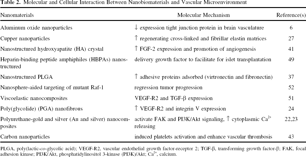
PLGA, poly(lactic-co-glycolic acid); VEGF-R2, vascular endothelial growth factor-receptor 2; TGF-β, transforming growth factor-β; FAK, focal adhesion kinase; PI3K/Akt, phosphatidylinositol 3-kinase (PI3K)/Akt; Ca2+, calcium.
It is also of interest to note that in conjunction with microvascular dysfunction, nanoparticle exposure also reduces nitric oxide (NO) activation through at least two functional distinct mechanisms that may mutually increase local reactive species (39). A report also bring the attention that PU-Au nanocomposites activate FAK and PI3K/Akt signaling pathway in ECs, which leads to proliferation and migration of ECs on a series of nanocomposites from polyurethane (PU) incorporated with various gold nanoparticles (23). Another report also suggested that the nanometric micelles on PU surface may interact with ECs and accelerate their migration by increasing cytoplasmic Ca2+ and stimulating the PI3K/Akt/eNOS signaling pathway (22). Indeed, carbon nanoparticles and microparticles have the ability to induce platelet activation and enhance vascular thrombosis, and this observation will be important for the pharmacological application (43). These reports lead to the conclusion that biomaterials can successfully integrate into a tissue's mechanical properties and material topography. The cellular molecular mechanism to a biomaterial may be enhanced in synthetic vascular conduits by mimicking the surface roughness that is created by the associated nanostructured extracellular matrix components of natural tissue (37). The proposed mechanism by which nanomaterials contribute to the modulation nof vascular tissue engineering is shown in Figure 1.

Schematic representation of the proposed mechanism by which nanomaterials contribute to the modulation of vascular tissue engineering. As a result of vascular tissue injuring, the nanomaterials (including nanoparticles, nanofibers, nanocarries, nanostructure pattern, nanosphere, and nanofluore) trigger molecular mechanism by virtue of their expression of tight junction protein, adhesion molecules (e.g., FAK, integrin V), signaling molecules (e.g., PI3K/Akt, eNOS, and Ca2+), growth factors (e.g., FGF-2, VEGF-R2, and TGF-β), and extracellular matrix (e.g., elastin, fibronectin, and vitronectin). The interaction of endothelial cells with injured tissue could lead to activation of signal molecules that may contribute to the recovery of vascular injury (e.g., increase the adhesion, migration, proliferation, angiogenesis), promotion of drug delivery (e.g., regulatory of cancer), and tracing of stem cell differentiation. These molecular effects of nanomaterials contribute to restored vascular tissue regeneration of the injured area and result in the induction of endothelialization.
Perspective on Nanotechnology in Manipulating Vascular Stem Cells
Although stem cells hold great potential for the vascular tissue engineering application, the development of advanced and novel method to track and guide transplanted stem cells should be addressed in more detail (48). For example, nanopatterns on the surface of a vascular graft with nanofilms can increase its adhesion for circulating endothelial progenitor cells (12). Another way to influence stem cell differentiation and transplantation is engineered nanometer-scale scaffolds (14). For instance, functional nanofiber scaffolds with different spacers modulate adhesion and expansion of cryopreserved umbilical cord blood hematopoietic stem/progenitor cells (7). This is especially noteworthy in the case of scaffold topography and cell—substrate interaction that regulate hematopoietic stem and progenitor cell (HSPC) proliferation and self-renewal in cytokine-supplemented expansion.
The formation of an endothelial cell layer on the luminal surface of vascular grafts is an important attribute to improve their potential for endothelialization. The formation of blood vessels is mainly due to the activity of EPCs (3). It is of interest to note that nanocomposite-containing bioactive peptides could promote adhesion, spreading, and formation of a confluent endothelial monolayer by circulated progenitor cells (1).
Application of Nanoparticles for Tracking Stem Cells
Many applications of new cellular magnetic labeling method to EPCs exhibited therapeutic potential for revascularization. One report indicated that when magnetic nanoparticle-incorporated EPCs were injected into a microfluidic channel by using a syringe pump it can control the flow rate of cells (25). Magnetic cell delivery and targeting offers a new opportunity for vascular tissue engineering and delivery of localized cell-based therapies. Using a magnetically tagged device at a site of artery injury is able to enhance EPC localization (28). This technology could be more adapted to trace cells in other organs and may provide a novel approach for the systemic injection of cell therapies in vascular tissue engineering application. When EPCs are labeled with mo-nocrystalline iron oxide nanoparticles (MIONs), it does not adversely change their viability and its migration ability in vitro, and this allowed successful detection of limited numbers of these cells in muscle (30). MION-labeled EPCs provides a noninvasive strategy to monitor the location of small signal molecules of these cells.
There is a technique using advanced ultra-high-field 7-Tesla (7T) MRI of nanoparticle-labeled transplanted human EPCs in porcine ischemic hearts for clinical application (45). Indeed, another report also pointed out that magnetically labeled EPCs transplanted for therapeutic neovascularization in myocardial ischemia can be visualized by MRI (54). It has been observed that self-synthesized superparamagnetic iron oxide particles (PLL-SPIO) can effectively improve EPC mobilization and proliferation (34). Therefore, it can be assumed that PLL-SPIO can serve as a tracker for implanted EPCs.
Nanobiomaterials for the Delivery of Vascular Genetic Molecules
Strategies for vascular conduit application have been focused on the delivery of angiogenic growth factors such as VEGF and FGF-2 to induce angiogenesis progress from the host vasculature (32,35). Nanofibrous materials made from bioabsorbable and biocompatible polymers are promising applications as tissue-engineered scaffolds (9). The nanocell comprising a nuclear nanoparticle within an extranuclear pegylated-lipid envelope is preferentially taken up by the tumor. The technology can be extended by using additional agents aimed to target multiple signaling pathways, such as hypoxia-inducible factor-1α (HIF-1α), as well as a temporal targeting of tumor cells (47). It was observed in a NOD/SCID mouse hind limb vascular injury model that nanofiber-expanded cells exhibited more therapeutic potential in blood flow restoration after further applying VEGF (16), PDGF-BB, and growth factor (11). Additionally, overexpression of proangiogenic growth factor can promote the cellular therapeutic efficiency in EPCs for ischemic disease (58). In summary, nanofiber-based ex vivo expansion technology can provide a novel therapeutic strategy for stem cells in vascular tissue engineering. Moreover, the immobilization of CD34 antibodies on a lumenal vascular stent enhanced recruitment of circulated cells and accelerated postimplantation endothelialization in vitro (44).
Future Prospects
The incorporation of vascular tissue engineering, stem cell application, and nanotechnology research can improve regenerative medicine for current clinical application. Recently, the application of stem cells to vascular tissue engineering has become a novel technology. Having developed a fully biological vascular graft, functional concerns about its biological compatibility and mechanical property have become the focus of research effort. Although the main goal of nanotechnology is to enhance the ability to repair vascular tissue, it is unclear how nanobiomaterials mimic the native properties of blood vessel grafts in a long-term implantation in vivo and this will be the important issue to be further pursued for a new therapy strategy. For example, development of genetic engineering methods (47,52) and the variety of cell types that are able to express signaling molecules (6,22,23,39), proteins (27,36), peptides (49), and growth factors (13,32,35,58) all show great promise for modification of vascular nanobiomaterials. However, a more advanced knowledge on the modulation of molecular mechanism will provide a medium closely mimicking the physiological environment within an engineered vascular graft. This may advance a better biocompatibility, ultimately providing more durable vascular nanobiomaterials. Alternatively, advanced nanobiomaterial fabrication and application will be essential for more functional vascular tissue in regenerative medicine. For example, modification of nanobiomaterials has been shown to have a lower degradation rate in vivo, and this promotes the host immune response (57). A concern remains regarding the trace amounts of potentially antigenic compounds of animal origin (lipids, DNA, glycosilation products) that have been reported to be present in various types of nanobiomaterial and may induce an inflammatory response at the implantation site (10,16).
A recent study has demonstrated that the tissue-specific matrix components cause significant different adhesion efficiency and phenotype (31,40) of vascular tissue, as well as endothelial cells. These observations suggest the need for more appropriate, tissue-specific nanobiomaterials from in vitro to in vivo study. Indeed, utilization of nanobiomaterials in clinical application may provide successful vascular tissue regeneration for the patient. It is worthy of consideration that circulating hematopoietic blood stem cells from bone marrow (53), umbilical cord blood (7), or peripheral blood (12) have a high proliferative and regenerative potential, and they may be an optimal and ideal cell source for mimicking in vivo endothelialization of synthetic nanobiomaterials. However, due to the heterogeneity of hematopoietic stem cell populations and the definition of stem cells surface molecules are still poorly identified, success of this approach depends on a better characterization and understanding of hematopoietic stem cell biology and property.
Conclusion
Nanotechnology has become an attractive method for vascular tissue engineering from the viewpoint of biology (i.e., stem cells, extracellular matrix, growth factors, and signaling molecules). A range of nanobiomaterials has been shown to provide an excellent biocompatibility for many processes that occur in the early stages of vessel repair and allow a long-term maintenance of neo-vascular tissue development. The endothelialization of synthetic nanobiomaterials is a promising therapeutic approach to create a nonthrombogenic barrier on surfaces, and could thereby improve the efficiency of vascular prosthesis of blood contacting devices. The design of athrombogenic nanosurfaces on vascular grafts and the development of nanopatterned biomimicking vascular grafts are already well established. The use of nanopatterned functionalized biomaterials for the expansion and directed differentiation of human stem cells into cells of vascular lineage is a fast evolving and promising technology. The development of novel methods of vascular tissue assembly and rapid vascular biofabrication, such as magnetic force-driven tissue engineering and simultaneous electrospinning of vascular scaffolds and living cells, represents probably the most important breakthroughs to date.
Based on the above findings, evidence strongly indicates the potential of nanotechnology in vascular tissue engineering for the development of successful and effective vascular tissue-engineered products. Immobilization of homing factor mimetic molecules onto synthetic nanobiomaterials to increase the production of EPCs from circulating blood opens a new therapeutic strategy in regenerative medicine. This concept is based on the understanding that every patient will be able to seed his implants with his own progenitor cells after implantation, and this technology may bring new therapeutic strategy for vascular tissue engineering.
For reasons mentioned above, the application of signal molecules can be incorporated into functional development of nanobiomaterials for regulation of stem cell mobilization of vascular biology. With the incorporation of nanotechnology, biomaterial surface modification and stem cell biology for developing a novel therapeutic strategy could be advanced for vascular tissue engineering.