Transforming growth factor- (TGF-) is a multifunctional cytokine that plays a vital role in regulating cell growth, differentiation and survival in various tissues. It participates in a variety of cellular processes, including cell apoptosis, cell migration and evasion, and plays a paradoxical role in tumor genesis and development. In the early stage of tumor, TGF- inhibits the occurrence of tumor by inhibiting cell proliferation and regulating cell apoptosis. In the advanced stage of tumor, TGF- promotes tumor development and affects prognosis by promoting cell survival and proliferation, cell migration and invasion, participates in immune escape, etc. In this article, we will review the paradoxical role of TGF- on the occurrence and development of oral squamous cell carcinoma.
Oral cancer is the most common malignant tumor in the head and neck region, and its incidence has been on the rise. According to the latest data from the International Agency for Research on Cancer, there were approximately 377,713 new cases and 177,757 deaths worldwide [1]. Oral squamous cell carcinoma (OSCC) accounts for 90% of all oral cancer cases. Despite the effectiveness of surgical treatment combined with postoperative radiotherapy and chemotherapy, the 5-year survival rate of oral cancer patients is still around 5060% due to tumor metastasis, recurrence, drug resistance, and other factors [2]. The survival rate of advanced-stage patients is even lower. Oral cancer can affect patients’ eating, swallowing, breathing, speaking, as well as their physical and mental health, causing significant health and economic burdens [3]. The etiology of oral cancer is not clear, with the most common risk factors being smoking, alcohol consumption, and other risk factors including systemic diseases such as diabetes, metabolic syndrome, chronic inflammation, and HPV virus infection [4, 5, 6]. Identifying the key molecules and pathways driving OSCC progression and elucidating the pathogenesis and regulatory mechanisms of oral cancer are important and urgent tasks.
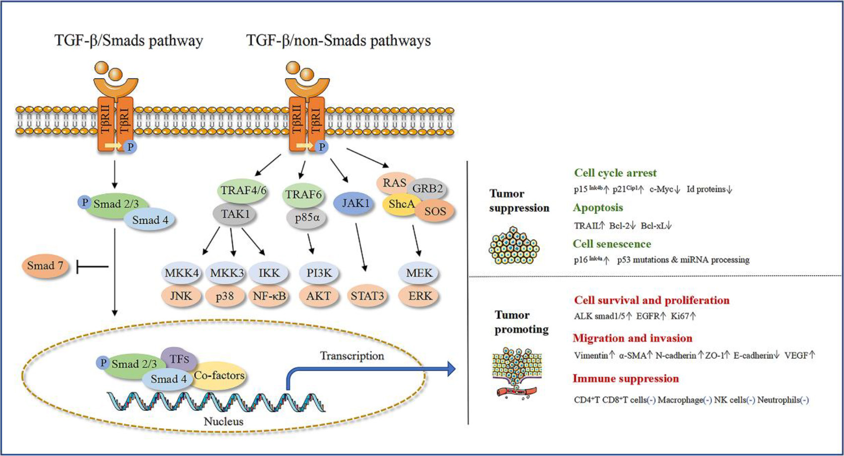
The transforming growth factor- (TGF- signaling pathway is an important transmembrane signaling pathway that is closely associated with various human diseases, including tissue fibrosis, cardiovascular disease, skeletal disease, and cancer [7]. Aberrant TGF- signaling is considered one of the pathways leading to OSCC carcinogenesis. Studies have shown that TGF- is significantly expressed in oral cancer tumor samples, especially in the tumor center and invasive front, and is associated with cancer invasion, metastasis, and poor prognosis [8, 9]. The TGF- signaling pathway is mainly composed of TGF- signaling molecules, transmembrane receptors (TRs), intracellular transcription factors, and corresponding target genes. TGF- has three highly homologous subtypes, namely TGF-1, TGF-2, and TGF-3, which are located on chromosomes 19q13, 1q41, and 14q24, respectively [10]. The transmembrane receptors are also divided into three types: TRI, TRII, and TRIII. The TGF- signaling molecule first forms a complex with TRII on the cell membrane surface, and then recruits and phosphorylates TRI (ALK5). The activated TRI continues to activate and phosphorylate downstream Smads proteins (Smad1/2/3/5/8 is receptor related Smads also named R-Smads, Smad4 is common Smads also named Co-Smads, and Smad6/7 is inhibitory Smads also named I-Smads). The activated R-Smads and Co-Smads form a polymer, enter the cell nucleus, interact with intracellular transcription factors, and achieve DNA binding and selective transcription, translation, and protein synthesis of target genes, thereby activating the typical TGF- signaling pathway [11]. In addition, TGF- can phosphorylate the linker region between the N-terminal MH1 (Madhomology 1) and C-terminal MH2 of the Smad2/3 protein, activating the non-typical TGF- signaling pathway [12] (Fig. 1). For example, TGF- can activate the MAPK kinases TAK1 and downstream JNK, p38 MAPK signaling, and NF-B signaling through the specific Smad E3 ligase (TRAF6) [13, 14], or promote the formation of TRI and p85 complex through the polyubiquitination of p85 by TRAF6, leading to the activation of downstream PI3K and AKT signals [15]. TGF- can also activate the STAT3 signal through the interaction between JAK1 and TRI [16], and activate the RAS pathway through TRI binding and phosphorylating adaptor protein ShcA, recruiting GRB2 and SOS to form the ShcA-GRB2-SOS complex, leading to the activation of downstream ERK1/2 [17]. At the same time, the TGF- signaling pathway is closely related to other signaling pathways, including the Wnt/-catenin, Hippo/YAP signaling pathways, which can also regulate the transmission of TGF-/Smad signaling pathway in various situations [18, 19].
TGF- was initially discovered in the 1980s to promote the proliferation of fibroblasts and anchorage-independent cell growth in vitro [20]. Subsequently, TGF- has been shown to inhibit the proliferation of various types of cells, including epithelial cells, endothelial cells, and hematopoietic cells [21]. Therefore, TGF- is considered to play a paradoxical role in cell proliferation and growth [22]. In the tumor microenvironment, TGF- can act as a tumor suppressor, maintaining cellular homeostasis and preventing early tumor deterioration. However, long-term abnormal activation can transform its function into promoting cell proliferation, migration, invasion, and immune evasion, thereby promoting tumor development [23, 24]. The seemingly opposite roles of TGF- in tumors are referred to as the “TGF- paradox”. There have been some reviews that mainly summarize the activation of the TGF pathway and its general mechanisms in systemic tumors or squamous cell carcinoma. However, there is no in-depth summary of its mechanisms in oral squamous cell carcinoma. This article summarized new research and the progress of TGF- in promoting and inhibiting OSCC development, providing a review and theoretical basis for exploring the occurrence and regulation mechanisms of oral cancer.
TGF- inhibits OSCC development
The double role of TGF- signaling in OSCC development. Figure note: TGF- signaling pathway can be activated through typical and non-typical TGF- signaling pathway. TGF- plays a double role on the development of oral squamous cell carcinoma. TGF- can inhibit the proliferation of OSCC cells and inhibit tumor occurrence by interfering with cell cycle, mediating cell apoptosis and cellular senescence effects, etc. TGF- also can promote the development of OSCC by promoting cell survival and proliferation, promoting cell migration and invasion, and participating in immune escape, etc. The figure is created by Adobe AI software.
TGF- is a typical anti-proliferative cytokine for many epithelial cells, including oral epithelial cells. TGF- can interfere with the normal cell cycle and inhibit cell proliferation, also mediate apoptosis and senescence, thereby inhibiting tumor development (Fig. 1). Among them, the effect of TGF- on inhibiting cell proliferation has been extensively studied, and its function is worthy of recognition. However, more research is needed to reveal the role of TGF- in regulating cell apoptosis and senescence.
TGF- can mediate cell cycle arrest in multiple ways
Firstly, TGF- can induce the expression of cyclin-dependent kinase inhibitors (CDKIs) in epithelial cells, such as p15Ink4b, p21Cip1, p57Kip2, etc. p15Ink4b belongs to the inhibitor of cyclin-dependent kinase 4 (INK4) family protein, which can specifically bind to CDK4 and CDK6 proteins, inhibiting the binding of cell cycle protein D and CDK4/6, thereby inhibiting cell cycle progression from G1 to S phase [25]. p21Cip1 and p57Kip2 belong to the CIP/KIP family protein, among which p21Cip1 can block the binding between cyclin E and CDK2, leading to cell cycle arrest in G1 phase [26]. The study by Nakamura et al. showed that TGF- can mediate cell cycle arrest by inducing the expression of p21Cip1 and p15Ink4b, thereby inhibiting the proliferation and differentiation of tongue epithelial cells [27]. There are also studies showing that TGF- requires the activation of the Notch signaling pathway to inhibit epithelial cell proliferation, and the two pathways synergistically regulate the cell cycle inhibitor p21Cip1 to inhibit epithelial cell proliferation [28]. Zhang et al. found that low concentrations of TGF- (0.5 ng/mL) can inhibit the proliferation of dental epithelial cells by activating typical TGF- signaling pathway through activating ALK5 receptor and the downstream Smad2/3 [22]. Liu et al. found that Smad7 deficiency can lead to upregulation of Smad2/3, resulting in excessive activation of the TGF- pathway, upregulation of p21Cip1expression, and downregulation of Cyclin D1 expression, leading to the inhibition of dental epithelial cell proliferation [29]. Another study showed that tongue squamous cell carcinoma cells (Ts-Smad4 cells) with high expression of Smad4 can upregulate p21Cip1 and MMP-2 expression under TGF- stimulation, thereby inhibiting the proliferation and reducing the metastasis of tongue squamous cell carcinoma cells [30].
TGF- can also induce cell cycle arrest through downregulating growth-stimulating proteins, especially the proliferative transcription factor c-Myc and inhibitors of differentiation (Id proteins). The downregulation of c-Myc expression is mediated by the TGF--induced repressor complex, which includes the Smad3-Smad4 complex, transcription factor p107, E2F4/5, and C/EBP (CCAAT/enhancer binding protein . Under TGF- stimulation, the repressor complex translocates to the nucleus and binds to the transcriptional inhibitory element on the c-Myc promoter, thereby inhibiting c-Myc transcription [31]. The reduction of c-Myc expression not only directly prevents cells from entering the proliferative state but also activates several important CDKIs promoter inhibitory genes, such as p21Cip1 and p15Ink4b, leading to cell cycle arrest [32]. Members of the Id proteins, including Id1, Id2, and Id3, can inhibit the process of cell differentiation and promote cell proliferation. The transcriptional inhibition of Id1, Id2, and Id3 is mediated by Smad signaling [33]. In epithelial cells, TGF- can directly promote the expression of the stress response factor ATF3 by activating Smad3, which directly inhibits the expression of Id1 [33]. Id2 can antagonize the anti-proliferative effect of p21Cip1, and TGF- can inhibit Id2 expression and induce p21Cip1 expression, mediating epithelial cell growth arrest [34].
In summary, the inhibition of oral squamous cell carcinoma cell proliferation by TGF- is mainly related to the upregulation of p21Cip1 and downregulation of c-Myc and Id proteins, but the specific mechanism still needs to be explored.
TGF- can induce cell apoptosis and senescence
Apoptosis is a genetically controlled autonomous and orderly death process that eliminates excessive or damaged cells during embryonic organogenesis and adult organ homeostasis, maintaining stable internal environment [35]. TGF- can induce apoptosis in liver cancer cells through the death receptor pathway (extrinsic pathway) or the mitochondrial pathway (intrinsic pathway) [36, 37]. The extrinsic apoptosis pathway is usually recognized by death receptors for tumor necrosis factor (TNF)-related ligands, including TNF-, FasL, and TNF-related apoptosis-inducing ligand (TRAIL), which activate caspase-3/7/8 to execute the apoptosis process [36]. The intrinsic apoptosis pathway is usually activated by intracellular cues such as p53 protein activation, and is regulated by the Bcl-2 (B-cell lymphoma 2) protein family. Its characteristic is the release of cytochrome c from mitochondria and the formation of apoptotic bodies, leading to the activation of caspase-3/7/8 to execute the apoptosis process [36, 38]. However, it’s still unknown TGF- inhibits cell apoptosis in oral squamous cell carcinoma through which mechanismMoreover, it is imperative to investigate the impact of TGF- on alternative forms of programmed cell death, such as pyroptosis and ferroptosis.To our knowledge,on the effect of TGF- on pyroptosis and ferroptosis is rarely related to squamous cell carcinoma cells but is mainly related to acute renal injury or attenuated cardiac fibrosis, lung cancer, etc. Perhaps TGF- has a similar effect on pyroptosis and ferroptosis in oral squamous cell carcinoma.Therefore, more research is needed to fill the gap in this area.
Cellular senescence is a stress response program that limits cell proliferation by inducing persistent and irreversible cell cycle arrest [39]. Cellular senescence can be triggered by telomere shortening (referred to as replicative senescence) or various cell stresses, such as activation of oncogenes, mitochondrial dysfunction, inactivation of tumor suppressor genes, endoplasmic reticulum stress, DNA damage, etc [40]. The cell cycle arrest associated with cellular senescence is one of the key mechanisms that limit the proliferation of epithelial tumor cells. Loss of genes such as p53 and p16Ink4a in late-stage malignant tumors leads to the occurrence of genetically unstable OSCC (GU-OSCC) [41]. Hassona et al. found that fibroblasts extracted from GU-OSCC showed high levels of cellular senescence, with an increased activity of senescence-associated -galactosidase (SA-Gal) and overexpression of p16Ink4a, which was induced by GU-OSCC-related tumor cells through ROS and TGF--dependent mechanisms [42]. The p53 tumor suppressor is also one of the most studied mediators of cellular senescence, and p53 gene mutations can promote tumor development. The p53 pathway and TGF- signaling intersect at multiple levels, such as direct interaction of p53 with Smad2 and Smad3, leading to transcriptional activation of genes containing p53 binding elements and TGF- response elements, mediating cell cycle arrest and senescence [43]. In addition, p53 and Smads can co-regulate miRNA expression, among which anti-proliferative miR-34a, miR-215, and miR-192 can regulate tumor cell senescence [44]. Although there is some evidence to suggest the role of TGF- in cell senescence, its specific mechanism remains to be studied.
TGF- promotes OSCC development
TGF- can promote the development of oral cancer by promoting cancer cell survival and proliferation, promoting epithelial-mesenchymal transition, stimulating tumor angiogenesis, and promoting immune escape, etc (Fig. 1). Among them, the research progress in promoting epithelial-mesenchymal transition and promoting immune escape is the most significant.
TGF- can promote cell survival and proliferation
TGF- can stimulate the activation of both pro-apoptotic and pro-survival signals in normal or malignant tumor cells, and the balance between the two will determine the cell’s fate. TGF- can promote the activation of various survival pathways, such as PI3K/Akt, Ras/ERK, NF-B, and JAK/STAT3, to counteract TGF--induced cell death [21]. A comprehensive cohort study integrating genomic and transcriptomic data from 9,125 tumor samples of 33 cancer types in The Cancer Genome Atlas (TCGA) database showed that 39% of tumors had alterations in TGF--related genes [45]. High expression of TGF- is associated with the survival, proliferation, migration, and invasion of oral cancer cells, as well as a significantly increased risk of lymph node metastasis, disease recurrence, and shortened survival in patients with pathological stage III-IV oral cancer, which can serve as a diagnostic or prognostic biomarker [8]. Zhang et al. showed that high concentrations of TGF-1 (5 ng/mL) can promote the proliferation of odontogenic epithelial cells through the activation of downstream noncanonical Smad1/5 signaling pathways by the ALK1/2-ALK5 receptor [22]. Activation of the TGF- pathway can also promote the in vitro proliferation of oral squamous cell carcinoma cells and the growth of tumors in mice, upregulate Ki67 expression, and promote the development of oral cancer [46]. In addition, a novel TRII mutation of TGF- (I227T/N236D) can promote the survival of oral squamous cell carcinoma by enhancing epidermal growth factor receptor(EGFR) signaling pathway, as well as cell migration and invasion [47].
TGF- can also promote the survival of OSCC cells by inducing autophagy [48]. Autophagy is a fundamental cellular process that degrades damaged organelles and dysfunctional proteins within cells, and recycles intracellular material for survival, thereby maintaining cellular and tissue homeostasis, especially under stress conditions (such as nutrient deprivation) to enable cell survival [49]. Autophagy is believed to protect OSCC cells from apoptosis, allowing them to survive under stressful conditions and avoid death caused by chemotherapy drugs [50]. In vitro studies have shown that TGF- can induce autophagy in normal fibroblasts, acting prior to cellular senescence, and can induce the transformation of normal fibroblasts into cancer-associated fibroblasts (CAFs), which then secrete growth factors that promote OSCC proliferation and invasion, promoting OSCC cell survival and invasion [51]. Another study suggests that TGF- can induce normal fibroblasts to convert into CAFs, which in turn secrete growth factors that lead to OSCC proliferation and invasion [52]. Studies have also shown that CAFs can promote the migration and invasion of oral squamous cell carcinoma cells by activating the TGF-/SOX9 pathway [53].
In summary, researchers have found that TGF- can promote the survival and proliferation of oral squamous cell carcinoma cells by activating noncanonical Smad1/5 signaling pathways or the EGFR signaling pathway. However, the study of TGF- and autophagy is still in its early stages, but given the important role of autophagy in maintaining cellular energy homeostasis and altering cellular signal output, exploring the interaction between TGF- signaling and autophagy in the tumor microenvironment would be highly meaningful.
TGF- can promote cell migration
Epithelial to mesenchymal transition (EMT) plays a key driving role in tumor cell migration and invasion. When epithelial-mesenchymal-like cells lose adhesion to the extracellular matrix, they undergo mesenchymal-to-amoeboid transition (MAT), which enhances the contractile ability of actin and forms cell membrane protrusions, thereby enhancing the movement of deformed cells [54]. During the process of EMT, the expression of epithelial cell-cell adhesion proteins such as E-cadherin and zonula occludens-1(ZO-1) is downregulated, while the expression of mesenchymal markers such as N-cadherin, vimentin, and -smooth muscle actin (-SMA) is upregulated [55]. TGF- can increase the expression of N-cadherin and vimentin and decrease the expression of E-cadherin by activating EMT transcription factors SNAIL, TWIST, and ZEB through downstream Smad proteins, thus promoting the EMT process [56]. Studies have also indicated that TGF- can promote the EMT process of OSCC cells by activating the STAT3 and downstream Malat1/miR-30a signaling pathways, upregulating N-cadherin and downregulating E-cadherin [57]. Bu et al. have shown that, TGF- can upregulate the expression of vimentin and downregulate the expression of E-cadherin in OSCC cells, inducing the EMT process and promoting the invasion and migration of OSCC cells [58]. Meng et al. have shown that the secretion of CXCL8 by OSCC cells binds to CXCR2 in bone mesenchymal stem cells (BMSCs), promoting their migration to OSCC, while TGF- secreted by BMSCs induces the EMT process of OSCC through the activation of downstream Ras/Raf/Erk signaling pathways, promoting their proliferation, migration, and invasion [59]. In addition, fibroblasts extracted from genetically unstable GU-OSCC tumor samples secrete TGF-, weakening the adhesion of tumor epithelial cells and promoting the EMT process of tumor cells [60].
Increasing evidence suggests that EMT is not a single, rigid process, but a multi-step process that passes through a partial EMT state (P-EMT), and the TGF--induced transition of epithelial cells to P-EMT is reversible [61]. Single-cell RNA-seq analysis shows that P-EMT plays an important role in head and neck cancer, and further in vitro analysis shows that TGF- can dynamically control the transition of cells between P-EMT and non-P-EMT states [62]. EMT can promote tumor cell dissemination, followed by mesenchymal-epithelial transition (MET) to promote the formation of new tumors at the site of dissemination, during which tumor cells undergo amoeboid-to-mesenchymal transition (AMT). Yokoyama et al. have shown that TGF-1 can promote the AMT process of CD44high oral squamous cell carcinoma cells by activating Erk1/2 and Cofilin-1 phosphorylation, downregulating the expression of miR-422a, and upregulating the expression of N-cadherin, thereby promoting the settlement of tumor cells at the site of dissemination [63].
Angiogenesis provides the necessary nutrients and oxygen for tumors, and TGF- promotes sustained angiogenesis, which is beneficial for tumor invasion and metastasis. TGF- indirectly promotes angiogenesis by upregulating the expression and activity of vascular endothelial growth factor (VEGF), basic fibroblast growth factor (BFGF), connective tissue growth factor (CTGF), and proteases [64]. TGF- can regulate VEGF expression not only through the canonical Smad2/3 pathway but also through the non-canonical AKT pathway [65]. Animal studies have found that TGF- can stimulate VEGF expression around the tumor necrosis area by synergizing with hypoxia-inducible factor-1 (HIF-1 through Smad3, promoting tumor invasion and metastasis [61]. In addition, under the synergistic action of TGF- and sine oculis homeobox homolog 1(SIX1) molecules, VEGF-C expression can be increased, promoting tumor lymphangiogenesis and lymph node metastasis [66]. TGF-1 can also activate the TRII receptor and downstream Smad2/3 intracellular signaling, causing OSCC-associated tumor-associated macrophages (TAMs) to secrete more VEGF, promoting angiogenesis in the tumor tissue and affecting the prognosis of OSCC patients [67].
In summary, recent studies pointed out that TGF- can promote EMT mainly by activating Smad proteins or the STAT3 signaling pathway. TGF- can also promote AMT by activating Erk1/2 and Cofilin-1 phosphorylation. Moreover, TGF- can regulate VEGF expression mainly through the Smad2/3 pathway, thus indirectly promoting tumor invasion and metastasis
TGF- can promote cell immune escape
The tumor microenvironment (TME) is a complex system composed of various cell components, such as fibroblasts, endothelial cells, and various immune cells, including T cells and natural killer (NK) cells, which have natural cytotoxic effects on tumor cells [68]. Dendritic cells present tumor antigens to T cells, while macrophages and neutrophils clear cell debris through phagocytosis. TGF- can affect the proliferation, differentiation, and survival of various immune cells (such as T cells, NK cells, macrophages, and neutrophils) in multiple ways, exerting anti-tumor immune effects [69].
TGF- has a significant inhibitory effect on T cell proliferation, activation, and effector function, and can directly suppress CD4+ and CD8+ T cells, and regulate the development and differentiation of immune inhibitory regulatory T cells (Treg cells) by regulating the Th1/Th2 balance of T helper cells [70, 71]. TGF- can inhibit CD4+ T cell differentiation by silencing the expression of two Th1-related transcription factors, T-BET and STAT-4 [72], and can also inhibit the proliferation of CD4+ T cells by suppressing interleukin-2 expression through Smad3, Smad4, and co-repressor TOB1 [73]. Another study showed that the TGF-/SMAD3 pathway limits the growth and proliferation of CD4+ T cells by relieving the impact of CD28 co-stimulation and leads to a decrease in mTOR signaling [74]. TGF- has also been shown to be an effective inhibitor of CD8+ T cells. TGF- can inhibit the proliferation and function of CD8+ T cells through the TGF-/Smad3 pathway [75]. Kondo et al. found that TGF- can inhibit the proliferation and function of OSCC antigen-specific cytotoxic CD8+ T cells. TGF-1 mRNA expression is significantly negatively correlated with the ratio of CD8+ T cells to Treg cells and is also significantly negatively correlated with Ki-67 expression in CD8+ T cells [76].
TGF- can directly inhibit macrophage differentiation and function, such as inhibiting the expression of macrophage inflammatory protein 1 and 2 (MIP1, MIP-2) and chemokine CXCL1, cytokine granulocyte-macrophage colony-stimulating factor (GM-CSF), as well as interleukins IL-1, IL-8, and IL-10 [77]. TGF- also has a widespread inhibitory effect on the development and function of NK cells. TGF- suppresses the expression of surface activating receptors on NK cells, including NKG2D and NKp30 [78]. TGF- can also inhibit the metabolic activity and proliferation of NK cells by inhibiting the mammalian target of rapamycin(mTOR) pathway [79]. Studies have also shown that TGF- drives the conversion of NK cells to type 1 innate lymphoid cells (ILC1s), thereby promoting tumor immune escape [80]. In addition, TGF-1 and IL-17A are significantly upregulated in OSCC patient tissue samples, and after co-infection with neutrophils, they can reduce the killing ability of neutrophils on tumor cells and promote immune escape [81].
In summary, recent studies have found that TGF- can directly suppress CD4+ and CD8+ T cells through Smad3/4 proteins. TGF- can also inhibit macrophage and NK cell proliferation and function. However, the mechanisms involved in immune escape are complex, and further research is needed to explore the TGF- effect on immune cells.
Conclusions and outlooks
Indeed, the dual role of TGF- in cancer is a complex and multifaceted phenomenon that involves multiple mechanisms and pathways. One key mechanism by which TGF- promotes tumor growth and metastasis is through its ability to induce EMT. TGF- can induce EMT by activating various signaling pathways (including the SMAD pathway, PI3K-Akt pathway, and MAPK pathway) and regulating the expression of various EMT-related genes and transcription factors. Another way in which TGF- promotes tumor growth and progression is through its regulation of the tumor microenvironment, such as inducing the expression of angiogenic factors, inhibiting T cells, macrophages, and other immune cells, and promoting immune escape. However, in the early stages of cancer, TGF- can induce cell cycle arrest and apoptosis in normal and early cancer cells, exerting a tumor-suppressive effect. In some cases, TGF- can also induce senescence in cancer cells, rendering them in an irreversible growth arrest state.
The dual role of TGF- in cancer is highly dependent on the tumor microenvironment and can vary depending on the stage and type of cancer as well as other factors (such as the patient’s genetic background and the presence of other signaling pathways or environmental factors). Therefore, developing effective TGF--targeted cancer therapies will require a deeper understanding of the underlying bidirectional regulatory mechanisms, which will help to better understand the impact on tumor metastasis and provide new treatment strategies for the prevention and treatment of cancer metastasis.
Funding
This study was supported by Science Foundation of Sichuan (2023NSFSC0558).
Author contributions
PENG Ruiting: Contributed to conception, interpretation of data, preparation of the manuscript.
HUANG Yun & HUANG Ping & LIU Linyi: Contributed to conception, preparation of the manuscript.
CHENG Lei: Contributed to conception, revision for important intellectual content.
PENG Xian: Contributed to revision for important intellectual content, supervision.
All authors gave their final approval and agree to be accountable for all aspects of the work.
Footnotes
Conflict of interest
The authors declare no conflict of interest.
References
1.
SungH.FerlayJ.SiegelR.L.LaversanneM.SoerjomataramI.JemalA. and BrayF., Global cancer statistics 2020: GLOBOCAN estimates of incidence and mortality worldwide for 36 cancers in 185 countries, CA Cancer J Clin71 (2021), 209–249. doi: 10.3322/caac.21660.
2.
MachielsJ.P.Rene LeemansC.GolusinskiW.GrauC.LicitraL. and GregoireV., E.E.B.E. a. secretariat@ehns.org, E.G.C.E. a. clinicalguidelines@esmo.org, E.E.B.E. a. info@estro.org, Squamous cell carcinoma of the oral cavity, larynx, oropharynx and hypopharynx: EHNS-ESMO-ESTRO Clinical Practice Guidelines for diagnosis, treatment and follow-up, Annals of Oncology: Official Journal of the European Society for Medical Oncology31 (2020), 1462–1475. doi: 10.1016/j.annonc.2020.07.011.
3.
RenZ.H.HuC.Y.HeH.R.LiY.J. and LyuJ., Global and regional burdens of oral cancer from 1990 to 2017: Results from the global burden of disease study, Cancer Commun (Lond)40 (2020), 81–92. doi: 10.1002/cac2.12009.
4.
MelloF.W.MeloG.PasettoJ.J.SilvaC.A.B.WarnakulasuriyaS. and RiveroE.R.C., The synergistic effect of tobacco and alcohol consumption on oral squamous cell carcinoma: a systematic review and meta-analysis, Clinical Oral Investigations23 (2019), 2849–2859. doi: 10.1007/s00784-019-02958-1.
5.
GanlyI.YangL.GieseR.A.HaoY.NossaC.W.MorrisL.G.T.RosenthalM.MigliacciJ.KellyD.TsengW.HuJ.LiH.BrownS. and PeiZ., Periodontal pathogens are a risk factor of oral cavity squamous cell carcinoma, independent of tobacco and alcohol and human papillomavirus, Int J Cancer145 (2019), 775–784. doi: 10.1002/ijc.32152.
6.
HuX.WuJ.XiongH.ZengL.WangZ.WangC.HuangD.ZhangT.PengY.ChenW.XiaK. and SuT., Type 2 diabetes mellitus promotes the proliferation, metastasis, and suppresses the apoptosis in oral squamous cell carcinoma, Journal of Oral Pathology and Medicine: Official Publication of the International Association of Oral Pathologists and the American Academy of Oral Pathology51 (2022), 483–492. doi: 10.1111/jop.13244.
7.
YuY. and FengX.H., TGF-beta signaling in cell fate control and cancer, Curr Opin Cell Biol61 (2019), 56–63. doi: 10.1016/j.ceb.2019.07.007.
8.
LuZ.DingL.DingH.HaoF.PuY.WangY.ChenS.YangY.ZhaoX.HuangX.ZhangL.WangZ.HuQ. and NiY., Tumor cell-derived TGF-beta at tumor center independently predicts recurrence and poor survival in oral squamous cell carcinoma, Journal of Oral Pathology and Medicine: Official Publication of the International Association of Oral Pathologists and the American Academy of Oral Pathology48 (2019), 696–704. doi: 10.1111/jop.12888.
9.
SantanaI.T.S.Dos SantosJ.N.A.de AlmeidaV.L.FerreiraW.N.S.SantosE.M.de Almeida FreitasR.PintoC.C.K.de Carvalho BarretoI.D. and de MatosF.R., Association of PON1, TNF-alpha and TGF-beta gene polymorphisms with prognosis in oral and oropharyngeal squamous cell carcinoma, Acta Odontol Scand79 (2021), 327–334. doi: 10.1080/00016357.2020.1850856.
10.
PengD.FuM.WangM.WeiY. and WeiX., Targeting TGF-beta signal transduction for fibrosis and cancer therapy, Mol Cancer21 (2022), 104. doi: 10.1186/s12943-022-01569-x.
11.
Vander ArkA.CaoJ. and LiX., TGF-beta receptors: In and beyond TGF-beta signaling, Cell Signal52 (2018), 112–120. doi: 10.1016/j.cellsig.2018.09.002.
12.
ZhangY.E., Non-smad signaling pathways of the TGF-beta family, Cold Spring Harb Perspect Biol9 (2017). doi: 10.1101/cshperspect.a022129.
13.
SorrentinoA.ThakurN.GrimsbyS.MarcussonA.von BulowV.SchusterN.ZhangS.HeldinC.H. and LandstromM., The type I TGF-beta receptor engages TRAF6 to activate TAK1 in a receptor kinase-independent manner, Nat Cell Biol10 (2008), 1199–1207. doi: 10.1038/ncb1780.
14.
YamashitaM.FatyolK.JinC.WangX.LiuZ. and ZhangY.E., TRAF6 mediates Smad-independent activation of JNK and p38 by TGF-beta., Mol Cell31 (2008), 918–924. doi: 10.1016/j.molcel.2008.09.002.
15.
HamidiA.SongJ.ThakurN.ItohS.MarcussonA.BerghA.HeldinC.H. and LandstromM., TGF-beta promotes PI3K-AKT signaling and prostate cancer cell migration through the TRAF6-mediated ubiquitylation of p85alpha, Sci Signal10 (2017). doi: 10.1126/scisignal.aal4186.
16.
TangL.Y.HellerM.MengZ.YuL.R.TangY.ZhouM. and ZhangY.E., Transforming growth factor-beta (TGF-beta) directly activates the JAK1-STAT3 axis to induce hepatic fibrosis in coordination with the SMAD pathway, J Biol Chem292 (2017), 4302–4312. doi: 10.1074/jbc.M116.773085.
17.
LeeM.K.PardouxC.HallM.C.LeeP.S.WarburtonD.QingJ.SmithS.M. and DerynckR., TGF-beta activates Erk MAP kinase signalling through direct phosphorylation of ShcA, EMBO J26 (2007), 3957–3967. doi: 10.1038/sj.emboj.7601818.
LuoK., Signaling cross talk between TGF-beta/Smad and other signaling pathways, Cold Spring Harb Perspect Biol9 (2017). doi: 10.1101/cshperspect.a022137.
20.
MosesH.L.RobertsA.B. and DerynckR., The discovery and early days of TGF-beta: A historical perspective, Cold Spring Harb Perspect Biol8 (2016). doi: 10.1101/cshperspect.a021865.
21.
ZhangY.AlexanderP.B. and WangX.F., TGF-beta family signaling in the control of cell proliferation and survival, Cold Spring Harb Perspect Biol9 (2017). doi: 10.1101/cshperspect.a022145.
22.
ZhangH.ZhanY.ZhangY.YuanG. and YangG., Dual roles of TGF-beta signaling in the regulation of dental epithelial cell proliferation, J Mol Histol (2020). doi: 10.1007/s10735-020-09925-1.
23.
WuF.WeigelK.J.ZhouH. and WangX.J., Paradoxical roles of TGF-beta signaling in suppressing and promoting squamous cell carcinoma, Acta Biochim Biophys Sin (Shanghai)50 (2018), 98–105. doi: 10.1093/abbs/gmx127.
24.
SeoaneJ. and GomisR.R., TGF-beta family signaling in tumor suppression and cancer progression, Cold Spring Harb Perspect Biol9 (2017). doi: 10.1101/cshperspect.a022277.
25.
SunW.YiD.ZhuL.ZengJ.LiuY.ChangJ.TengJ.ZhangY.DongY.PanX.ChenY.ZhouY.LaiM.ZhouQ.LiuJ.ChenB. and MaF., RUNX1 overexpression triggers TGF-beta signaling to upregulate p15 and thereby blocks early hematopoiesis by inducing cell cycle arrest, Stem Cell Res60 (2022), 102694. doi: 10.1016/j.scr.2022.102694.
26.
EngelandK., Cell cycle regulation: p53-p21-RB signaling, Cell Death and Differentiation29 (2022), 946–960. doi: 10.1038/s41418-022-00988-z.
27.
NakamuraS.KamakuraT. and OokuraT., Tongue epithelial KT-1 cell-cycle arrest by TGF-beta associated with induction of p21(Cip1) and p15 (Ink4b), Cytotechnology61 (2009), 109–116. doi: 10.1007/s10616-010-9251-7.
28.
NiimiH.PardaliK.VanlandewijckM.HeldinC.H. and MoustakasA., Notch signaling is necessary for epithelial growth arrest by TGF-beta, J Cell Biol176 (2007), 695–707. doi: 10.1083/jcb.200612129.
29.
LiuZ.ChenT.BaiD.TianW. and ChenY., Smad7 regulates dental epithelial proliferation during tooth development, Journal of Dental Research98 (2019), 1376–1385. doi: 10.1177/0022034519872487.
30.
WangX.SunW.ZhangC.JiG.GeY.XuY. and ZhaoY., TGF-beta1 inhibits the growth and metastasis of tongue squamous carcinoma cells through Smad4, Gene485 (2011), 160–166. doi: 10.1016/j.gene.2011.06.023.
31.
ChenC.R.KangY.SiegelP.M. and MassagueJ., E2F4/5 and p107 as Smad cofactors linking the TGFbeta receptor to c-myc repression, Cell110 (2002), 19–32. doi: 10.1016/s0092-8674(02)00801-2.
32.
FengX.H.LiangY.Y.LiangM.ZhaiW. and LinX., Direct interaction of c-Myc with Smad2 and Smad3 to inhibit TGF-beta-mediated induction of the CDK inhibitor p15(Ink4B), Mol Cell63 (2016), 1089. doi: 10.1016/j.molcel.2016.08.027.
33.
KangY.ChenC.R. and MassagueJ., A self-enabling TGFbeta response coupled to stress signaling: Smad engages stress response factor ATF3 for Id1 repression in epithelial cells, Mol Cell11 (2003), 915–926. doi: 10.1016/s1097-2765(03)00109-6.
34.
KowanetzM.ValcourtU.BergstromR.HeldinC.H. and MoustakasA., Id2 and Id3 define the potency of cell proliferation and differentiation responses to transforming growth factor beta and bone morphogenetic protein, Mol Cell Biol24 (2004), 4241–4254. doi: 10.1128/MCB.24.10.4241-4254.2004.
35.
Ketelut-CarneiroN. and FitzgeraldK.A., Apoptosis, Pyroptosis, and Necroptosis-Oh My! The Many Ways a Cell Can Die, J Mol Biol434 (2022), 167378. doi: 10.1016/j.jmb.2021.167378.
36.
ZhangK.ZhangM.LuoZ.WenZ. and YanX., The dichotomous role of TGF-β in controlling liver cancer cell survival and proliferation, J Genet Genomics (2020). doi: 10.1016/j.jgg.2020.09.005.
37.
Sanchez-CapeloA., Dual role for TGF-beta1 in apoptosis, Cytokine Growth Factor Rev16 (2005), 15–34. doi: 10.1016/j.cytogfr.2004.11.002.
38.
BakhshayeshM.ZakerF.HashemiM.KatebiM. and SolaimaniM., TGF- beta1-mediated apoptosis associated with SMAD-dependent mitochondrial Bcl-2 expression, Clin Lymphoma Myeloma Leuk12 (2012), 138–143. doi: 10.1016/j.clml.2011.12.001.
39.
OgrodnikM.SalmonowiczH.JurkD. and PassosJ.F., Expansion and cell-cycle arrest: Common denominators of cellular senescence, Trends Biochem Sci44 (2019), 996–1008. doi: 10.1016/j.tibs.2019.06.011.
40.
Munoz-EspinD. and SerranoM., Cellular senescence: from physiology to pathology, Nat Rev Mol Cell Biol15 (2014), 482–496. doi: 10.1038/nrm3823.
41.
NatarajanE.SaebM.CrumC.P.WooS.B.McKeeP.H. and RheinwaldJ.G., Co-expression of p16(INK4A) and laminin 5 gamma2 by microinvasive and superficial squamous cell carcinomas in vivo and by migrating wound and senescent keratinocytes in culture, Am J Pathol163 (2003), 477–491. doi: 10.1016/s0002-9440(10)63677-2.
42.
HassonaY.CirilloN.LimK.P.HermanA.MelloneM.ThomasG.J.PitiyageG.N.ParkinsonE.K. and PrimeS.S., Progression of genotype-specific oral cancer leads to senescence of cancer-associated fibroblasts and is mediated by oxidative stress and TGF-beta, Carcinogenesis34 (2013), 1286–1295. doi: 10.1093/carcin/bgt035.
43.
ElstonR. and InmanG.J., Crosstalk between p53 and TGF-beta Signalling, J Signal Transduct2012 (2012), 294097. doi: 10.1155/2012/294097.
44.
BraunC.J.ZhangX.SavelyevaI.WolffS.MollU.M.SchepelerT.OrntoftT.F.AndersenC.L. and DobbelsteinM., p53-Responsive micrornas 192 and 215 are capable of inducing cell cycle arrest, Cancer Research68 (2008), 10094–10104. doi: 10.1158/0008-5472.CAN-08-1569.
45.
KorkutA.ZaidiS.KanchiR.S.RaoS.GoughN.R.SchultzA.LiX.LorenziP.L.BergerA.C.RobertsonG.KwongL.N.DattoM.RoszikJ.LingS.RavikumarV.ManyamG.RaoA.ShelleyS.LiuY.JuZ.HanselD.de VelascoG.PennathurA.AndersenJ.B.O’RourkeC.J.OhshiroK.JogunooriW.NguyenB.N.LiS.OsmanbeyogluH.U.AjaniJ.A.ManiS.A.HousemanA.WiznerowiczM.ChenJ.GuS.MaW.ZhangJ.TongP.CherniackA.D.DengC.ResarL., N. Cancer Genome Atlas ResearchWeinsteinJ.N.MishraL. and AkbaniR., A pan-cancer analysis reveals high-frequency genetic alterations in mediators of signaling by the TGF-beta superfamily, Cell Syst7 (2018), 422–437 e427. doi: 10.1016/j.cels.2018.08.010.
46.
PengR.T.SunY.ZhouX.D.LiuS.Y.HanQ.ChengL. and PengX., Treponema denticola promotes OSCC development via the TGF-beta signaling pathway, Journal of Dental Research101 (2022), 704–713. doi: 10.1177/00220345211067401.
47.
SonH.K.KimD.LimY.KimJ. and ParkI., A novel TGF-beta receptor II mutation (I227T/N236D) promotes aggressive phenotype of oral squamous cell carcinoma via enhanced EGFR signaling, BMC Cancer20 (2020), 1163. doi: 10.1186/s12885-020-07669-5.
48.
Pena-OyarzunD.ReyesM.Hernandez-CaceresM.P.KretschmarC.MorselliE.Ramirez-SarmientoC.A.LavanderoS.TorresV.A. and CriolloA., Role of Autophagy in the Microenvironment of Oral Squamous Cell Carcinoma, Front Oncol10 (2020), 602661. doi: 10.3389/fonc.2020.602661.
49.
YuL.ChenY. and ToozeS.A., Autophagy pathway: Cellular and molecular mechanisms, Autophagy14 (2018), 207–215. doi: 10.1080/15548627.2017.1378838.
50.
Abd El-AzizY.S.LeckL.Y.W.JanssonP.J. and SahniS., Emerging role of autophagy in the development and progression of oral squamous cell carcinoma, Cancers (Basel)13 (2021). doi: 10.3390/cancers13246152.
51.
TanM.L.ParkinsonE.K.YapL.F. and PatersonI.C., Autophagy is deregulated in cancer-associated fibroblasts from oral cancer and is stimulated during the induction of fibroblast senescence by TGF-beta1, Scientific Reports11 (2021), 584. doi: 10.1038/s41598-020-79789-8.
52.
KellermannM.G.SobralL.M.da SilvaS.D.ZecchinK.G.GranerE.LopesM.A.KowalskiL.P. and ColettaR.D., Mutual paracrine effects of oral squamous cell carcinoma cells and normal oral fibroblasts: induction of fibroblast to myofibroblast transdifferentiation and modulation of tumor cell proliferation, Oral Oncology44 (2008), 509–517. doi: 10.1016/j.oraloncology.2007.07.001.
53.
HagaK.YamazakiM.MaruyamaS.KawaharadaM.SuzukiA.HoshikawaE.ChanN.N.FunayamaA.MikamiT.KobayashiT.IzumiK. and TanumaJ.I., Crosstalk between oral squamous cell carcinoma cells and cancer-associated fibroblasts via the TGF-beta/SOX9 axis in cancer progression, Transl Oncol14 (2021), 101236. doi: 10.1016/j.tranon.2021.101236.
54.
DongreA. and WeinbergR.A., New insights into the mechanisms of epithelial-mesenchymal transition and implications for cancer, Nat Rev Mol Cell Biol20 (2019), 69–84. doi: 10.1038/s41580-018-0080-4.
55.
LamouilleS.XuJ. and DerynckR., Molecular mechanisms of epithelial-mesenchymal transition, Nat Rev Mol Cell Biol15 (2014), 178–196. doi: 10.1038/nrm3758.
56.
XieF.LingL.van DamH.ZhouF. and ZhangL., TGF-beta signaling in cancer metastasis, Acta Biochim Biophys Sin (Shanghai)50 (2018), 121–132. doi: 10.1093/abbs/gmx123.
57.
WangY.WuC.ZhangC.LiZ.ZhuT.ChenJ.RenY.WangX.ZhangL. and ZhouX., TGF-beta-induced STAT3 overexpression promotes human head and neck squamous cell carcinoma invasion and metastasis through malat1/miR-30a interactions, Cancer Lett436 (2018), 52–62. doi: 10.1016/j.canlet.2018.08.009.
58.
BuJ.Q. and ChenF., TGF-beta1 promotes cells invasion and migration by inducing epithelial mesenchymal transformation in oral squamous cell carcinoma, Eur Rev Med Pharmacol Sci21 (2017), 2137–2144.
59.
MengL.ZhaoY.BuW.LiX.LiuX.ZhouD.ChenY.ZhengS.LinQ.LiuQ. and SunH., Bone mesenchymal stem cells are recruited via CXCL8-CXCR2 and promote EMT through TGF-beta signal pathways in oral squamous carcinoma, Cell Prolif53 (2020), e12859. doi: 10.1111/cpr.12859.
60.
CirilloN.HassonaY.CelentanoA.LimK.P.ManchellaS.ParkinsonE.K. and PrimeS.S., Cancer-associated fibroblasts regulate keratinocyte cell-cell adhesion via TGF-beta-dependent pathways in genotype-specific oral cancer, Carcinogenesis38 (2017), 76–85. doi: 10.1093/carcin/bgw113.
61.
HaoY.BakerD. and Ten DijkeP., TGF-beta-mediated epithelial-mesenchymal transition and cancer metastasis, Int J Mol Sci20 (2019). doi: 10.3390/ijms20112767.
62.
PuramS.V.ParikhA.S. and TiroshI., Single cell RNA-seq highlights a role for a partial EMT in head and neck cancer, Mol Cell Oncol5 (2018), e1448244. doi: 10.1080/23723556.2018.1448244.
63.
YokoyamaS.ShigeishiH.MurodumiH.SakumaM.KatoH.HigashikawaK.OhtaK.SugiyamaM.TakechiM., TGF-beta1 induces amoeboid-to-mesenchymal transition of CD44(high) oral squamous cell carcinoma cells via miR-422a downregulation through ERK activation and Cofilin-1 phosphorylation, Journal of oral pathology and medicine: official publication of the International Association of Oral Pathologists and the American Academy of Oral Pathology50 (2021), 155–164. doi: 10.1111/jop.13113.
64.
CourauT.Nehar-BelaidD.FlorezL.LevacherB.VazquezT.BrimaudF.BellierB. and KlatzmannD., TGF-beta and VEGF cooperatively control the immunotolerant tumor environment and the efficacy of cancer immunotherapies, JCI Insight1 (2016), e85974. doi: 10.1172/jci.insight.85974.
65.
PakK.H.ParkK.C. and CheongJ.H., VEGF-C induced by TGF- beta1 signaling in gastric cancer enhances tumor-induced lymphangiogenesis, BMC Cancer19 (2019), 799. doi: 10.1186/s12885-019-5972-y.
66.
LiuD.LiL.ZhangX.X.WanD.Y.XiB.X.HuZ.DingW.C.ZhuD.WangX.L.WangW.FengZ.H.WangH.MaD. and GaoQ.L., SIX1 promotes tumor lymphangiogenesis by coordinating TGFbeta signals that increase expression of VEGF-C, Cancer Research74 (2014), 5597–5607. doi: 10.1158/0008-5472.CAN-13-3598.
67.
SunH.MiaoC.LiuW.QiaoX.YangW.LiL. and LiC., TGF-beta1/TbetaRII/Smad3 signaling pathway promotes VEGF expression in oral squamous cell carcinoma tumor-associated macrophages, Biochem Biophys Res Commun497 (2018), 583–590. doi: 10.1016/j.bbrc.2018.02.104.
68.
BatlleE. and MassagueJ., Transforming Growth Factor-beta Signaling in Immunity and Cancer, Immunity50 (2019), 924–940. doi: 10.1016/j.immuni.2019.03.024.
69.
SanjabiS.OhS.A. and LiM.O., Regulation of the Immune Response by TGF-beta: From Conception to Autoimmunity and Infection, Cold Spring Harb Perspect Biol9 (2017). doi: 10.1101/cshperspect.a022236.
70.
DahmaniA. and DelisleJ.S., TGF-beta in T Cell Biology: Implications for Cancer Immunotherapy, Cancers (Basel)10 (2018). doi: 10.3390/cancers10060194.
71.
ThomasD.A. and MassagueJ., TGF-beta directly targets cytotoxic T cell functions during tumor evasion of immune surveillance, Cancer Cell8 (2005), 369–380. doi: 10.1016/j.ccr.2005.10.012.
72.
GorelikL.ConstantS. and FlavellR.A., Mechanism of transforming growth factor beta-induced inhibition of T helper type 1 differentiation, J Exp Med195 (2002), 1499–1505. doi: 10.1084/jem.20012076.
73.
McKarnsS.C.SchwartzR.H. and KaminskiN.E., Smad3 is essential for TGF-beta 1 to suppress IL-2 production and TCR-induced proliferation, but not IL-2-induced proliferation, J Immunol172 (2004), 4275–4284. doi: 10.4049/jimmunol.172.7.4275.
74.
DelisleJ.S.GirouxM.BoucherG.LandryJ.R.HardyM.P.LemieuxS.JonesR.G.WilhelmB.T. and PerreaultC., The TGF-beta-Smad3 pathway inhibits CD28-dependent cell growth and proliferation of CD4 T cells, Genes Immun14 (2013), 115–126. doi: 10.1038/gene.2012.63.
75.
McKarnsS.C. and SchwartzR.H., Distinct effects of TGF-beta 1 on CD4+ and CD8+ T cell survival, division, and IL-2 production: a role for T cell intrinsic Smad3, J Immunol174 (2005), 2071–2083. doi: 10.4049/jimmunol.174.4.2071.
76.
KondoY.SuzukiS.TakaharaT.OnoS.GotoM.MiyabeS.SugitaY.OgawaT.ItoH.SatouA.TsuzukiT.YoshikawaK.UedaR. and NagaoT., Improving function of cytotoxic T-lymphocytes by transforming growth factor-beta inhibitor in oral squamous cell carcinoma, Cancer Science112 (2021), 4037–4049. doi: 10.1111/cas.15081.
77.
MaldonadoL.A.G.NascimentoC.R.Rodrigues FernandesN.A.SilvaA.L.P.D’SilvaN.J., Jr., Influence of tumor cell-derived TGF-beta on macrophage phenotype and macrophage-mediated tumor cell invasion, Int J Biochem Cell Biol153 (2022), 106330. doi: 10.1016/j.biocel.2022.106330.
78.
LeeJ.C.LeeK.M.KimD.W.HeoD.S., Elevated TGF-beta1 secretion and down-modulation of NKG2D underlies impaired NK cytotoxicity in cancer patients, J Immunol172 (2004), 7335–7340. doi: 10.4049/jimmunol.172.12.7335.
79.
VielS.MarcaisA.GuimaraesF.S.LoftusR.RabilloudJ.GrauM.DegouveS.DjebaliS.SanlavilleA.CharrierE.BienvenuJ.MarieJ.C.CauxC.MarvelJ.TownL.HuntingtonN.D.BartholinL.FinlayD.SmythM.J. and WalzerT., TGF-beta inhibits the activation and functions of NK cells by repressing the mTOR pathway, Sci Signal9 (2016), ra19. doi: 10.1126/scisignal.aad1884.
80.
GaoY.Souza-Fonseca-GuimaraesF.BaldT.NgS.S.YoungA.NgiowS.F.RautelaJ.StraubeJ.WaddellN.BlakeS.J.YanJ.BartholinL.LeeJ.S.VivierE.TakedaK.MessaoudeneM.ZitvogelL.TengM.W.L.BelzG.T.EngwerdaC.R.HuntingtonN.D.NakamuraK.HolzelM. and SmythM.J., Tumor immunoevasion by the conversion of effector NK cells into type 1 innate lymphoid cells, Nat Immunol18 (2017), 1004–1015. doi: 10.1038/ni.3800.
81.
TaoY.QinchaoT.XueruC.WanF.ZhuoqianZ.WanqianH. and FeixinL., TGF-beta1 and IL-17A comediate the protumor phenotype of neutrophils to regulate the epithelial-mesenchymal transition in oral squamous cell carcinoma, Journal of Oral Pathology and Medicine: Official Publication of the International Association of Oral Pathologists and the American Academy of Oral Pathology (2020). doi: 10.1111/jop.13122.