
Abstract
BACKGROUND:
Colorectal cancer (CRC) is the most common malignant tumor of the gastrointestinal tract with unfavorable prognosis. Therefore, novel biomarkers that may be used for new diagnostic strategies and drug-targeting therapy should be developed.
OBJECTIVES:
To investigate the expression of miR-29b in CRC and its association with ETV4 and cyclin D1 expression. Moreover, the current work aims to investigate the association between them and the clinicopathological features of CRC.
METHODS:
The expression of miR-29b and ETV4 (by qRT-PCR) and ETV4 and cyclin D1 (immunohistochemistry) was investigated in 65 cases of colon cancer and surrounding healthy tissues.
RESULTS:
MiR-29b down-regulated and ETV4 and Cyclin D1 up-regulated significantly in colon cancer tissues compared to normal nearby colonic tissues. In addition, significant associations between ETV4 and cyclin D1 expressions and progressive stage and lymph node (LN) metastasis (
CONCLUSION:
Down-regulation of miR-29b and over-expression of ETV4 and cyclin D1 may be utilized as early diagnostic marker for development of colon cancer. ETV4 and cyclin D1 correlate with poor prognostic indicators and considered as a possible target for therapy in colon cancer.
Keywords
Abbreviations
Introduction
Colorectal cancer (CRC) is the third prevalent form of cancer and among the tumors of the gastrointestinal tract [1]. Despite the recent advances in treatment, the CRC frequency is continuously increasing. Metastasis is indicator to bad prognosis and leads to most mortalities [2]. When combined, these epidemiological characteristics call for novel biomarkers to be adopted. The identification of aberrantly expressed and functionally involved genes in CRC pathogenesis has to be a priority in the perspective of the developing novel approaches for early detection and therapy.
MicroRNAs (miRs) are a class of small, non-coding RNAs, single-strand RNA. Approximately 22 nucleotides in length regulate target gene expression by binding to the 3’ post-transcriptional untranslated region [3] and play a critical role in human cancers and other health disorders [4]. Increasing evidence has proved that by focusing on several signaling pathways, miRNAs play a significant role in controlling cancer. Additionally, there is a connection between the development of numerous human malignancies and miRNA expression profiles [5].
Three miRNAs, miR-29a, miR-29b, and miR-29c, make up the miRNA-29 family; miR-29b is the most highly expressed and is found at two genomic loci. Moreover, miR-29b is a multifunctional miRNA participating in different types of cancers, such as glioblastoma [6], endometrial carcinoma [7] breast cancer [8], gastric [9], ovarian [10], hepatocellular [11] and prostate [12]. However, the molecular mechanisms through which the dysregulation of miR-29b contributes to the diagnosis and prognosis of CRC remain to be further elucidated.
ETS translocation variant 4 (ETV4) is a transcription factor that shares conserved ETS DNA-binding domains, enabling it to binds to targeting gene promoter dictated by specific sequence elements bearing a conserved 5’-GGAA/T-3’ motif [13]. Its increased expression has been noted in several cancers, including colorectal, breast, ovarian, prostate, gastric, and breast [14]. A worse prognosis is linked to the overexpression of ETV4 in advanced and more aggressive tumor types [15]. Additionally, ETV4 has been linked to cancer, epithelial to mesenchymal transition, and increased motility and invasiveness [16]. Cyclin D1 is essential for cell cycle enhancement and its progression from G1- to S-phase [14]. Previous reports highlighted the mechanisms of miR-29b’s anti-oncogenic function and the genes it targets including ETV4 in experimental studies using CRC cell lines. That is why ETV4 supposed to be a new target of miR-29b [17].
The main objective of this study was to investigate the levels of miR-29b, ETV4, and cyclin D1 in CRC tissues in Egyptian patients, coupled with investigating the correlation between these markers. A further aim was to investigate the association between their levels and the clinicopathological characteristics of CRC patients.
Subjects and methods
This study included 80 cases of CRC; the primary diagnosis was performed in Surgery and Medical Oncology Department outpatient clinics from August 2019 to June 2021. Cases included in the study were diagnosed through a full history, clinical examination, and tests (routine CBC, liver functions, renal functions computed tomography, pelvic-abdominal ultrasonography, colonoscopy, and tissue biopsy, CEA). In this study, only patients of colon cancer of conventional type and cases with complete data and adequate biopsy material were included (Inclusion Criteria). Cases needed upfront chemotherapy and or radiotherapy and cases of special type or with inadequate biopsy material were excluded (Exclusion Criteria).
All cases of rectal cancer (15 patients) were also excluded from the study as they were not candidates for upfront surgery (need upfront chemotherapy and/or radiotherapy). After confirming the diagnosis and the workup was completed, patients were referred to the Surgery Department of Zagazig University Hospitals for suitable surgical procedures.
Sixty-five cases of colon cancer were enrolled in this work and admitted to undergo resection in the Surgery Department of Zagazig University Hospitals from August 2019 to June 2021.
At surgery time, fresh colonic tissues were taken and divided into 2 pieces. The first was frozen immediately and kept at
The sequence of the different primers utilized in the study
The sequence of the different primers utilized in the study
Representative sections from the tumor were taken in the Pathology Department Faculty of Medicine, Zagazig University, and the adjacent normal tissue were used as control. Specimens were evaluated for type, grade, lymphovascular invasion (LVI), tumor budding (TB), and lymph node ratio (LNR). Tumors are staged according to American Joint Committee on Cancer (AJCC) tumor node metastasis (TNM) staging system [19]. The gene expressions were done in the Department of Medical Biochemistry and Molecular Biology, Faculty of Medicine, Zagazig University. Approval from the Institutional Research Board was taken by number (ZU-IRB # 9514).
The extraction of the RNA & PCR
Colonic tissues were homogenized with a power homogenizer until the disappearance of tissue clumps. From colon cancer tissues, total RNA with miRNAs extracted using the miRNeasy Mini Kit (Qiagen, Hilden, Germany). Using cDNA Synthesis Kit (iNtRON Biotechnology, Seongnam, Korea) reverse transcription of RNA wasdone. Real-time PCR was performed by Mx3005PTM (Stratagene, La Jolla, CA, USA). We follow this technique: initial denaturation and activation of polymerase for 15 min at 95
MiR-29b expression was normalized to U6, while ETV4 expression was standardized to GADPH as an internal reference [17]. The cycle threshold (Ct) values were estimated. The level of expression of the studied genes was normalized by calculating the
Demographic and clinical-pathologic characteristics of studied group
Sd: Standard deviation.
The association between the clinicopathological characteristics of studied colon cancer cases and miRNA 29-b
Immunohistochemistry
Paraffin-embedded colon cancer tissues were hatched with the following antibodies: anti-ETV4 antibody (1:100; ab189826; Abcam) and anti-cyclin D1 monoclonal antibody (sc-8396, 1:80, Santa Cruz Biochemicals, CA, USA) following pressure antigen retrieval (Target Retrieval Solution, pH 6.1; Dako, Carpinteria, CA, USA). All slides examined by 2 pathologists for evaluation of immunohistochemistry stain.
The positive cells for ETV4 were counted (defined as moderate-to-strong nuclear immunoreactivity), positive nuclear expression in over 50% of the cells in the tumor was scored as high expression, while cytoplasmic expression and nuclear expression in less than 50% of the tumor cells were scored as low expression [20].
Concerning immunohistochemical staining of cyclin D1, the percentage of nuclear staining of cyclin D1 was reported as follows: 0, less than 5%; 1, 5–25%; 2, 26–50%; 3, 51–75%; and 4, more than 75%. The coloring scheme was scaled from 0 to 3, where 0 was negative, 1 was weak, 2 was moderate, and 3 was strong. The final expression score was calculated as follows: ‘
Data analysis was performed using the SPSS software (Statistical Package for the Social Sciences) version 28. Quantitative variables were described using their means and standard deviations or median and range according to the data type. Categorical variables were described using their absolute frequencies and compared using the chi-square test. To compare two sets of ordinal 14 data, chi-square for trend test was used. Kolmogorov-Smirnov and Levene tests were also used to verify assumptions for parametric tests. To compare quantitative data between 2 groups, the Mann-Whitney test (for not normally distributed data) was used. Moreover, the Kruskal Wallis test was used to compare not normally distributed variables within more than two groups. To assess the strength and direction of correlation between two continuous variables, Spearman rank correlation coefficients (for not normally distributed data) were used. The ROC curve was used to determine the best cutoff of certain quantitative parameters in diagnosing certain health problems. The level of statistical significance was set at P below 0.05, and a highly significant difference was present if
The association between the clinicopathological characteristics of studied colon cancer cases and immunohistochemical expression of ETV4 and Cyclin D1
The performance of Cyclin D1 and ETV-4 in the diagnosis of colon cancer
The performance of miR29-b in the diagnosis of colon cancer
Immunohistochemical Expression of ETV4 in A. Normal colon (low expression with focal scattered staining x200 HPF), B. well differentiating colon cancer (low nuclear positivity), C. high grade colon cancer showing high nuclear expression of ETV4 (x400 HPF).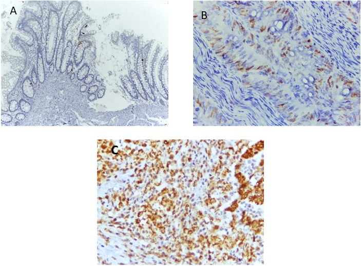
Immunohistochemistry for cyclin D1 with different pattern A. Normal colon (no staining) (x200), B. well differentiating colon cancer (no staining), C. colon cancer (low nuclear expression), D. a case of colon cancer with high nuclear expression of Cyclin D1 (b,c,d x400 HPF).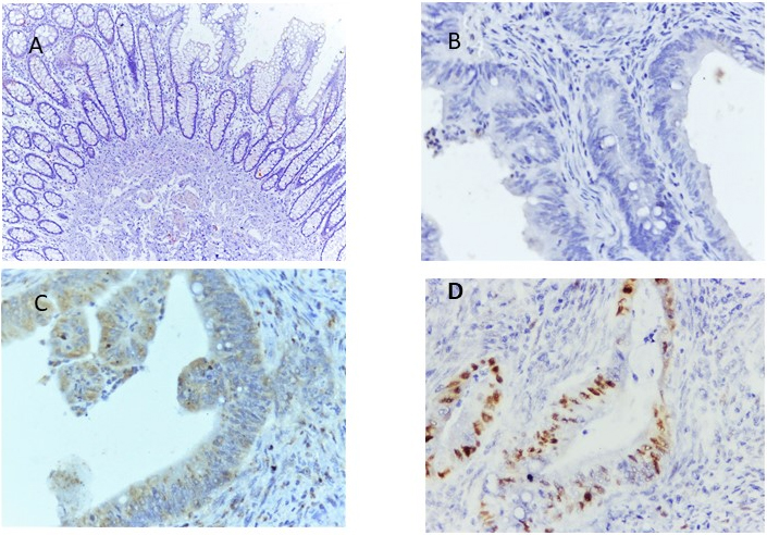
ROC curve demonstrating the validity of miRNA 29-b in the diagnosis of colon cancer.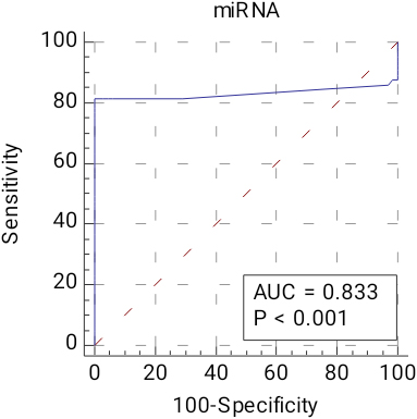
ETV4 and miRNA29b have a strong negative connection, as seen by the scatter dot.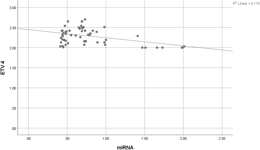
Association between cyclin D1 and miRNA29b.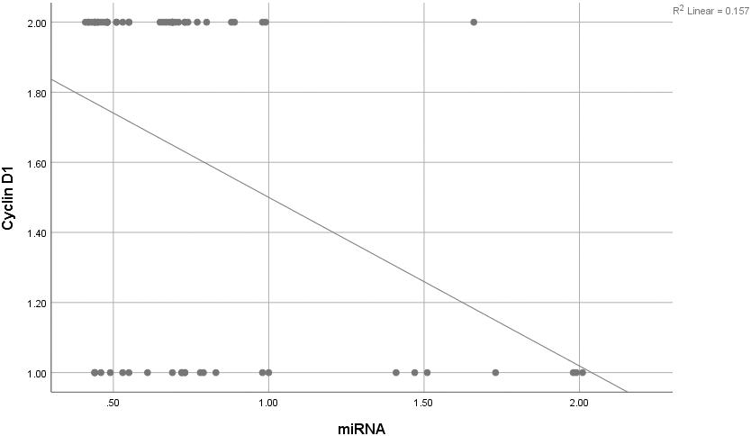
In this study, 65 patients with primary colon cancer were included; 39 males (60%) and 26 females (40%), with mean age
MiR-29b was significantly down-regulated (
MiR29b and ETV4 miRNA expressions had a significant negative correlation (
Immunohistochemical expression of ETV4 (Table 4 and Fig. 1) showed statistically significant associations with high grade, LVI, LNR, TB, LVI, TILs, and advanced TNM stage.
The relationship between the immunohistochemistry of cyclin D1 with the clinicopathologic characteristics of the studied cases is presented in Table 4 and Fig. 2. The data reveal a statistically significant connection between the expression of cyclin D1 and indicators of progression in the form of LVI, LNR, TB, and TNM stages (
Diagnostic role of cyclin D1 and ETV4 in colon cancer (Table 5): High cyclin D1 can diagnose colon cancer with 61.5% sensitivity, 96.9% specificity, 95.2% PPV71.6% NPV, and overall accuracy of 79.2%. High ETV can diagnose colon cancer with 93.9% sensitivity, 55.4% specificity, 67.8% PPV90% NPV, and overall accuracy of 74.6%
In colon cancer, the mean relative expression of miRNA-29b and ETV4 compared to control were (0.581
As shown in Table 6 and Fig. 3, the best miRNA 29-b cutoff for colorectal carcinoma diagnosis is 0.965, with an area under the curve of 0.833, a sensitivity of 81.5%, specificity of 93.8%, a positive predictive value of 98.3%, a negative predictive value of 83.6%, and accuracy of 87.7%.
Discussion
The screening methods for CRC are not easy to be used routinely. That is why many patients are undiagnosed tile the progressive stage, which affects the outcome of these patients. Colon cancer is a heterogeneous disease, and lines of therapy should be personalized. Furthermore, the increasing incidence of drug resistance among the colon cancer population supports the increasing need to find markers that help diagnose cases in the early stage and new targeted molecules [22].
The expression of miRNAs showed marked deregulation in many cancer types [23]. Their roles in various biological processes, such as cell differentiation and proliferation, have been studied [2]. Many gene expressions are under miRNAs control, post-transcriptionally, by interacting with the 3’UTR of specific target mRNAs [24]. It is hypothesized that miRNAs control cancer cell invasion and metastasis by suppressing the expression of their targets [25]. Although many researchers have studied the role of miR-29b in colon cancer, its role is still uncertain, and studying its association with ETV4 and cyclin D1 is still essential.
MiR-29b suppresses tumors by controlling oncogenes, such as T-cell leukemia/lymphoma 1 (TCL1), (Mcl-1), B-Myb, and CDC42 [26]. A previous study on CRC cell lines found a link between ETV4 expression and miR 29b [17].
First, our RT-qPCR results revealed that miR-29b expression was lower in colon cancer tissues than in nearby normal tissues. This finding was consistent with a previous study that reported decreased expression of miR-29b in colon cancer cell lines and tissue samples. They also demonstrated that miR-29b inhibits tumor growth and metastasis in colorectal cancer by down-regulating Tiam1 expression and inhibiting epithelial-mesenchymal transition [2].
Additionally, Basati et al. showed that colon cancer patients had considerably lower serum levels of miR-194 and miR-29b than control volunteers. Furthermore, the advanced TNM stages had an inverse relationship with the blood levels of the miRNAs in the patients. These findings imply that the effects of miR-29b may vary greatly depending on the target in each type of cancer and may be tumor-specific [27]. Another study found that miR-29b is typically down-regulated in colon cancer tissue, indicating that miR-29b may play a tumor-suppressive role in the emergence of colon cancer. It has been shown that miR-29b can act as a tumor suppressor to stop cancer cells from proliferating [28].
The current study reported that miR-29b expression was inversely related to the degree of TNM staging. More importantly, decreased miR-29b was associated with higher incidence of LNM, higher grade, and advanced stage in colon cancer patients.
In terms of ETV4 expression, it was detected that it is overexpressed in colon cancer. This finding was consistent with Fonseca et al. [29] and Lee et al. [30], who discovered that overexpression of ETV4 promoted colon cancer cell proliferation.
Previous experimental studies reported that knockdown of ETV4 inhibit the proliferative and invasive capacities of malignant CRC cells. Moreover, targeting of ETV4 by a certain molecule affects the downstream targets with inhibition of the ability of cancer cells to migrate and represents a future work to create a novel therapeutic strategy to treat metastatic prostate and breast cancers [30, 31].
Zeng et al. demonstrated that ETV4 regulate cell cycle genes and Wnt/catenin signalling, which was associated with an aggressive type of gastrointestinal stromal tumor [32]. Additionally, ETV4 has been linked to matrix metalloproteinase overexpression in colorectal cancer, with subsequent potentiation of invasion and cell proliferation [33]. This explains the significant correlation between ETV4 expression, LVI invasion, and metastasis capacity.
Leng et al. [17] discovered that ETV4 was a target gene of miR-29b in colon cancer cell lines. They also stated that the increased miR-29b may down-regulate ETV4 expression, and these results are consistent with the negative association between miR-29b and ETV4 miRNA expressions. These findings are intriguing because they could illuminate new mechanisms underlying miR-29b’s anti-cancer function.
Previous investigations indicated that cyclin D1 over-expression was not seen in normal mucosa, and only 15% of cases of adenoma exhibited it [21]. Similar findings were found in the current investigation since cyclin D1 expression was only present in 3.1% of the normal colonic tissue next to the tumor, strengthening its involvement in the early detection of colon cancer and supporting its contribution to the carcinogenic process.
It was noted that the overexpression of cyclin D1 was substantially correlated with grade, stage, and lymphatic vascular invasion in our study, which is consistent with prior research by Palaiologos et al. [34], who reported that no correlation between cyclin D1 and either stage or grade [21]. Thus, the etiology and metastasis of CRC are significantly influenced by the up regulation of cyclin D1. This may be explained by its regulation of the cell cycle, which disrupts the normal cell cycle and, if overexpressed, promotes the growth and spread of cancer [35].
A reverse relationship between cyclin D1 and miR-29b was found in this study. This makes sense because miR-29 controls the expression of cyclin D1, and the inhibition of the latter eliminates the cyclic pattern of cyclin D1 expression [36].
As far as the authors know, no previous studies investigated the cyclin D1 association with miR-29b in colon cancer.
The oncogenic effect of cyclin D1 occurs by controlling different substrates as Rb, the loss of Rb is consequent event for cyclin D1 mutation or amplification. Many therapeutic agents induce cyclin D1 degradation; cyclin D1 degradation by knockdown of USP2, a deubiquitylating enzyme that specifically targets cyclin D1found to inhibit the proliferation of cancer cells [37].
Although targeting cyclin D1 is difficult because it does not possess enzymatic activity, its catalytic partners CDK4/CDK6 can be targeted. As requisite functional partner kinases of cyclin D1, suppression of CDK4/CDK6 activity successfully blocks cyclin D1-mediated cell cycle progression, making these protein kinases attractive therapeutic targets [38].
Conclusion
Colon cancer is associated with up-regulation of ETV4, cyclin D1 overexpression and a down-regulation of miR-29b-3. Additionally, miR-29b expression was markedly reduced with progression of the tumor stage. Therefore, miR-29b expression may serve as a diagnostic indicator for early detection of colon cancer.
Author contributions
Conception: Hala Mosaad, Hanim M Abdelnour and Mona Mostafa Ahmed.
Interpretation or analysis of data: Mostafa M. Elaidy and Ola M Elfarargy.
Preparation of the manuscript: Mona Mostafa Ahmed and Hanim M Abdelnour.
Revision for important intellectual content: Mostafa M. Elaidy and Ola M Elfarargy.
Supervision: Hala Mosaad and Mai Mohamed Abdelwahab.