
Abstract
BACKGROUND:
The Notch signaling regulates numerous cell growth, differentiation, and death. However, the expression pattern of its ligand Delta-like 4 (DLL4) in tumors is still uncertain.
OBJECTIVE:
In the present study, we examined DLL4 expression in colorectal cancer as well as assessed its role as a prognostic indicator in the present study.
METHODS:
DLL4 expression was examined by immunohistochemistry in 289 surgically resected specimens of colorectal cancer and adjacent normal tissues. The relationship between DLL4 expression and clinicopathological characteristics was analyzed. The association of DLL4 expression with the patients’ overall survival rate was assessed by Kaplan-Meier and Cox proportional-hazards regression.
RESULTS:
Increased DLL4 level was detected in colorectal cancer compared with that of normal tissues. Elevated DLL4 level in colorectal cancer was associated with increased body mass index of patients. Moreover, increased DLL4 level was also found to be correlated with tumor invasion, metastases and unfavorable clinical outcom of patients.
CONCLUSIONS:
DLL4 level is increased in colorectal cancer, especially in patients with increased body mass index, indicating potential involvement of obesity-related tumorigenesis and development. It might also serve as a novel molecular marker to predicate outcome of patients.
Introduction
Colorectal cancer (CRC) is one the most common and malignant tumor globally, accounting for roughly 1.36 million new cases and 694,000 deaths per year [1, 2]. In addition, the incidence rates of CRC are rapidly increasing in Eastern Asia, including China, Japan, and South Korea [3, 4]. This type of unfavorable trend is thought to be, at least partly, due to an increase of obesity rate [5, 6, 7]. Despite recent advances in early screening, surgical resection technique and adjuvant therapy, tumor recurrence and mortality rate is still unfavorable among patients with CRC [8, 9]. Since molecular pathogenesis is heterogeneous in CRC, molecules involved in cancer progression and metastasis may help to identify patients at high risk of relapse and predict their prognosis as well as response to certain therapy [10, 11].
The Notch signaling regulates numerous cell growth, differentiation, and death during normal embryogenesis and in the adult [12, 13]. The Notch system consists of four transmembrane receptors Notch 1–4 and five ligands including Jagged 1, Jagged 2, Delta-like-1, 3 and 4 [14, 15]. The binding of Notch ligand of receptor initiates a series of proteolytic cleavages and finally activates signaling transduction to regulate cell fate decisions [16, 17]. Among Notch ligands, DLL4 has raised extensive interests since it is required for normal vascular development and is closely associated with tumor carcinogenesis [18, 19, 20]. It has been reported that DLL4 expression was elevated in several kinds of tumors such as pancreatic ductal adenocarcinoma, clear cell renal cell carcinoma, and nasopharyngeal carcinoma [21, 22, 23]. However, till now, the potential function of DLL4 in human CRC is still uncertain. Recent studies have proved that the impact of DLL4-induced Notch signaling is to restrain the angiogenic response triggered by vascular endothelial growth factor (VEGF) in the regulation of angiogenesis [24, 25]. As adipose tissue in obese patients is also an endocrine organ that can secrete various adipokines including VEGF, and induce proinflammatory impact to promote vascular injury [26, 27, 28, 29, 30]. Considering obesity has been confirmed to be a risk factor in carcinogenesis of CRC and other human malignancies [31, 32, 33, 35, 35]. Whether DLL4 is involved in CRC tumorigenesis and progression is still to be determined.
In present study, we investigated the expression of DLL4 in clinical CRC specimens and evaluated its association with body mass index (BMI) and clinical outcome of patients.
Patients and methods
Study cohort and clinical specimens
Colorectal carcinoma specimens and adjacent noncancerous tissues were collected from 289 sequential patients diagnosed with CRC (surgery was performed from January 2009 to December 2010 at the First Affiliated Hospital of Xi’an Jiaotong University, and Fourth Military Medical University, Xi’an, China). Patients meeting the following criteria were excluded: received neoadjuvant chemotherapy before surgery; diagnosed with additional cancers; suffered from serious systemic complications. Clinicopathologic information such as gender, age, tumor location and size, differentiation status, depth of invasion, lymph node and distant metastasis, and TNM stage were collected at enrollment. The patients were followed-up every 3 months to update their disease condition, life quality and survival status. All tissue specimens were obtained within 10 min after surgical removal and fixed in 10% formalin and embedded in paraffin for histological sections. Informed consent was obtained from all patients participated in the study. Approval was obtained for the use of all human tissue from the local research ethics committee (2019G-136).
Assessment of BMI and tumor location
To ensure the consistency of BMI values, the weight (kilograms) and height (meters) of the patients were measured and recorded at uniform time points relative to surgery by trained staff. BMI was categorized according to the World Health Organization (WHO) classification for Asian populations as normal weight (18.5 kg/m
Immunohistochemistry
Immunohistochemistry was performed using the avidin-biotin-peroxidase method on all CRC tissue specimens. All histological sections were deparaffinized in xylene and dehydrated through a gradient concentration of alcohol before endogenous peroxidase activity was blocked with 0.5% H
Statistical analysis
The Mann Whitney test was used to evaluate associations between two continuous and categorical variables. Kruskal Wallis test was used to compare two or more categorical variables of equal or different sample sizes. Survival analyses included disease-free survival and overall survival. Disease-free survival rate was the percentage of individuals in the treatment group who were likely to be free of the signs and symptoms of a disease after a specified duration of time. Overall survival rate referred to the percentage of people in a group who were alive after a length of time. Patients were censored in survival analyses according to the date last seen by a doctor. Survival curves for the entire study population were generated using Kaplan-Meier method, and differences in disease-free survival (DFS) and overall survival (OS) were evaluated by log-rank test. For multivariate survival models, a Cox regression analysis was used to identify independent prognostic factors. All statistical analyses were carried out using SPSS Statistics (version 22.0). The two-sided
Results
DLL4 immunohistochemical staining in CRC
DLL4 expression was examined by immunohistochemistry in all the 289 cases of surgically resected specimens of CRC and adjacent normal tissues. Immunohistochemistry staining of DLL4 was mainly located at the cytoplasm and cell membrane of cell (Fig. 1). Positive staining of DLL4 was detected in 221 samples of CRC. While only 38 cases of normal control tissues showed positive staining of DLL4. The positive rate of DLL4 in CRC, 77% (221 out of 289), was significantly increased (
DLL4 immunohistochemistry of colorectal cancer tissues. A. Strong positive DLL4 staining; B. Moderate positive DLL4 staining; C. Weak positive DLL4 staining; D. Negative DLL4 staining.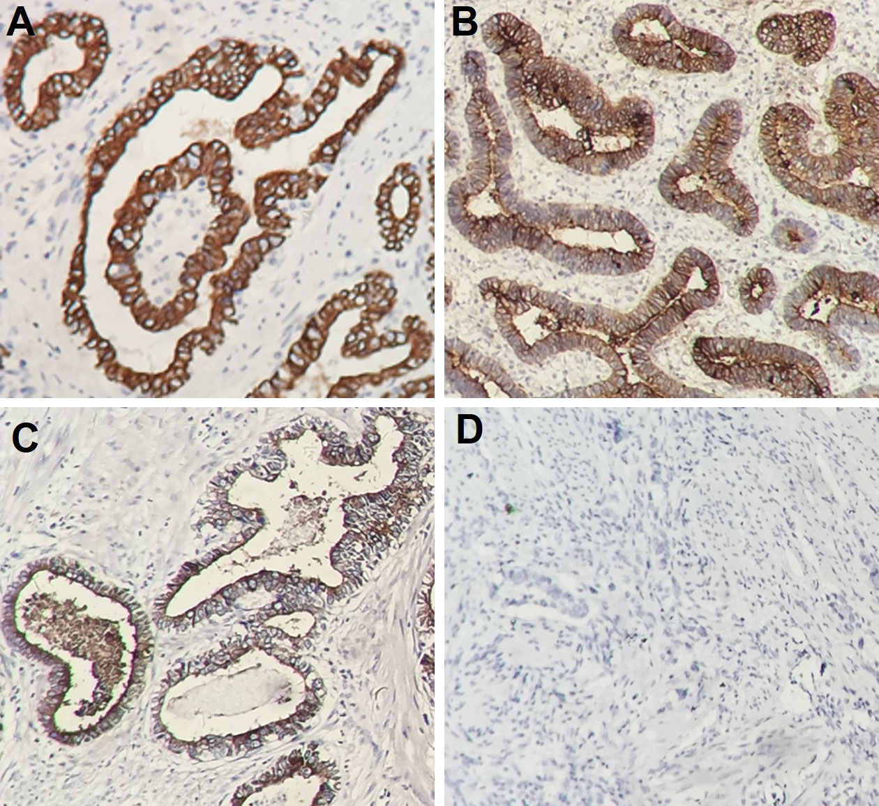
As DLL4 expression was found to be increased in CRC compared with that in normal tissue. We further analyzed the association of DLL4 level with clinicopathological characteristics of tumor in order to explore its potential biological function. Results indicated that DLL4 expression in CRC was significantly related to differentiation status, depth of invasion, BMI, lymph node metastasis and TNM stage (
Statistical results of DLL4 immunohistochemical staining
Statistical results of DLL4 immunohistochemical staining
Association of DLL4 and clinical factors with disease-free survival
Association of DLL4 and clinical factors with overall survival
Kaplan-Meier curves for disease-free survival of colorectal cancer patients categorised by DLL4 expression level.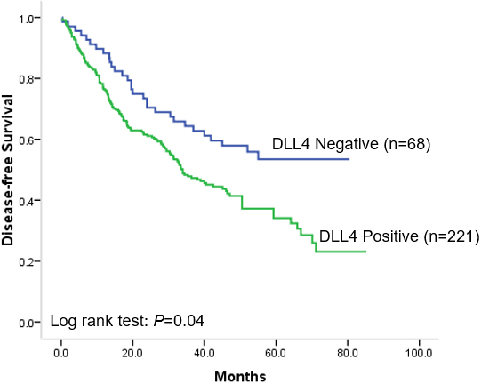
Kaplan-Meier curves for overall survival of colorectal cancer patients categorised by DLL4 expression level.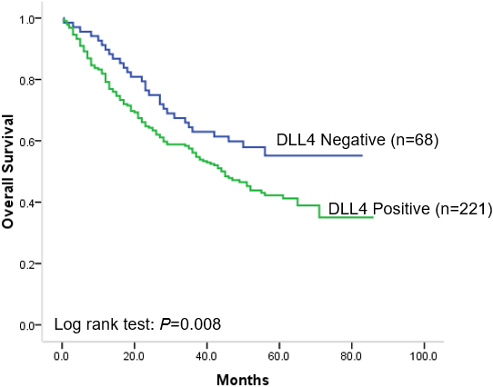
Median follow-up time of all recruited patients was 51 months (95% CI: 39–63 months). Among the 289 CRC patients, clinical recurrences or death from cancer occurred in 166 patients (57.4%). And 154 patients (53.3%) died until the last follow-up. Thus, disease-free survival (DFS) rate and overall survival (OS) rate were 42.6% and 46.7% respectively. Kaplan-Meier postoperative survival curves were drew to evaluate the association of survival with DLL4 expression. Results showed that high DLL4 expression was significantly associated with poor DFS (
Discussion
CRC remains one of the major health issues worldwide due to its high incidence and mortality rate [2]. Nowadays, clinicians divide patients into groups according to tumor invasion, lymph node metastasis and distant metastasis and then give them corresponding treatment based on the clinical stage. However, it often fails to predict clinical outcome for a single patient in intermediate groups due to the heterogeneity and complexion of tumor [11, 36]. Therefore, clinicians still cannot make a distinction between node-positive patients of low recurrence risk and node-negative patients of high risk, which leads to implementation of debilitating and ineffective adjuvant chemotherapy frequently. With the rapid development of molecular medicine, we have become increasingly aware of the interconnections between molecular pathogenesis, prognosis, and therapy response. A classical diagnosed cancer could be sorted into several subtypes based on the combined analysis of gene expression profiles, which is promising for the identification of patients at high risk of recurrence and having good response to certain therapy regimens [37]. Thus, it is of great importance to find effective molecular prognostic markers for individualized cancer management.
In the present study, we investigated the expression of DLL4 in CRC and evaluated its association with clinical outcome of patients. Results indicated that DLL4 expression was remarkably increased in neoplastic epithelial cells of CRC compared with adjacent normal colorectal tissue from the same individual. According to the evaluation of staining, positive DLL4 was more frequently to be detected in tumors from patients with increased BMI, suggesting that DLL4 might be involved in obesity-related tumor carcinogenesis and progression. As recent investigations also revealed that increased BMI is associated with response of immunotherapy in progressive malignancies, DLL4 might also be a potential molecular marker for immunotherapy sensitivity [38]. In addition, increased DLL4 expression was positively correlated with deep invasion of tumor (
The Notch signaling is a conserved ligand-receptor pathway which modulates cell fate. The cancer cell expresses high levels of DLL4 could activate Notch signal on the adjacent epithelial cell. Therefore, over-expressed DLL4 by the tumor cell could facilitate tumor cell proliferation and promote tumor growth. Consistently, DLL4 suppression was found to inhibit tumor growth in a variety of murine tumor xenograft models such as lung cancer. For the role of DLL4 in CRC, previous investigations found that increased DLL4 expression mainly localized at endothelial cells adjacent to tumor cells [39]. Our results indicated that CRC epithelial cell also expressed increased DLL4. In consistent with our study, several investigations also found increased DLL4 expression pattern in CRC. However, the sample size of these investigations was limited and its association with tumor progression was still contradictive [40, 41]. In our study, a large size group of patients who underwent curative CRC resection was included. DLL4 expression was evaluated by immunohistochemistry. This method is relatively common, uncomplicated and cheap, and operative specimen can be achieved before the implantation of adjuvant chemotherapy, making it applicable for routine DLL4 detection and clinical decision making.
Conclusion
In conclusion, our study provides convincing evidence that expression of DLL4 was elevated in CRC, and DLL4 was an independent prognostic marker for those patients besides TNM stage. The findings may have significant clinical implications, helping doctors to identify CRC patients at high risk of relapse and make more rational chemotherapy decisions.
Footnotes
Acknowledgments
The present study is supported by grants from the National Natural Science Foundation of China (Nos. 81201927, 81672460 and 82173337), Innovation Talent Promotion Plan of Shaanxi (Nos. 2017KJXX-07) and Key Research and Development Program of Shaanxi Province (Nos. 2019SF-012). The funders had no role in the design of the study, the collection, analysis, or interpretation of the data, the writing of the manuscript, or the decision to submit the manuscript for publication. There are no potential conflicts of interest to disclose.
Author contributions
Conception: Dake Chu.
Interpretation or analysis of data: Zixi Zhang, Xiao Li, Gai Li, He Qiu, Jingyi Yang, Xin Bu, Shaojun Zhu.
Preparation of the manuscript: Xiaowen Guo, Yan Lu, Min Jiao, Xue Chen, Chengxue Dang, Weizhong Wang.
Revision for important intellectual content: Dake Chu.
Supervision: Dake Chu.