
Abstract
OBJECTIVE:
We performed differential gene screening for lymphatic metastasis of hypopharyngeal carcinoma by gene sequencing. We also aimed to investigate the expression and clinicopathological significance of the screened gene in hypopharyngeal carcinoma lymphatic metastasis.
METHODS:
The clinicopathological characteristics of 98 patients with hypopharyngeal carcinoma were collected to make survival analysis by Kaplan-Meier & log-rank test. Six cases of tumor tissues from patients with or without lymphatic metastasis were used for gene sequencing of differentially expressed genes. The most frequently differently expressed genes were validated by RT-PCR and Western blot in another 20 patients diagnosed for hypopharyngeal carcinoma. A total of 70 cases of hypopharyngeal carcinoma tumor tissues and normal tissues were investigated to examine the immunohistochemical expression and to explore the prognostic value by Kaplan-Meier & log-rank test and Cox’s test.
RESULTS:
Lymphatic metastasis has been proved to cause a reduction in postoperative survival of patients with hypopharyngeal carcinoma. The results of gene sequencing analysis showed that Raf-1 was a differentially expressed gene in lymphatic metastasis of hypopharyngeal carcinoma. Moreover, the expression of Raf-1 was significantly up-regulated in tumor tissues of lymphatic metastasis patients compared to non-lymphatic metastasis tumor tissues and normal tissues. Meanwhile, Raf-1 had been verified to be an independent risk factor affecting the prognosis of hypopharyngeal carcinoma.
CONCLUSIONS:
For the first time, we investigated Raf-1 as an independent prognostic risk factor of lymphatic metastasis in hypopharyngeal carcinoma. It suggests that Raf-1 may serve as an important potential biomarker in preventing and diagnosing lymphatic metastasis in patients with hypopharyngeal carcinoma and improving the prognosis of patients.
Introduction
Hypopharyngeal carcinoma is a highly malignant head and neck tumors. According to the location of tumor, hypopharyngeal carcinoma can be divided into three types: pyriform fossa, posterior pharyngeal and postcricoid carcinoma [25]. Hypopharyngeal carcinoma accounts for 0.8
Relevant studies had confirmed that cervical lymph node metastasis of hypopharyngeal carcinoma is an important factor affecting prognosis [35]. It was reported that the 3-year and 5-year survival rates of patients with cervical lymph node metastasis were 40.0% and 28.1%, respectively, while the 3-year and 5-year survival rates of patients without cervical lymph node metastasis were 96.2% and 77.3%, respectively [2, 21]. Patients with metastatic lymph nodes larger than 3 cm in diameter and with multiple positive lymph nodes had a worse prognosis than those with single positive lymph nodes [23]. A multivariate analysis of lymph node factors affecting the prognosis of hypopharyngeal carcinoma showed that diameter, number of metastatic lymph nodes and extra invasion were independent risk factors for the prognosis of hypopharyngeal carcinoma [9]. Therefore, by observing and analyzing the factors related to cervical lymph node metastasis, exploring the influence of the factors related to lymph node metastasis on prognosis, and formulating relevant treatment strategies to improve the therapeutic effect of hypopharyngeal carcinoma are urgent problems to be solved clinically.
Gene sequencing expression profiling had been extensively applied in clinical research and bioinformatics analysis as the major biomarkers tool for a long time. Gene sequencing allows the largescale identification and comprehensive analysis of gene expression profiles and mutation mapping. This technology represents a mature, efficient and high-throughput transcriptomic analysis approach for validation of biomarkers, discovery of gene functions, and development of new targeted drugs [36]. In recent years, series of biomarkers and gene expression profile-based predictive models had already been established for human disease diagnosis, prognostic evaluation, and clinical treatment guidance [7, 31, 33, 37]. Meanwhile, microarray analysis of head and neck tumors had identified specific gene expression signatures associated with expression changes in head and neck squamous cell carcinoma tissue compared to normal tissue [28]. However, gene sequencing of lymphatic metastasis of hypopharyngeal carcinoma has not been reported and relevant experiments are required to be examine.
The Raf family of protein kinases, which is one class of Raf effectors, contains A-Raf, B-Raf and C-Raf. C-Raf (which is more often called Raf-1), and participates in Raf/MEK/ERK signaling pathway. It transmits extracellular signals into the nucleus through cell membrane receptors, thereby mediating the expression of intracellular specific proteins and participating in the regulation of cell proliferation, differentiation, apoptosis, autophagy and other functions [13, 19, 26]. EGFR/Raf/MEK/ERK signaling pathway which mediated by epidermal growth factor receptor (EGFR) is a classic anti-tumor therapy approach [30]. However, the expression and mechanism of Raf-1 in lymphatic metastasis of tumors, especially hypopharyngeal carcinoma, have not been explored yet.
The current study, we verified that lymphatic metastasis seriously influenced the postoperative survival of hypopharyngeal carcinoma. Through gene sequencing, we screened out Raf-1 as a differentially expressed gene affecting lymph node metastasis of hypopharyngeal carcinoma, and further examined the expression and prognostic value of Raf-1 in hypopharyngeal carcinoma. It indicates that Raf-1 can be used as a potential biomarker for diagnosis of lymphatic metastasis of hypopharyngeal carcinoma, evaluation of patients’ prognosis, and guidance of clinical targeted therapy.
Methods
Patients and samples
The patients were screened from the First Affiliated Hospital of Chongqing Medical University between 2012 and 2019 for laryngopharyngectomy. The inclusion criteria were as follows: (1) Pathological diagnosis was hypopharyngeal squamous cell carcinoma. (2) Preoperative cervical ultrasound, enhanced CT or MRI examination was performed. (3) The data of admission and follow-up were complete. (4) The patient underwent laryngopharyngectomy for the first time. Exclusion criteria are as follows: (1) Preoperative radiotherapy, chemotherapy, or other anti-tumor targeted therapy was received. (2) Combined with other types of carcinoma. (3) Distant metastasis of the tumor was detected. Finally, the clinical data of 98 patients with hypopharyngeal carcinoma who met the standard were collected and counted. Informed consent for surgery and related experiment was obtained from each subject before any treatment. The experiment was reviewed and approved by the ethics committee of Chongqing Medical University, and conformed with The Code of Ethics of the World Medical Association. The fresh tumor tissues and adjacent normal tissues required were taken from the isolated tissues resected during the operation. The tissue composition, pathological stage, differentiation degree and lymphatic metastasis of the specimens were identified by two experienced pathologists. Some samples were treated with liquid nitrogen and stored at
Analysis of influence factors of hypopharyngeal carcinoma
According to the clinicopathological characteristics of patients with hypopharyngeal carcinoma collected previously (Table 1), survival curve was drawn by Kaplan-Meier & log-rank test to analyze the influence of each clinicopathological feature on the overall survival of hypopharyngeal carcinoma patients.
The clinicopathological features of patients with hypopharyngeal carcinoma
The clinicopathological features of patients with hypopharyngeal carcinoma
Total RNA was extracted from 3 cases tumor tissues of hypopharyngeal carcinoma patients with lymphatic metastasis and another 3 cases tumor tissues of patients without lymphatic metastasis. RNA was extracted using RNA extraction kit (TaKaRa, Japan) and was determined using denaturing gel electrophoresis to confirm that total RNA was not contaminated with DNA and the RNA was not degraded. The RNA samples which passed the quality test were reverse transcribed into cDNA. Random segments of DNA were broken up into a few hundred bases, with specific joints at each end. After denaturation of DNA segment into single strand, it combined with joint primer on sequencing channel to form bridge structure. Four fluorescent labeled dNTP were added for PCR amplification. Then, image could be read by the software. Illumina Custom software and Mev software was used for analysis of differential gene expression. The difference was statistically significant when FDR
Total-RNA extraction and RT-PCR
A total of 10 pairs of tumor tissues and corresponding adjacent normal tissues from hypopharyngeal carcinoma patients with lymphatic metastasis and another 10 pairs of tissues from patients without lymphatic metastasis were collected. Total RNA was extracted by using RNA extraction kit (TaKaRa, Japan), and was reverse transcribed into cDNA using PrimeScript RT reagent Kit (TaKaRa, Japan). GAPDH served as an internal reference for RNA integrity. The primer sequences designed by PrimerBank were as follows: Raf1-F: 5’-GGGAGCTTGGAAGACGATCAG- 3’, Raf1-R: 5’-ACACGGATAGTGTTGCTTGTC-3’, GAPDH-F: 5’-CAGCGACACCCACTCCTC-3’, GA- PDH-R: 5’-TGAGGTCCACCACCCTGT-3’. RT-PCR was performed in anannealing temperature of 55
Protein extraction and Western blotting analysis
Protein was extraction from 14 pairs of tumor tissues and corresponding adjacent normal tissues (including 7 lymphatic metastasis patients and another 7 non-lymphatic metastasis ones) by using total protein extraction kit (KeyGen BioTECH, China). The protein lysates were then loaded into 12% sodium dodecyl sulphate-polyacrylamide gels, and then transferred onto polyvinylidene difluoride (PVDF) membranes. After blocking for one hour, membranes were incubated with primary antibody and stored overnight at 4
Immunohistochemistry
Immunohistochemistry was performed on 4
The immunohistochemistry results were analyzed separately by two experienced pathologists and scored considering both the intensity of staining and the proportion of tumor cells with an unequivocal positive reaction. Slides which incubated with Phosphate buffer saline (PBS) instead of primary antibody were selected as negative controls. According to the staining results, the expression of Raf-1 was scored as either positive expression (stained brown, tumor cells
Analysis of prognostic risk factors of hypopharyngeal carcinoma
According to the immunohistochemistry results, Kaplan-Meier & log-rank test and COX’s univariate and multivariate analysis were used to examine the association between Raf-1 and other clinicopathological features, and to evaluate independent risk factors of prognosis in patients with hypopharyngeal carcinoma.
Statistical analysis
The data of patients were imported to SPSS 22.0. Kaplan-Meier & log-rank analysis and COX’s univariate and multivariate analysis were performed by SPSS to evaluate the prognostic factors. The expression of Raf-1 in immunohistochemistry was evaluated by chi-square test. One-Way ANOVA test was used for analysis involving three group of samples in RT-PCR. Protein expression level in Western blotting was analyzed by Image J. The results of gene sequencing were analyzed by Illumina Custom software and Mev software. For all tests, differences were considered statistically significant when
Results
Lymphatic metastasis affected the survival of hypopharyngeal carcinoma patients
Through the Kaplan-Meier & log-rank test of the clinicopathological features of hypopharyngeal carcinoma patients, lymphatic metastasis has been verified to be an important factor affecting the survival rate of patients with hypopharyngeal carcinoma. Compared to hypopharyngeal carcinoma patients without lymphatic metastasis, survival rates in patients with lymphatic metastasis were significantly reduced (15% reduction in 3-year overall survival and 25% reduction in 5-year overall survival), and the difference was statistically significant (
Correlations between lymphatic metastasis and overall survival in patients with hypopharyngeal carcinoma. The horizontal axis represents survival time (months), and the vertical axis represents the percent survival. The upper line shows the survival rate of patients without lymphatic metastasis, while the bottom line represents for patients with lymphatic metastasis.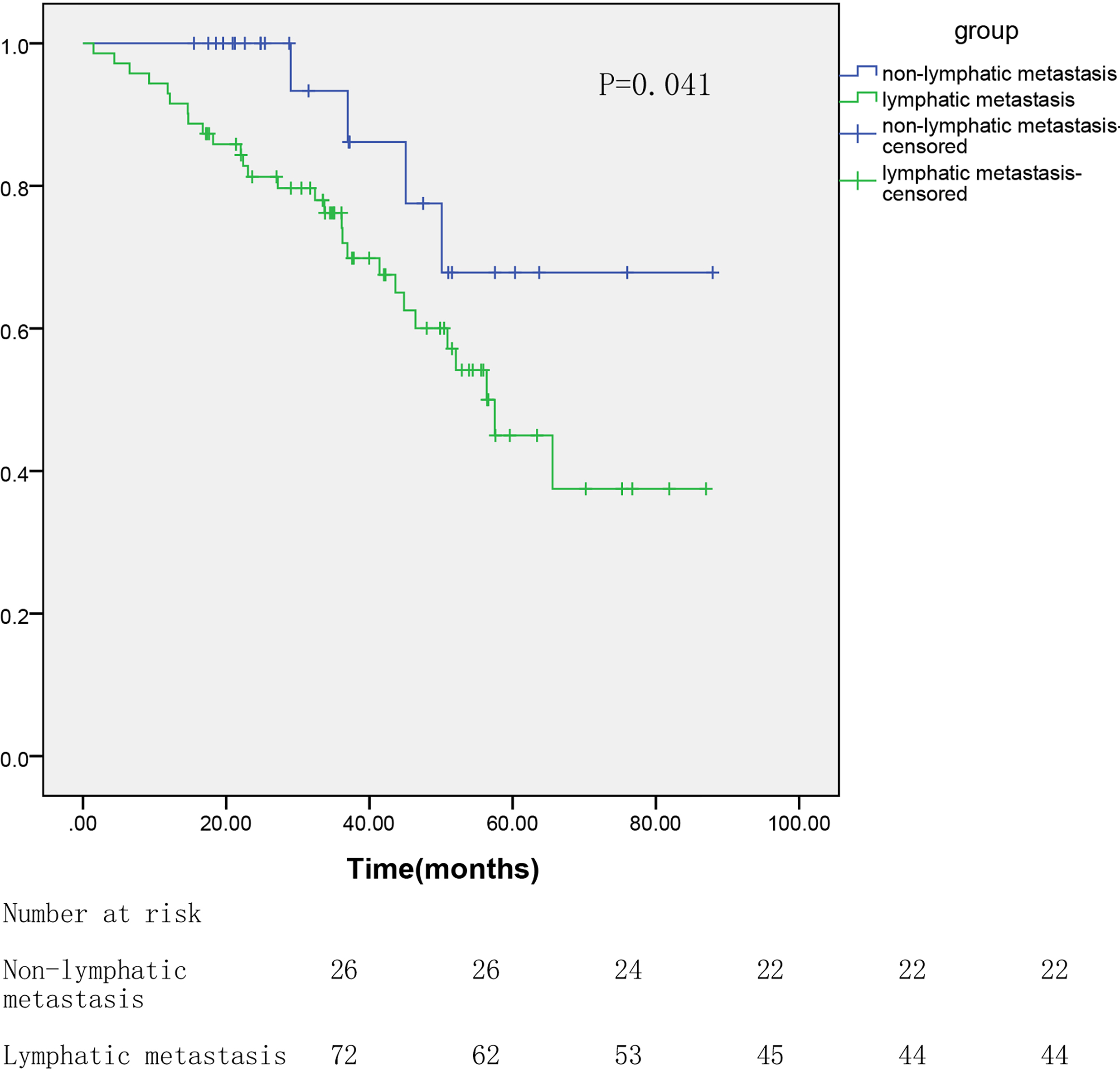
Heatmap of gene sequencing shows differentially expressed genes. For each gene (row), red indicated a higher expression and green a lower one relative to the average level of expression of that gene across the samples (columns). The labels at the bottom are the number of the samples. N: Tumor tissue without lymphatic metastasis. L: Tumor tissue with lymphatic metastasis.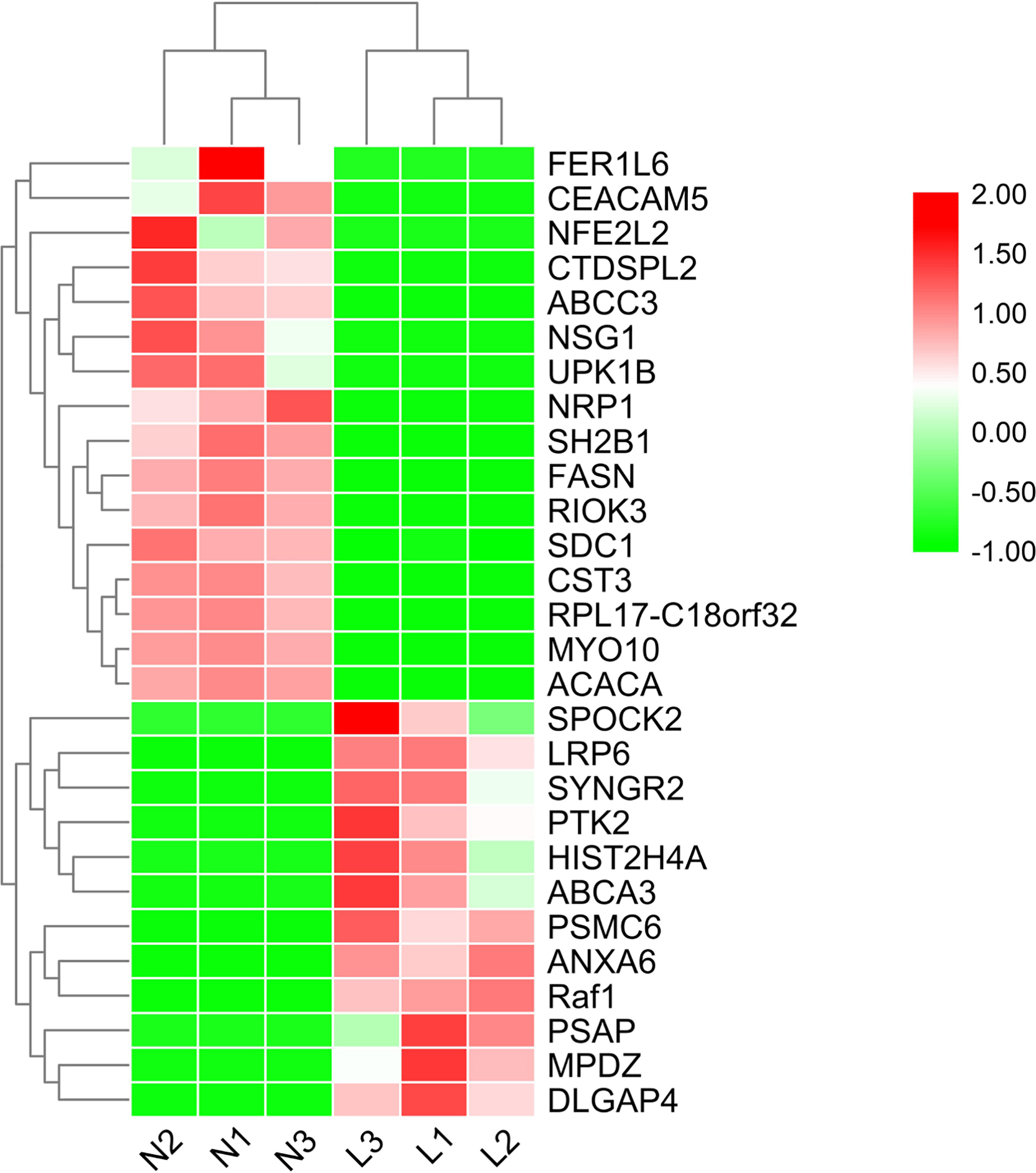
Scatter diagram of the differentially expressed genes: the horizontal axis represents gene expression in the non-lymphatic metastasis group, while the vertical axis represents the expression in the lymphatic metastasis group. Red points indicated higher expression genes (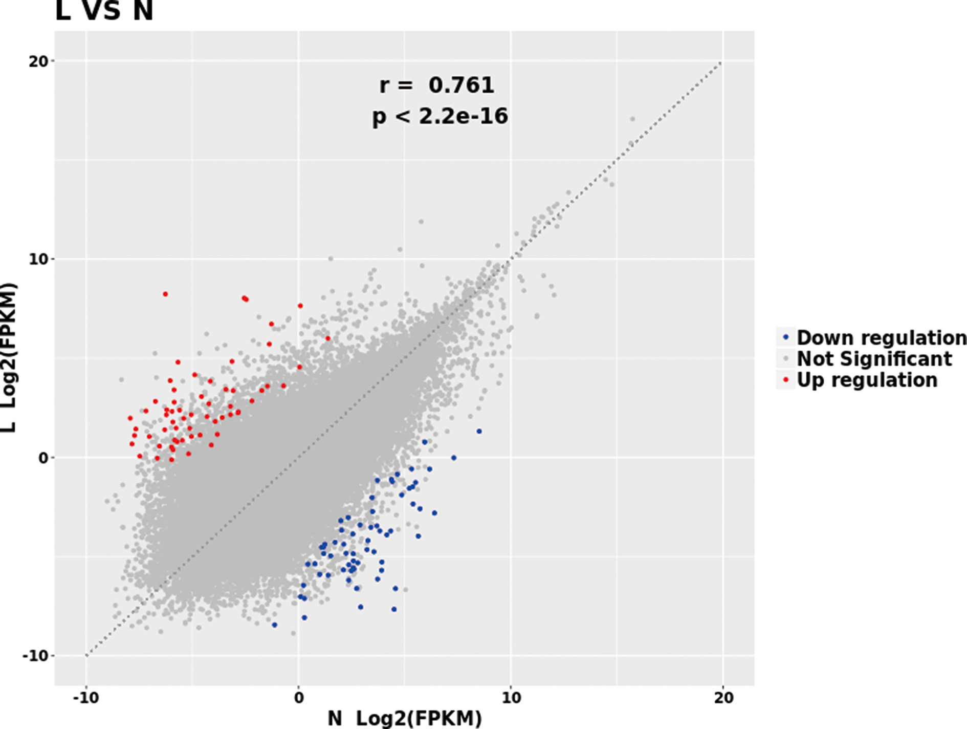
Gene sequencing of the differential gene expression was performed on 3 hypopharyngeal carcinoma tumor tissues of patients with lymphatic metastasis and another 3 cases of patients without lymphatic metastasis. Among the 126595 genes analyzed, 419 genes showed statistically significant differences in the expression between lymphatic metastasis group and non-lymphatic metastasis group (
The mRNA expression of Raf-1 in lymphatic metastasis patients was significantly up-regulated
We expanded the sample cases to further verify the expression of the differentially expressed genes and to find appropriate biomarkers. A total of 20 pairs of tumor tissues (including 10 tissues from lymphatic metastasis patients and another 10 from non-lymphatic metastasis patients) and corresponding adjacent normal tissues were used for RT-PCR. Normal tissues served as a control group. As was presented in Fig. 4, the mRNA expression of Raf-1 in lymphatic metastasis group was significantly higher than that in the non-lymphatic metastasis group and the normal group, and the difference was statistically significant (
The mRNA expression level of Raf-1 in RT-PCR. The horizontal axis represents the different groups including lymphatic metastasis patients’ tumor tissues, non-lymphatic metastasis patients’ tumor tissues and normal tissues. The vertical axis represents the relative mRNA expression level of Raf-1. 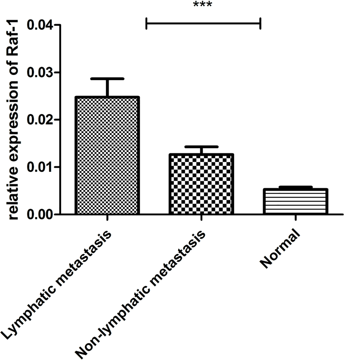
As shown in Fig. 5a, protein band signals reflected the protein expression level. The results were basically consistent with RT-PCR: the expression of Raf-1 in tumor tissues of lymphatic metastasis patients was significantly higher than other two groups. Protein expression levels were further evaluated by Image J (Fig. 5b), and the difference was statistically significant (
Identification of Raf-1 expression in immunohistochemistry
According to the pathologists’ score, the immunohistochemistry results were classified as positive and negative. The expression of Raf-1 in different groups was shown in Fig. 6. The expression of Raf-1 in tumor tissues of lymphatic metastasis patients showed strongly positive, while the expression in tumor tissues of non-lymphatic metastasis patients and normal tissues showed negative most of the time. More importantly, we found that the positive expression of Raf-1 was significantly up-regulated in tumor tissues of lymphatic metastasis patients (39/49, 79.6%) when compared to the tumor tissues of non-lymphatic metastasis patients (7/21, 33.3%) (Table 2).
Expression of Raf-1 protein level by Western blot assay. a. 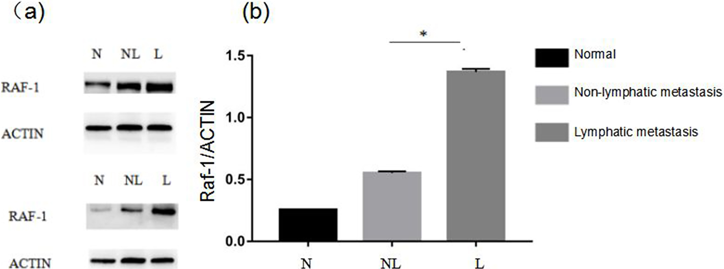
The immunohistochemical results of Raf-1 were as follows. A. Positive immunohistochemistry in tumor tissues from patients with lymphatic metastasis. B. Negative immunohistochemistry in tumor tissues from patients without lymphatic metastasis. C. Negative immunohistochemistry in normal tissues. D. Phosphate buffer saline (PBS) instead of primary antibody incubation was selected as negative control.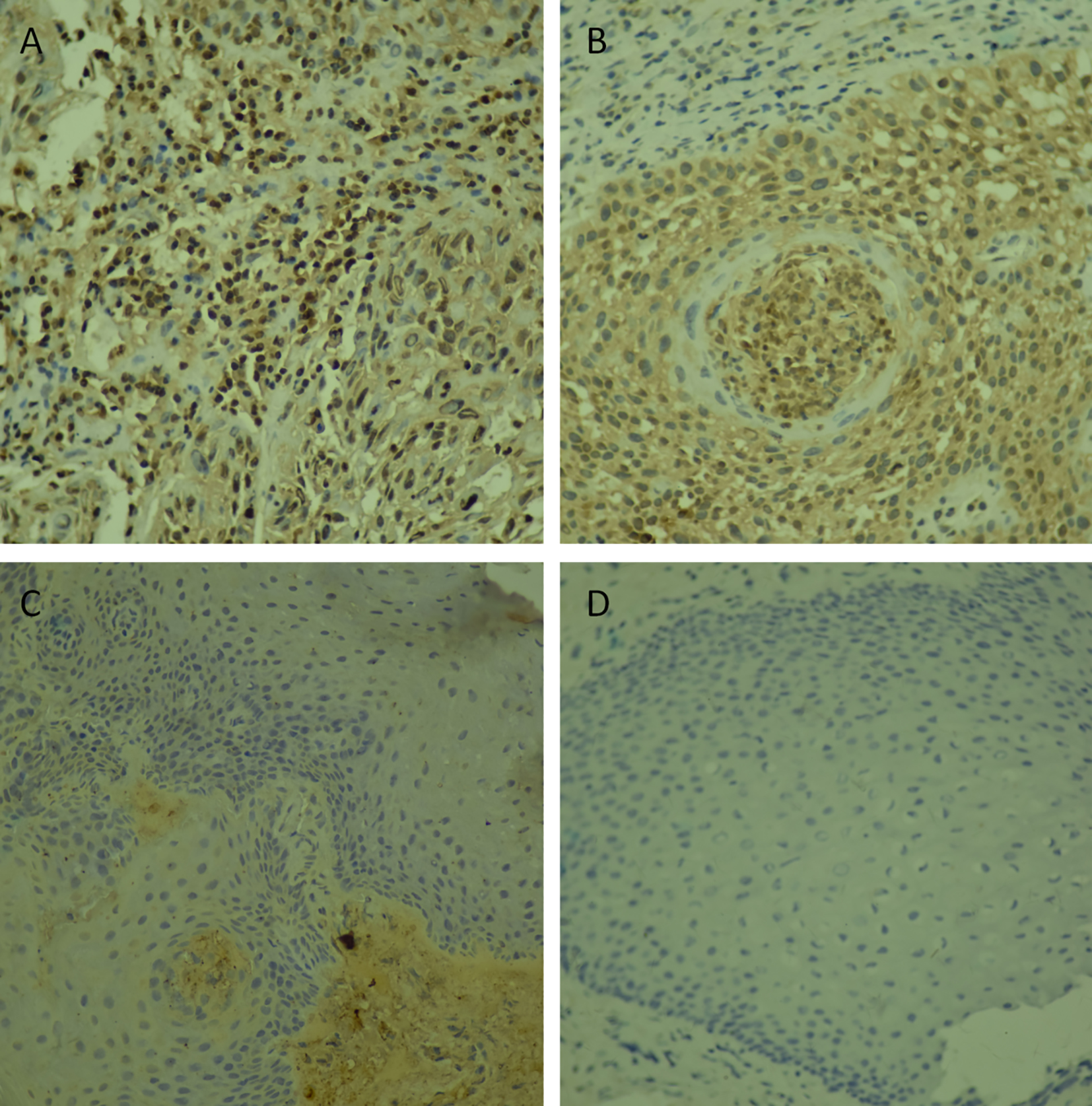
The immunohistochemical expression of Raf-1 in patients with lymphatic metastasis and patients without lymphatic metastasis
According to Kaplan-Meier & log-rank test and COX’s univariate and multivariate analysis, we found that lymphatic metastasis and expression of Raf-1 were both independent risk factors for prognosis in patients with hypopharyngeal carcinoma compared to other clinicopathological features (HR
Univariate and multivariate Cox regression analyses of overall survival in hypopharyngeal carcinoma patients
Univariate and multivariate Cox regression analyses of overall survival in hypopharyngeal carcinoma patients
Correlations between Raf-1 expression and overall survival in patients with hypopharyngeal carcinoma. The horizontal axis represents survival time (months), and the vertical axis represents the percent survival. The upper line shows the survival rate of patients with negative expression of Raf-1, while the bottom line represents for patients with positive expression of Raf-1.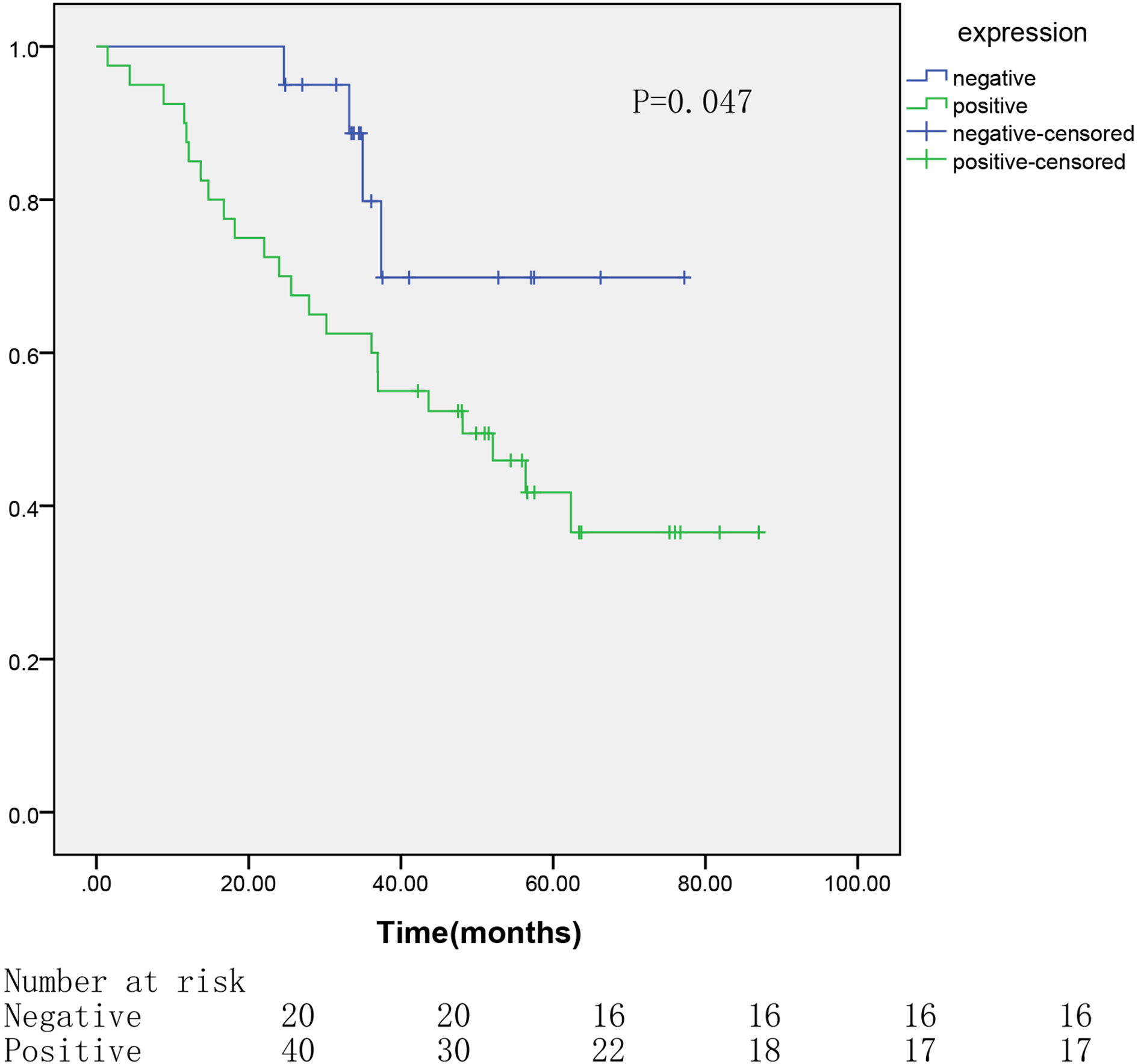
The anatomical structure of the hypopharynx is relatively special, which starts from below the hyoid extension line, and ends at the lower edge of the annular cartilage connected to the esophagus. This special structure makes it difficult for patients with hypopharyngeal carcinoma to be diagnosed in the early time, which leads to the advanced clinical stage, high frequency of lymphatic metastasis, and poor prognosis [32]. To improve the prognosis of patients, advancements had been achieved in the treatment of hypopharyngeal carcinoma including induction chemotherapy, concurrent chemoradiotherapy and salvage total laryngectomy. Especially for advanced hypopharyngeal carcinoma patients with lymph node metastasis, according to the situation of preoperative induction chemotherapy, the decision to supplement concurrent chemoradiotherapy or radical surgery has been recognized as a comprehensive treatment. In addition, postoperative adjuvant chemoradiotherapy is also a necessary treatment currently [18]. However, there was no significant change in survival rate due to improvements in treatment [16]. It suggests there may be other risk factors that seriously affect the patient’s prognosis.
Lymph node metastasis is the most common form of tumor metastasis. Infiltrating tumor cells penetrate the lymphatics and exfoliate, then tumor cells are carried along with the lymph into the confluence of lymph nodes, and grow the same tumor from this center. It has been reported that the cervical lymph node metastasis rate of patients with hypopharyngeal carcinoma is 60%
Genome-wide analysis using gene sequencing is a new gene function research technique developed in recent years, which can be used to rapidly detect gene differential expression, genome expression profile, DNA sequence polymorphism, pathogenic gene or disease-related biomarkers on a large scale. A number of large-scale microarray data have been set for public reference, such as CMAP, CEBS, DrugMatrix and TG-GATEs [14, 17, 34]. In this study, we used gene sequencing to extensively search for differentially expressed genes in lymphatic metastasis of hypopharyngeal carcinoma. Through preliminary screening, 419 differentially expressed genes were identified (including 200 high expression genes and 219 low expression genes). Then, RT-PCR will be used for further screening to select biomarkers associated with lymphatic metastasis in hypopharyngeal carcinoma.
Among the differentially expressed genes screened from gene sequencing, we selected the top 10 genes with the greatest differences in expression (including Raf-1, ANXA6, FASN, CST3, etc.), then the expression differences were examined by RT-PCR. According to the results, the expression of Raf-1 in tumor tissues of lymphatic metastasis patients was much higher than tumor tissues of non-lymphatic metastasis patients and normal tissues. Compared to other differentially expressed genes, expression differences of Raf-1 was the most significant. Further more, we further verified the expression of Raf-1 by Western blotting and immunohistochemistry. The experimental results were basically consistent with RT-PCR. Moreover, the analysis of Kaplan-Meier & log-rank test and COX’s test revealed that expression of Raf-1 is an independent risk factor affecting prognosis of hypopharyngeal carcinoma. It suggests that Raf-1 may serve as an important potential biomarker in preventing and diagnosing lymphatic metastasis in patients with hypopharyngeal carcinoma and improving the prognosis of patients.
Raf-1 serves as an important member of Raf/MEK/ ERK signaling pathway, has been reported to be up-regulated in several cancers and correlated with tumor progression [3, 4, 27]. However, to our knowledge, whether the expression of Raf-1 is involved in the development and progression of lymphatic metastasis remains unclear now. Thus, the purpose of this study is to screen for differentially expressed genes in lymphatic metastasis of hypopharyngeal carcinoma and evaluate the expression and prognostic value of the genes. Finally, it may contribute to find out the prevention biomarkers of lymphatic metastasis and the targets of clinical treatment. Meanwhile, the functional mechanism of Raf-1 in lymphatic metastasis also remains further intensive study.
Footnotes
Acknowledgments
We appreciate Department of Pathology, The First Affiliated Hospital of Chongqing Medical University for technical assistance and accurate diagnosis. This study was supported by the Natural Science Foundation of Key Projects of Chongqing China (Grant Number. cstc2012jjB10015).
Conflict of interest
We declare no conflict of interest, the authors contributed equally to this work.