
Abstract
BACKGROUND:
The PANDAR, a novel identified long non-coding RNA, is previously reported to function as oncogene in various cancers including breast cancer. the study aims to explore the role of lncRNA PANDAR for cell proliferation and invasion of breast cancer, and its underlying mechanism.
METHODS:
The expression of lncRNA PANDAR in 65 pairs of breast cancer tissues and adjacent normal tissues was detected by quantitative Real-time polymerase chain reaction (qRT-PCR) assay. The association between lncRNA PANDAR expression and clinical factors of breast cancer was analyzed. Cell proliferation, cell colony formation and cell invasion assays were performed to detect the effects of lncRNA PANDAR expression tumor proliferation and invasion abilities. The western blot analysis was also performed to detected the EMT related makers expression of E-cadherin, Vimentin, MMP2 and MMP9.
RESULTS:
We demonstrated that lncRNA PANDAR expression was higher in breast cancer tissues and cells compared with adjacent normal tissues and the normal mammary epithelial cell line, respectively. Higher lncRNA PANDAR expression positively associated with lymph node metastasis and advanced clinical stage in patients. In vitro, we demonstrated that knockdown of lncRNA PANDAR significantly suppressed cell proliferation, cell colony formation and cell invasion ability in breast cells. Furthermore, we verified that knockdown of lncRNA PANDAR dramatically inhibited cell epithelial-mesenchymal transition (EMT) pathway by downregulating Vimentin, MMP2 and MMP9 expression, but upregulating E-cadherin expression in breast cancer.
CONCLUSIONS:
Our results proved that PANDAR may serve as potential target of breast cancer treatment.
Introduction
Breast cancer is common diagnosed malignancy in female worldwide [1]. Although significant developments including surgery and chemotherapy have been improved in recent years, tumor relapse and metastasis for patients at advanced stage cause large mortality [2, 3]. To identify effective therapeutic targets for breast cancer is urgently needed.
Long non-coding RNAs (lncRNAs) are non-coding RNA transcripts that are longer than 200 nucleotides [4]. Recent studies have documented that lncRNAs act as crucial regulators of numerous physiological and pathological processes in various cancers including cell proliferation, cell differentiation, cell apoptosis, cell invasion and tumor metastasis [5, 6, 7]. LncRNA PANDAR (promoter of CDKN1A antisense DNA damage-activated RNA) displays a major role for regulating tumor progression in some cancers. For example, low expression of long noncoding RNA PANDAR predicts a poor prognosis of non-small cell lung cancer and affects cell apoptosis by regulating Bcl-2 [8]. Up-regulation of long non-coding RNA PANDAR is associated with poor prognosis and promotes tumorigenesis in bladder cancer [9]. LncRNA PANDAR were reported to be higher in breast cancer and promote cell proliferation by impacting p16 (INK4A) expression [10]. Moreover, lncRNA PANDAR expression associated with tumor epithelial-mesenchymal transition (EMT) process. Such as, suppression of lncRNA PANDAR impaired migration and invasion capacity in vitro partly by affecting EMT process in cholangiocarcinoma [11]. However, the role of PANDAR in breast cancer progression still needed to be explored.
In the study, we demonstrated that PANDAR was higher in breast cancer tissues and cells. Knockdown of PANDAR inhibited cell proliferation, cell invasion and EMT signaling by regulating EMT-related markers expression. Therefore, our results proved that PANDAR may serve as potential target of breast cancer treatment.
The association of clinicopathological factors with lncRNA PANDAR expression in 65 breast cancer patients
The association of clinicopathological factors with lncRNA PANDAR expression in 65 breast cancer patients
ER
Patient tissue samples
A total of 65 paired of breast cancer tissue samples and adjacent normal tissue samples were obtained from patients who received the surgical resection between January 2012 and November 2014 at Sichuan Academy of Medical Sciences and Sichuan Provincial People’s Hospital. None of patients had received any therapy before surgery. Consent form was signed from all patients in the study. The study was approval from the Research Ethics Committee of Sichuan Academy of Medical Sciences and Sichuan Provincial People’s Hospital. After surgery, tissue samples were immediately stored in liquid nitrogen. The clinicopathological characteristics of patients were shown in Table 1.
Cell lines culture
Breast cancer cell lines including MCF-7, MDA-MB-231, MDA-MB-436, SK-BR-3 cells and the normal mammary epithelial cell line (MCF-10A) were purchased from the Shanghai Institute of Biochemistry and Cell Biology (Shanghai, China). All of cells were cultured in Dulbecco’s modified Eagle’s medium (DMEM) supplemented with 10% fetal bovine serum (FBS) (Gibco; Thermo Fisher Scientific, Inc., Waltham, MA, USA) at 37
LncRNA PANDAR expression is significantly upregulated in breast cancer tissues. (A) LncRNA PANDAR expression was detected in 65 paired of breast cancer tissue sample and adjacent normal tissue samples by using qRT-PCR. GAPDH expression was used as an internal control. (B) The association of lncRNA PANDAR expression with lymph node metastasis was shown. (C) The association of lncRNA PANDAR expression with clinical stage was shown. (D) LncRNA PANDAR expression was detected in MCF-7, MDA-MB-231, MDA-MB-436, SK-BR-3 cells and the normal mammary epithelial cell line (MCF-10A) by using qRT-PCR. GAPDH expression was used as an internal control. *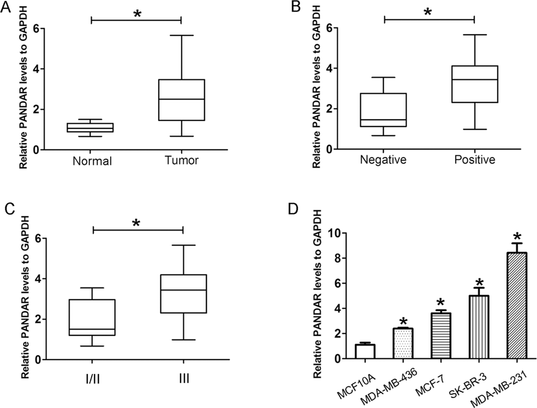
Knockdown of lncRNA PANDAR reduces cell invasion ability in breast cancer cells. (A)–(B) The relative expression of lncRNA PANDAR were detected by qRT-PCR after SK-BR-3 and MDA-MB-231 cells were transfected with siRNA#1, siRNA#2 or si-NC. Relative PANDAR levels to GAPDH. (C)–(D) Cell invasion number was calculated after SK-BR-3 and MDA-MB-231 cells were transfected with siRNA#1, siRNA#2 or si-NC. *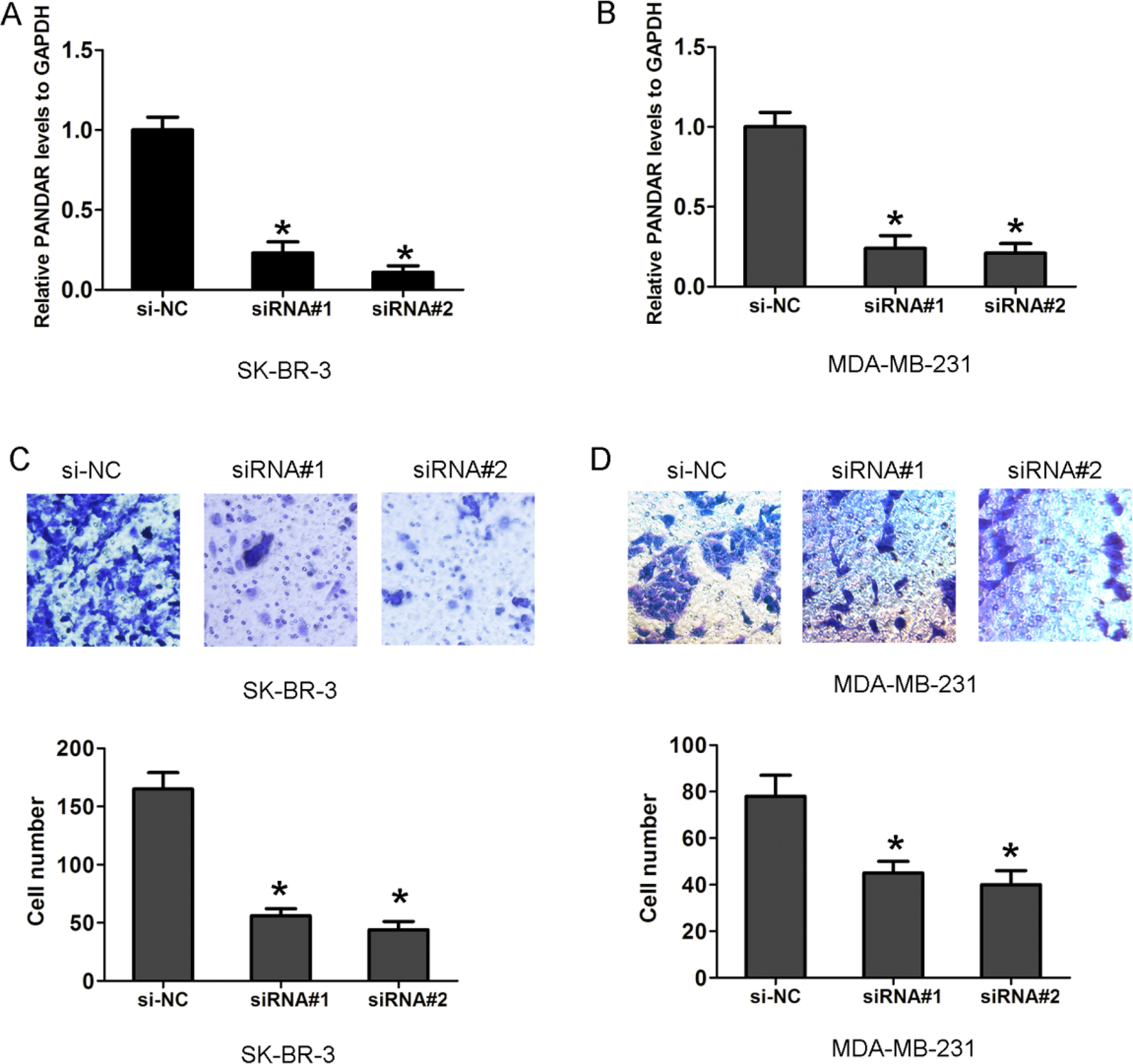
Knockdown of lncRNA reduces cell proliferation ability in breast cancer cells. (A)–(B) CCK8 assay were performed after SK-BR-3 and MDA-MB-231 cells were transfected with siRNA#1, siRNA#2 or si-NC. (C)–(D) Cell colony forming number was calculated after SK-BR-3 and MDA-MB-231 cells were transfected with siRNA#1, siRNA#2 or si-NC by performing cell colony forming assay. The cells were cultured until most colonies comprised more than 50 cells, *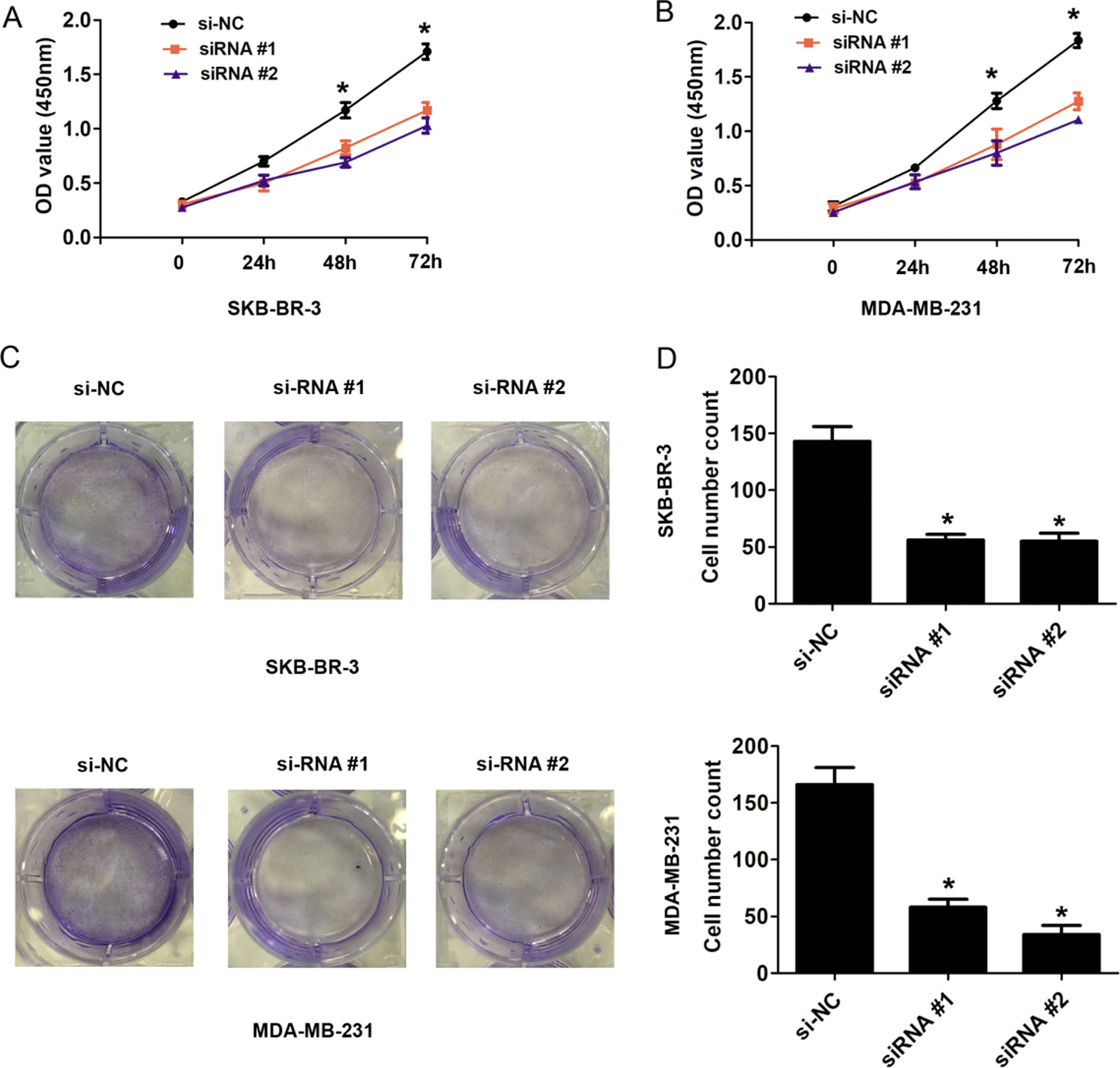
Knockdown of lncRNA affects cell EMT pathway by regulating EMT pathway related protein expression levels in breast cancer cells. (A) The expression of E-cadherin, Vimentin, MMP2 and MMP9 was detected and quantitative analysis using western blot analysis after SK-BR-3 cells were transfected with siRNA#1, siRNA#2 or si-NC. (B) The expression of E-cadherin, Vimentin, MMP2 and MMP9 was detected and quantitative analysis using western blot analysis after MDA-MB-231 cells were transfected with siRNA#1, siRNA#2 or si-NC. *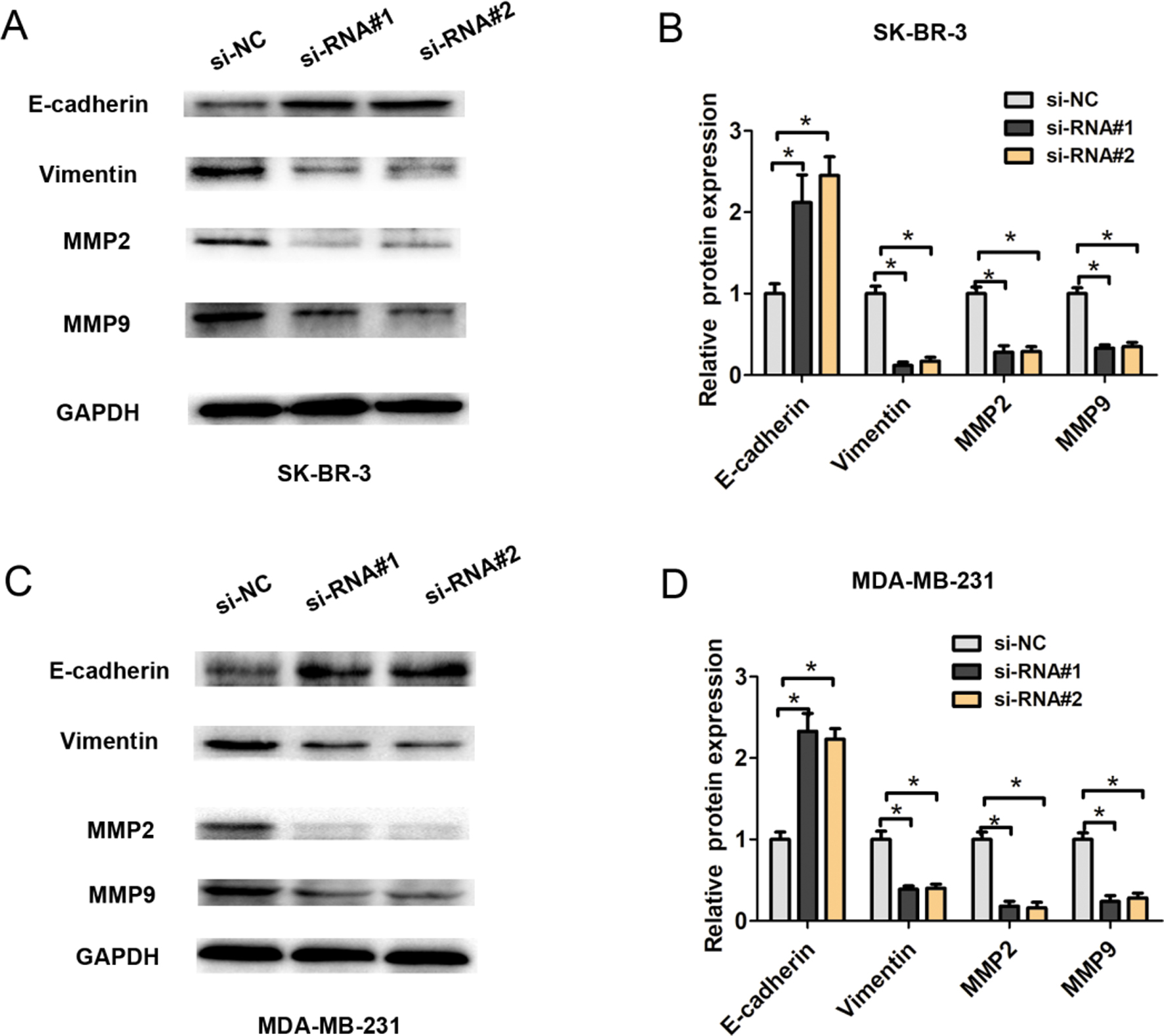
Total RNA was extracted from tissues and cells by TRIzol reagent (Invitrogen, Carlsbad, CA, USA) and RNA was reversed into cDNA through reverse transcription kits (Takara Bio, Tokyo, Japan). The relative mRNA expression was determined using qRT-PCR by SYBR
Cell transfection
The two siRNAs targeting PANDAR were designed and constructed based on PANDAR sequence by Gene- chem Biotech (Shanghai, China). The target sequence for PANDARwere as follow: si-NC: 5’-GTAGT-GAG ATGCAGATGATAA-3’, siRNA#1: 5’-GCAATCTAC AACCTGTCTT-3’, siRNA#2: 5’-TTTCGAACGGAA CAGAGACUUAUACAGATT-3’.Cell transfection was performed using Lipofectamine 2000 (Invitrogen, Car- lsbad, CA, USA) reagent according to manufacturer’s protocol. Cells were harvested after 48 h after transfection.
Transwell invasion assay
Cell invasion were detected using transwell chambers (Corning Incorporated, Corning, NY, USA) and pre-coated with Matrigel (BD Biosciences, San Jose, CA, USA). 1
Cell proliferation assay
Cell proliferation ability was evaluated by Cell Counting Kit-8 (CCK8) (Dojindo, Kumamoto, Japan) assays in breast cancer cells according to the manufacturer’s protocol. Briefly, 3000 transfected cells/well were plated in 96-well plates in triplicate. Cells were cultured for 0 until 72 h. Then, 10
Cell colony forming assay
Cells were treated with si-NC, siRNA #1 or siRNA #2 and then cultured for 7 days at 37
Western blot analysis
Total protein was extracted from transfected breast cancer cells after 48 h using radioimmunoprecipitation assay (RAPA) lysis buffer (Santa Cruz Biotechnology, Inc., Dallas, TX, USA). Protein concentrations were detected using BCA protein assay reagent kit (Thermo Fisher Scientific, Inc., Carlsbad, CA, USA). The equal protein was separated on 10% SDS-polyacrylamide gels and transferred to PVDF membranes (EMD Millipore, Billerica, MA, USA). Then, the membranes were treated with antibodies including E-cadherin, Vimentin, MMP2, MMP9 and GAPDH overnight at 4
Statistical analysis
Statistical analysis was performed using SPSS 19.0 (SPSS, Inc., Chicago, IL, USA). Data are presented as the mean
Results
LncRNA PANDAR expression is significantly upregulated in breast cancer tissues
The expression of lncRNA PANDAR was detected in 65 paired of breast cancer tissue samples and adjacent normal tissue samples by using qRT-PCR. GAPDH expression was used as an internal control. The results showed that lncRNA PANDAR expression were significantly upregulated compared with adjacent normal tissues (Fig. 1A,
Knockdown of lncRNA reduces cell invasion ability in breast cancer cells
In order to investigate the effects of lncRNA PANDAR on cell invasion, the two siRNAs targeting lncRNA PANDAR were used to downregulate the expression of lncRNA PANDAR in SK-BR-3 and MDA-MB-231 cells (Fig. 2A and B). The transwell invasion assays results showed that knockdown of lncRNA PANDAR in SK-BR-3 and MDA-MB-231 cells reduced cell invasion number compared with the si-NC groups (Fig. 2C and D). The results indicated that downregulation of lncRNA PANDAR suppressed the invasive ability of SK-BR-3 and MDA-MB-231 cells.
Knockdown of lncRNA inhibits cell proliferation ability in breast cancer cells
Furthermore, we explore the effects of lncRNA PANDAR expression on cell proliferation and cell colony forming ability, we transfected two siRNAs targeting lncRNA PANDAR into SK-BR-3 and MDA-MB-231 cells. The CCK8 cell proliferation assays results showed that knockdown of lncRNA PANDAR in SK-BR-3 and MDA-MB-231 cells suppresses cell capacity compared to the si-NC groups (Fig. 3A and B). In addition, the cell colony forming assays results showed that knockdown of lncRNA PANDAR in SK-BR-3 and MDA-MB-231 cells decreased cell colony forming number compared to the si-NC groups (Fig. 3C and D). These results indicated that downregulation of lncRNA PANDAR suppressed the cell proliferation ability of SK-BR-3 and MDA-MB-231 cells.
Knockdown of lncRNA affects cell EMT process by regulating EMT-related makers expression
As knockdown of lncRNA PANDAR could inhibit breast cancer cell invasion, we speculated that lncRNA PANDAR could affect cell EMT process in breast cancer. We selected the siRNA#1 and siRNA#2 for knockdown experiments due to higher efficiency for lncRNA PANDAR in SK-BR-3 and MDA-MB-231 cells. The western blot analysis was used to detect the expression of E-cadherin, Vimentin, MMP9 and MMP2 after knockdown of lncRNA PANDAR in SK-BR-3 and MDA-MB-231 cells. The results demonstrated that knockdown of lncRNA PANDAR significantly reduced the expression of Vimentin, MMP2 and MMP9 compared to si-NC group in SK-BR-3 and MDA-MB-231 cells, but upregulating the E-cadherin expression (Fig. 4A–D). Thus, we demonstrated that knockdown of lncRNA PANDAR may affect cell EMT pathway by regulating EMT-related markers expression in breast cancer cells.
Discussion
Accumulating evidence has verified that lncRNAs could play crucial roles in tumor proliferation, migration, invasion and metastasis in various malignances including breast cancer [12]. Recently, increasing studies about lncRNA PANDAR in cancer progression were reported. Increased expression of lncRNA PANDAR predicts a poor prognosis in gastric cancer [13]. Up-regulation of long non-coding RNA PANDAR is associated with poor prognosis and promotes tumorigenesis in bladder cancer [9]. The high expression of long non-coding RNA PANDAR indicates a poor prognosis for colorectal cancer and promotes metastasis by EMT pathway [14]. Upregulated long noncoding RNA PANDAR predicts an unfavorable prognosis and promotes tumorigenesis in cholangiocarcinoma [15]. Silencing of PANDAR exerted tumor suppressive effect via reducing cell proliferation, colony-forming ability, inducing cell cycle G0/G1 arrest and apoptosis in pancreatic ductal adenocarcinoma [10]. Thus, these results indicated that lncRNA PANDAR expression acted as regulator in tumor progression.
In the study, our results demonstrated that lncRNA PANDAR expression was significantly upregulated in breast cancer tissues compared to adjacent normal tissues. Statistical analysis results showed that higher PANDAR expression was significantly associated with lymph node metastasis and advanced TNM staging, which indicated that lncRNA PANDAR expression showed a significant clinical significance. CCK8 and cell colony formation assays showed that knockdown of lncRNA PANDAR suppressed cell proliferation in breast cancer. Furthermore, knockdown of lncRNA PANDAR reduces cell invasion ability and EMT pathway by downregulating Vimentin, MMP2 and MMP9 expression, but upregulating E-cadherin expression. In previous study, lncRNA PANDAR was reported to regulate the G1/S transition of breast cancer cells by suppressing p16 (INK4A) expression [10]. Suppression of lncRNA PANDAR impaired migration and invasion capacity in vitro partly by affecting EMT process in cholangiocarcinoma [11]. We speculated that lncRNA PANDAR may affect EMT process by regulating EMT-related makers expression. The underlying mechanism for lncRNA PANDAR affecting cell proliferation and EMT will be investigated in the future. Such as, lncRNA PANDAR whether regulates cell proliferation and invasion by affecting some signaling pathway including Wnt/
In conclusion, we showed that lncRNA PANDAR expression was upregulated in breast cancer tissues and cells. Furthermore, we demonstrated that lncRNA PANDAR knockdown inhibited cell proliferation, cell invasion ability and EMT signaling pathway in breast cancer. Thus, these results provide an important clinical and therapeutic value of lncRNA PANDAR for breast cancer.
Footnotes
Acknowledgments
This study was supported by The Science and Technology Project of The Health Planning Committee of Sichuan (17ZD021).
Conflict of interest
The authors declare that they have no conflict of interest.