
Abstract
OBJECTIVE:
To investigate the correlations of expression of vascular endothelial growth factor (VEGF) in gastric cancer tissues of patients and magnetic resonance imaging (MRI) features with clinical tumor-node-metastasis (TNM) staging and lymph node metastasis, and to analyze the diagnostic value of MRI features for preoperative TNM staging and lymph node metastasis of patients with gastric cancer, and the roles of VEGF in tumor development and metastasis.
METHODS:
A total of 120 gastric cancer patients treated in our hospital from May 2015 to July 2017 were selected as objects of study. The VEGF protein expressions in gastric cancer tissues of patients with different TNM staging and lymph node metastasis degrees were detected using immunohistochemical method, and the correlations of VEGF protein expression with TNM staging and lymph node metastasis were analyzed. Before operation, MRI was used to predict TNM staging and lymph node metastasis of all patients, and prediction results were compared with postoperative pathological diagnosis results. At the same time, the differences in lymph node apparent diffusion coefficient (ADC), long diameter and short diameter, relative ADC of primary lesion (rADCp) and relative ADC of muscle (rADCm) were compared and analyzed between lymph node metastasis group and non-lymph node metastasis group.
RESULTS:
The VEGF expression in patients with stage-N3 gastric cancer was about 7 times that in patients with stage-N0 gastric cancer, and it was increased with the increased degree of lymph node metastasis (
CONCLUSION:
The VEGF protein expression in gastric cancer tissues is positively correlated with TNM staging and lymph node metastasis in patients. The preoperative prediction results of MRI are well consistent with postoperative pathological results, and MRI features are correlated with lymph node metastasis in patients, which has an important guiding significance for the diagnosis and treatment of gastric cancer.
Introduction
Gastric cancer is one of the most common malignant tumors in clinical practice. At present, with the changes in people’s life style and eating habits, the incidence rate of gastric cancer has been increased year by year. According to the survey of International Agency for Research on Cancer in 2012, the morbidity and mortality rates of gastric cancer ranked fifth and third in malignant tumors, respectively, seriously threatening the life and health of patients [1, 2]. In China, the morbidity and mortality rates of gastric cancer account for about 50% globally, and China is a high-incidence country of gastric cancer, so clinical prevention and treatment are particularly important [3]. The most important biological characteristics of malignant tumors are invasion and metastasis of cancer cells, which are also important causes of death of patients. Similarly, whether lymph node metastasis or distant metastasis occurs in gastric cancer patients, and the infiltration depth of tumor cells are important factors affecting the prognosis and survival time of patients. Studies have shown that the tumor growth and metastasis is a very complex biological process, but nutrients are also needed to support such a process, among which the formation of tumor vessels plays an important role under the joint coordination of a number of anti-angiogenic factors and pro-angiogenic factors [4, 5]. Vascular endothelial growth factor (VEGF) is a kind of multi-functional cell regulatory factor that affects the formation and growth of tumor vessels, which is closely related to the tumor invasion, metastasis and clinicopathological prognosis [6]. Clinical data show that the accurate evaluation of tumor-node-metastasis (TNM) staging and lymph node metastasis in gastric cancer patients before operation has an important guiding significance for clinical diagnosis and treatment of gastric cancer. Magnetic resonance imaging (MRI) has advantages of non-invasiveness, high speed and no need of contrast injection, and it can be used to evaluate the tumor morphology more accurately with clear images, so it has been gradually applied in predicting the clinical staging and diagnosis of gastric cancer [7]. In this paper, therefore, the correlations of TNM staging and lymph node metastasis of gastric cancer patients with MRI features and VEGF expression were investigated, hoping to provide a reference for the clinical diagnosis and treatment of gastric cancer.
Materials and methods
General materials
A total of 120 gastric cancer patients treated in our hospital from May 2015 to July 2017 were selected as objects of study, including 78 males and 42 females aged 45–73 years old with an average of (59.3
Methods
Immunohistochemical method
All of the above patients were treated with radical gastrectomy for gastric cancer. All gastric cancer tissue specimens resected during operation were embedded in paraffin, and para-carcinoma normal tissues were used as controls. Tissues were sliced for comparative studies. The staining test was performed using rabbit anti-human VEGF monoclonal antibody (Product ID: ZA-0509, Jiangsu AbZyme Biotechnology Co., Ltd.) and LSAB kit (Diagnostic System Laboratories) strictly according to the instructions of kit. The above-mentioned rabbit anti-human VEGF monoclonal antibody needed to be diluted at 1:100, and the secondary antibody that played a role as biomarker needed to be diluted at 1:10000. After the staining test and color development using developing agent for 5 min, tissue specimens were re-stained via hematoxylin and sealed, and brown yellow-stained cells were positive. Finally, the proportion of area of positive gastric cancer cells in that of reference cells in para-carcinoma normal tissues was analyzed [8].
MRI examination method
All patients were fasted at 8–12 h before test, drank 300 mL warm water at 30 min before test, received intramuscular injection of 10 mg anisodamine for treatment, and then took 500 mL warm water orally, filling the patient’s gastrointestinal tract moderately. MRI was performed using 1.5T superconducting MRI scanner (Shenzhen BASDA Medical Apparatus Co., Ltd.), 8-channel phased array coil was used as the imaging coil, and coronal plane and cross section as the main scanning planes, and oblique coronal plane scan could be performed additionally if necessary. Specific scanning sequences are as follows: diffusion weighted imaging (DWI) (TE 74.9 ms, TR 1000 ms), T
Evaluation methods
TNM staging method
According to the infiltration depth and metastasis, TNM staging includes tumor infiltration depth (T), lymph node metastasis (N) and distant metastasis (M). Based on the different depth of tumor infiltration, tumors were subdivided as follows: tumor cells located in the mucous layer (Tis), tumor cells located in the submucous layer (T1), tumor cells infiltrating into the serosal layer or muscular layer (T2), tumor cells penetrating the serosal layer (T3), and tumor cells extending into the duodenum and esophagus, or invading into adjacent tissues (T4). At least 15 lymph nodes were taken from specimens for pathological analysis, and then tumors were subdivided as follows based on the lymph node metastasis: no lymph node metastasis (N0), regional metastasis of 1–6 lymph nodes (N1), regional metastasis of 7–15 lymph nodes (N2), and regional metastasis of more than 15 lymph nodes (N3). Based on the distant metastasis, tumors were subdivided as follows: no distant metastasis confirmed pathologically (M0), abdominal aortic, mesenteric and pancreatic lymph node metastases (M1) [9].
A 59-year-old male patient with stage-T3/N2/M0 gastric adenocarcinoma. 1: High signal in DWI. 2: Lesions are significantly enhanced in LAVA dynamic enhanced scan. 3: Lymph nodes show high signal in DWI.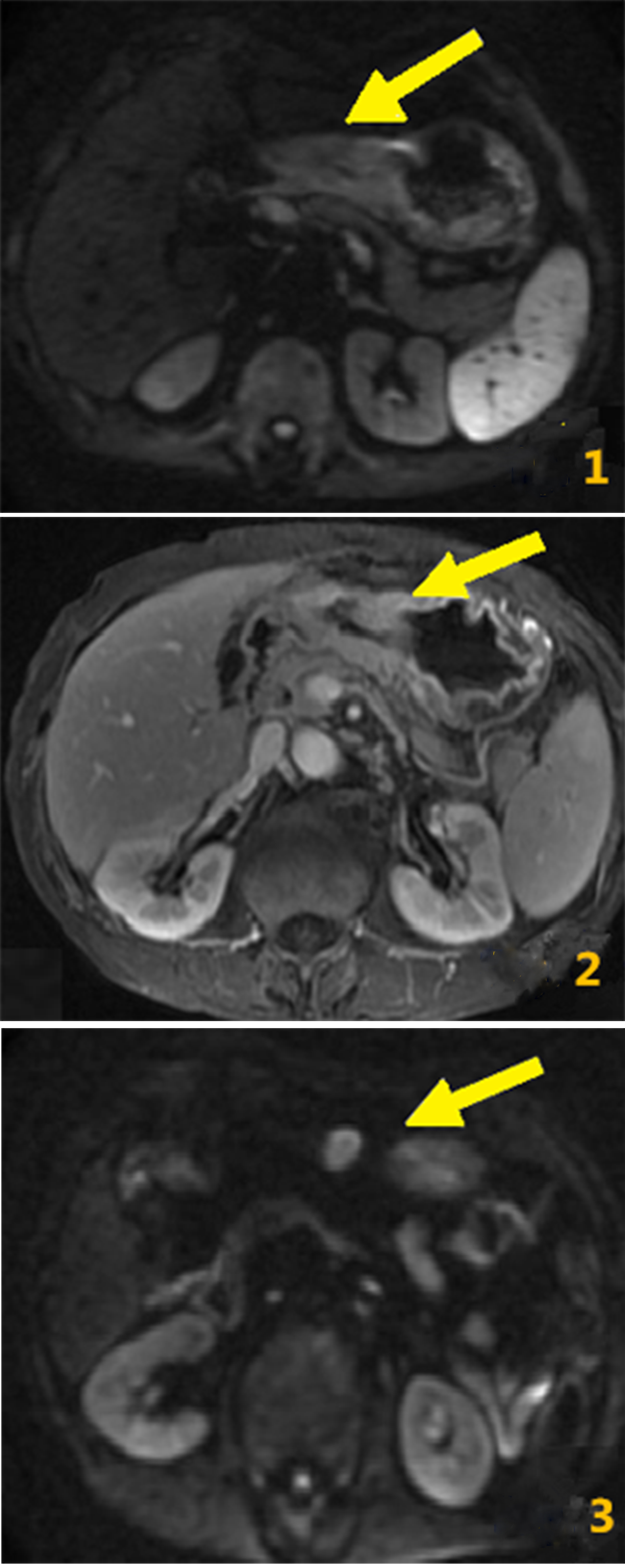
Analyses of VEGF expressions in gastric cancer tissues of patients with different TNM staging
MRI images of all the above patients were analyzed and processed by the same senior expert from the Department of Imaging of our hospital using Materialise Mimics 17.0 medical software. The pathological T staging of tumor was evaluated preoperatively, the apparent diffusion coefficient (ADC) of primary lesions was determined, and lymph nodes with a length of about 5 mm were selected as objects of study from the T
VEGF is expressed in the cytoplasm of cancer cells.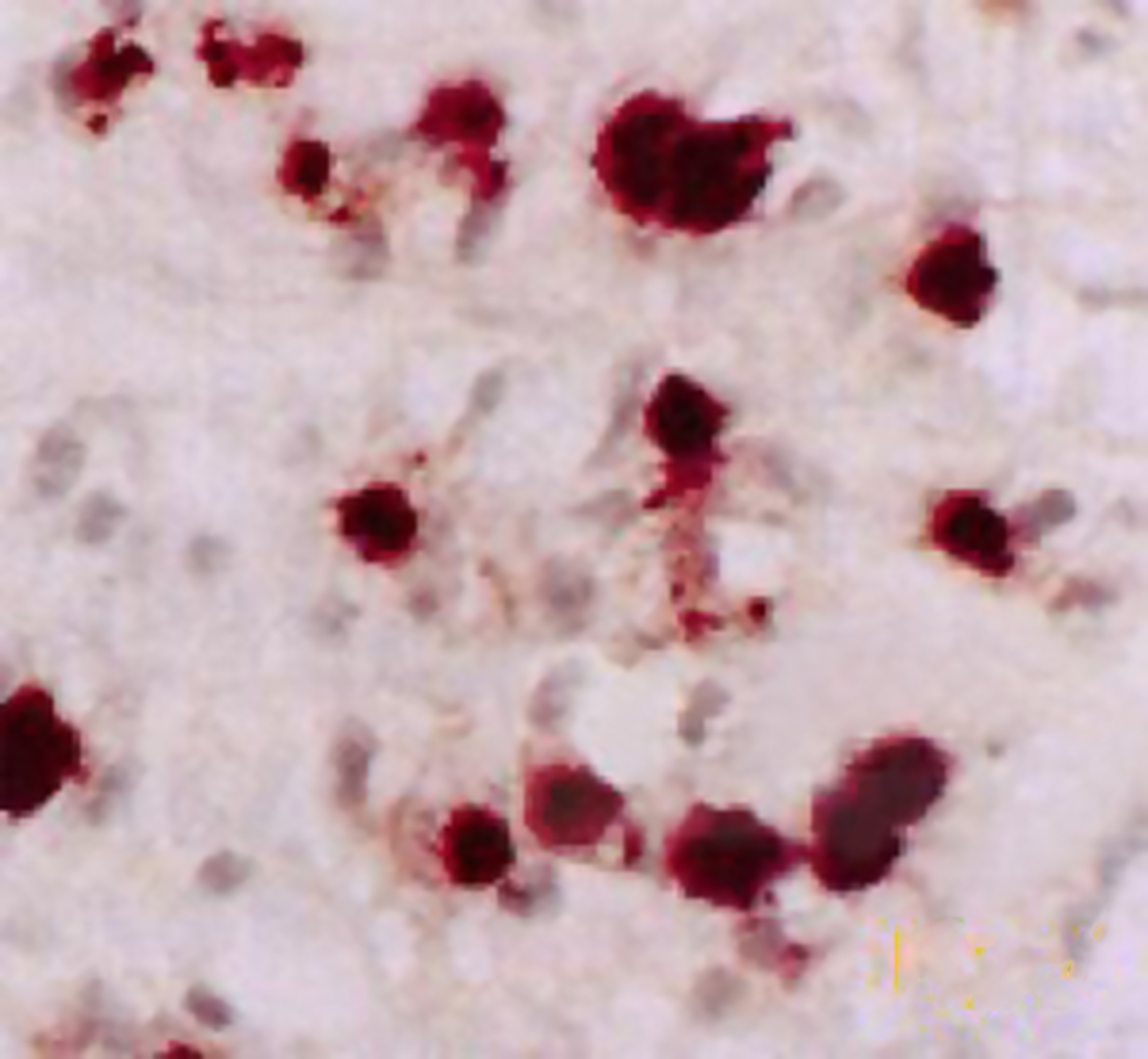
Correlation analyses of VEGF expression in gastric cancer tissues with TNM staging and lymph node metastasis
Analysis of consistency of preoperative prediction results of MRI for TNM staging with postoperative pathological results
The special statistical software Statistical Product and Service Solutions (SPSS) 20.0 was used to sort out and analyze the above data. Measurement data were presented as (
Comparisons of MRI parameters between lymph node metastasis group and non-metastasis group
Comparisons of MRI parameters between lymph node metastasis group and non-metastasis group
MRI manifestations and VEGF expression in gastric cancer
In DWI, tumors showed diffusion-limited high signal, and lesions showed uniform or non-uniform enhancement. In LAVA dynamic enhanced scan, the full-thickness gastric wall signal was enhanced, the delayed-phase enhancement was obvious, and lymph nodes showed diffusion-limited nodular high signal (Fig. 1). VEGF in gastric cancer tissues was mainly expressed in the cytoplasm of cancer cells, showing brown granules (Fig. 2).
Analyses of VEGF expressions in gastric cancer tissues of patients with different TNM staging
The VEGF expression in gastric cancer tissues of patients was increased with the increased degree of lymph node metastasis (
Correlation analyses of VEGF expression in gastric cancer tissues with TNM staging and lymph node metastasis
The VEGF expression in gastric cancer tissues was positively correlated with the infiltration depth, lymph node metastasis and distant metastasis of tumor cells in patients (Table 2).
Analysis of consistency of preoperative prediction results of MRI for TNM staging with postoperative pathological results
The prediction results of MRI for TNM staging of patients before operation were compared with postoperative pathological results, and it was found that there was better consistency (Kappa
Comparisons of MRI parameters between lymph node metastasis group and non-metastasis group
ADC, rADCp and rADCm in gastric cancer patients without lymph node metastasis were significantly higher than those in patients with lymph node metastasis, but the short diameter and long diameter were obviously shorter than those in patients with lymph node metastasis, and the differences were statistically significant (
Discussion
Gastric cancer is the most common malignant tumor in clinic. The metastasis and invasion of tumor cells are important causes of death, and the formation of lymphatic vessels and blood vessels among tumor cells is also an important link causing tumor metastasis. Therefore, studying related factors affecting the formation of lymphatic vessels and blood vessels is important for the clinical diagnosis and treatment, which can reduce the mortality rate and prolong the survival time of gastric cancer patients [10]. The molecular weight of VEGF gene is about 40 kD with a length of about 28 kb. VEGF gene is located in the short arm of human chromosome 6 and can be expressed as platelet-derived growth factor family protein, which is a kind of multi-functional cell regulatory factor, including 5 kinds of proteins [placental growth factor (PLGF), VEGF-D, VEGF-C, VEGF-B and VEGF-A] [11, 12]. In this paper, VEGF-C and VEGF-A proteins were detected, and the latter one is currently a vascular growth factor with the best effect and highest specificity in clinical practice, which can induce rapid mitosis and enhance differentiation function of endothelial cells, resulting in production of a large number of new blood vessels in patients, and greatly increasing the permeability of new blood vessels. In addition, studies have shown that VEGF-A can also further enhance the formation and growth of lymphatic vessels in patients, so that the structure and function of blood vessels and lymphatic vessels in patients can be complete, accelerating the metastasis and invasion of tumor cells [13]. VEGF-C, a vascular growth factor with homology to VEGF-A, has the effects of promoting cell proliferation and differentiation and enhancing permeability of lymphatic vessels, which can also form VEGFR-3-VEGF-C channel with VEGFR-3 to expand lymphatic vessels in patients, increase the contact area of lymphatic vessels with peripheral tumor cells, and increase the probability of tumor cells of passing through the lymphatic vessels for metastasis [11]. In addition, studies have shown that VEGF-C can also promote the fusion of lymphatic vessels and new lymphatic vessels in patients and realizes tumor cell metastasis. Furthermore, it can also promote the fusion of new lymphatic vessels and blood vessels, so that tumor cells can pass through the blood vessels and lymphatic vessels for systemic metastasis [12]. VEGF-C can also increase the contact area of blood vessels with tumors, and enhance the permeability of vascular wall, so that tumor cells can pass through the blood vessels more easily for metastasis [13]. The clinical TNM staging of gastric cancer can reflect the distant metastasis, lymph node metastasis and infiltration depth in patients in systematic and comprehensive manners. Data in this study showed that in patients in stage Tis-T4, N0-N3 and M0-M1, the expression level of VEGF protein was increasingly higher with the increase of infiltration depth and aggravation of distant metastasis and lymph node metastasis, and the VEGF protein expression showed positive correlations with TNM staging and lymph node metastasis, which has a certain guiding significance for the preoperative staging and diagnosis of patients.
MRI has the advantages of no radiation, high resolution, high scanning speed, etc., and it has been more and more widely used in the visceral organs due to the rapid development of respiratory gating technology [14]. Results of this study showed that there was better consistency of the preoperative prediction results of MRI for TNM staging of patients with postoperative pathological results (Kappa
In conclusion, the VEGF protein expression in gastric cancer tissues is positively correlated with TNM staging and lymph node metastasis in patients. The preoperative prediction results of MRI for TNM staging of patients are well consistent with postoperative pathological results, and MRI features are correlated with lymph node metastasis in patients, which has an important guiding significance for the diagnosis and treatment of gastric cancer.
Footnotes
Acknowledgments
Totally Laparoscopic Uncut Roux-en-Y VS Billroth II combined Braun for Racial Distal Gastrectomy to prevent postoperative bile reflux: The Study Protocol for a Multi-Randomized Controlled Trial. GSWSKY2017-36.
Conflict of interest
None.