
Abstract
BACKGROUND:
Despite the major advances in the treatment, the overall survival of osteosarcoma remains poor. MicroRNAs (miRNAs) are involved in tumorigenesis and progression though modulating their target genes. In the present study, the roles of miR-1285-3p in osteosarcoma was investigated.
METHODS:
Microarray profiling was applied to distinguish the up and down regulated microRNAs in osteosarcoma. Quantitative real-time PCR (qRT-PCR) assay was performed to detect the expression of miR-1285-3p and YAP1 expression. MTT and transwell assays were carried out to determine the cells proliferation and invasion respectively. Moreover, dual luciferase reporter assay was performed to evaluate the binding efficiency between miR-1285-3p and the 3’UTR of YAP1.
RESULTS:
MiR-1285-3p was down regulated in osteosarcoma tissues and cell lines and the reduction of miR-1285-3p expression predicted a poor overall survival of osteosarcoma patients. Ectopic expression of miR-1285-3p inhibited osteosarcoma cell proliferation, colony formation and invasion. In addition, YAP1 was further demonstrated as a direct target of miR-1285-3p. Moreover, overexpression of YAP1 reversed the inhibitory effects of miR-1285-3p on osteosarcoma cells proliferation and invasion.
CONCLUSIONS:
MiR-1285-3p which was low expressed in osteosarcoma inhibited the proliferation and invasion of osteosarcoma cells via direct targeting YAP1. These results suggested that miR-1285-3p might be a potential therapeutic targets and biomarker in osteosarcoma.
Introduction
Osteosarcoma, accounting for 20–35% of malignant bone tumors, represents a primary malignant bone tumor predominantly affecting children and adolescents [1, 2]. Despite major advances in treatment over the past decades, such as wide tumor excision, adjuvant chemotherapy and radiotherapy, the survival rate of patients with osteosarcoma remains poor [3]. Although recent advances in molecular biology have provided insight into the molecular pathogenesis of osteosarcoma, the exact molecular mechanisms underlying osteosarcoma are still not clarified [4]. Therefore, it is urgent to elucidate novel molecular targets which is facilitate to develop alternative therapeutic and diagnosis strategies for improving clinical outcome of patients with osteosarcoma.
MicroRNAs (miRNAs) which belong to a group of short non-coding RNA molecules consisting of about 18–22 nucleotides play critical roles in the pathogenesis of human diseases by binding to the 3
In the present study, we explored the potential functions of miR-1285-3p in osteosarcoma. We found that miR-1285-3p was down-regulated in osteosarcoma tissues which was significantly associated poor prognosis in osteosarcoma patients, and miR-1285-3p suppressed the proliferation and invasion of osteosarcoma cells in vitro. Moreover, we further demonstrated that Yes associated protein 1 (YAP1) was the direct target gene of miR-1285-3p. Therefore, our outcomes suggested that miR-1285-3p might be a promising therapeutic targets and biomarker in osteosarcoma.
Materials and methods
Patients and samples
Surgically resected paired osteosarcoma tumor tissues and corresponding non-cancerous bone tissue samples were collected from 120 primary osteosarcoma patients at Huai’an First People’s Hospital. All the tissues were immediately stored in liquid nitrogen until use. This study was approved by the Research Ethics Committee of Huai’an First People’s Hospital and written informed consent was obtained from all of the patients. All the patients did not receive any perioperative chemotherapy before surgery. The clinicopathological information of the patients was in Table 2. The clinical stage of these osteosarcoma patients was classified according to the sixth edition of the TNM classification of the Union for International Cancer Control (UICC).1
Add detail.
Human osteosarcoma cancer cell lines, Saos-2, U2OS, MG63 and HOS, and osteoblast hFOB1.19 were purchased from the Cell Bank of Type Culture Collection of Chinese Academy of Sciences (Xuhui, Shanghai, China). All the cell lines were maintained in Dulbecco’s Modified Eagle’s Medium (DMEM, Life Technologies, Carlsbad, CA, USA) supplemented with 10% fetal bovine serum (FBS, Life Technologies, Carlsbad, CA, USA), 100 U/ml penicillin, and 100
Cells transfection
The miR-1285-3p mimics and control mimics were synthesized by GenePharma (Pudong, Shanghai, China) and were transfected into the cells with a final oligonucleotide concentration of 20 nmol/L. The overexpression of YAP1 plasmids were constructed by GeneChemCo., Ltd. (Pudong, Shanghai, China). All of the cell transfections were carried out with Lipofectamine 2000 (Invitrogen, Carlsbad, CA, USA) according to the manufacturer’s instructions. Briefly, MG63 or U2OS cells were planted into 12-well plates and maintained at 37
RNA extraction and quantitative real-time PCR (qRT-PCR)
Total RNA was extracted from tissue samples or cell lines using TRizol reagent (Invitrogen, Carlsbad, CA, USA) and the miRNA was purified with miRNeasy mini kit (Qiagen, Hilden, Germany). For mRNA detection, 2
The primers for PCR
The primers for PCR
MiRNA profiling was performed using Exiqon mercury LNA
Cell proliferation assay
Cell proliferation was analyzed using 3-[4, 5-dim- ethylthiazol-2-yl]-2, 5 diphenyltetrazolium bromide (MTT; Sigma-aldrich, St. Louis, MO, USA) assay according to the manufacture’s protocol. Briefly, MG63 cells or U2OS cells were first transfected with corresponding miRNA mimics (NC mimics or miR-1285-3p mimics) or/and plasmids (YAP1 overexpressing plasmids). Subsequently, the treated MG63 cells or U2OS cells were collected and separately seeded into 96-well plates (at a density of 1
Colony formation assay
Cells were plated in 6-well plates (1000 cells/well) followed by incubation at 37
Transwell invasion assay
The invasion capabilities of MG63 and U2OS cells were measured by the number of cells invading Matri- gel-coated transwell chambers (Millipore, Billerica, MA, USA). Briefly, the miRNA mimics or/and plas- mids-transfected MG63 or U2OS cells (200
Dual luciferase assay
Dual luciferase reporter assay was performed to evaluate the binding efficiency between miR-1285-3p and the 3’UTR of YAP1. Briefly, MG63 or U2OS cells (1
Western blot analysis
Cells after treatment with corresponding miRNA mimics or plasmids were washed twice with phosphate-buffered saline (PBS) solution and lysed using lysis buffer (Beyotime, Pudong, Shanghai, China) for 30 min on ice. Bradford protein assay kit (Bio-Rad, USA) was used to detect the protein concentrations of the lysates. Standard methods were carried out to perform western blot assay. Briefly, proteins were separated by 10% SDS-PAGE and then transferred to polyvinylidene fluoride (PVDF) membranes (Millipore, Darmstadt, Hessen, Germany). Subsequently, proteins were blocked with 5% non-fat dried milk for 1 h and probed overnight at 4
Correlation of miR-1285-3p expression with clinicopathological features of osteosarcoma
Correlation of miR-1285-3p expression with clinicopathological features of osteosarcoma
Univariate and multivariate analyses for overall survival in osteosarcoma patients
Data was analyzed using SPSS 13.0 (statistical package for the Social Sciences Version 13.0, SPSS Inc., Chicago, IL, USA). All data from three independent experiments were presented as the mean
Expression levels of miR-1285-3p in osteosarcoma tissues and cell lines. (A) Heatmap of the differential expression microRNAs between osteosarcoma tissues and normal tissues. (B) Relative expression levels of miR-1285-3p in osteosarcoma tissues and normal tissues (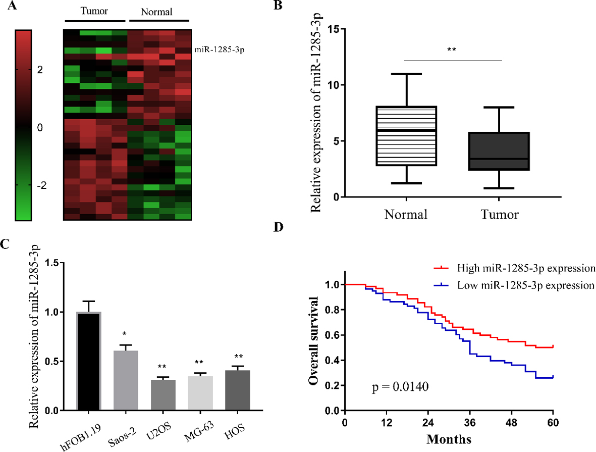
Ectopic expression of miR-1285-3p inhibited osteosarcoma cell proliferation and invasion. (A and B) Relative expression levels of miR-1285-3p in MG63 and U2OS cells. (C and D) The proliferation rates of MG63 and U2OS cells detected by MTT assay. (E-G) Representative images of the colony formation assays and statistical analysis of MG63 and U2OS cell colony number (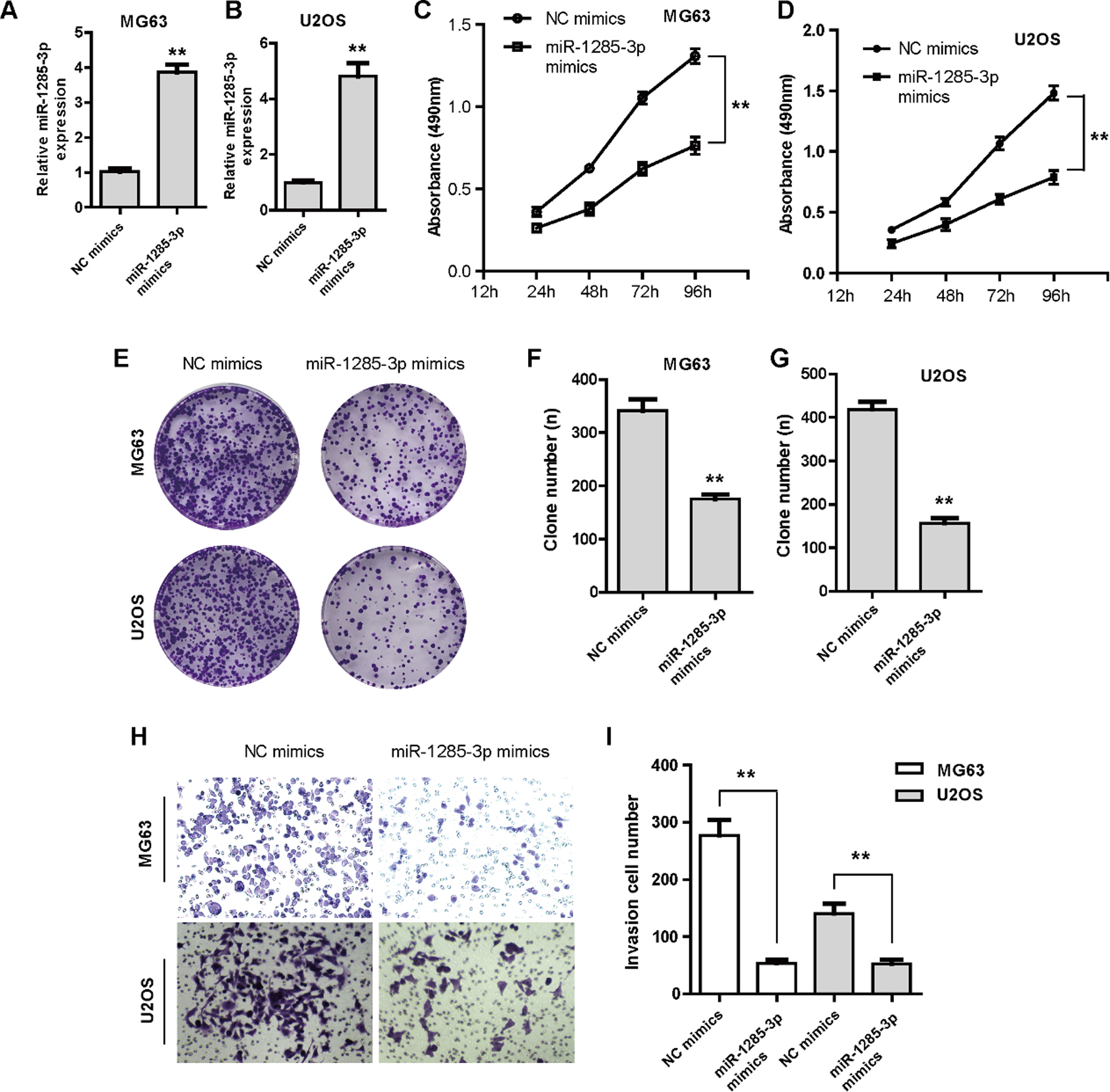
miR-1285-3p directly targeted YAP1. (A) miR-1285-3p binding site in YAP1 3’-UTR predicted by bioinformatics tool “miRBD”. (B and C) Dual-luciferase reporter assay showed that miR-1285-3p reduced luciferase activity of MG63 and U2OS cells transfected with YAP1 3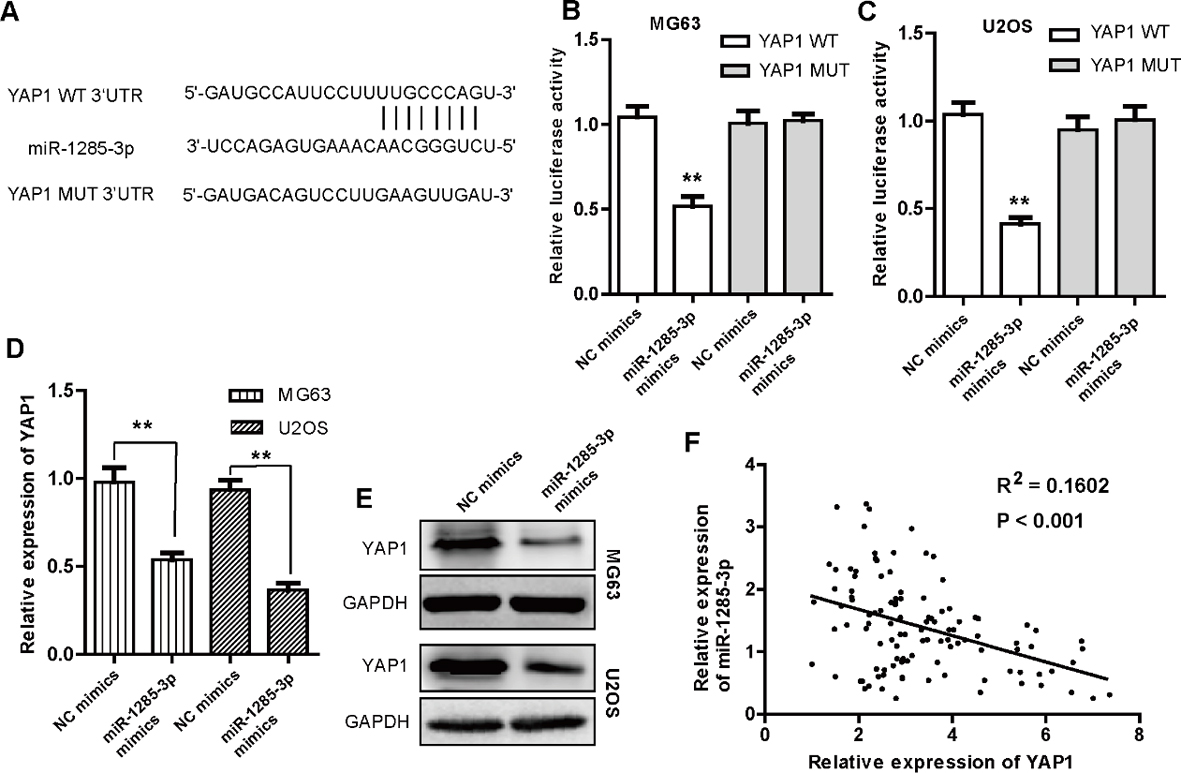
Overexpression of YAP1 abrogated the tumor suppressive roles of miR-1285-3p. (A) Relative mRNA expression levels of YAP1 in both MG63 and U2OS cells in different groups. (B and C) MTT assays showed the proliferation rates of MG63 and U2OS cells transfected with NC mimics, miR-1285-3p or co-transfected with miR-1285-3p and YAP1 overexpression plasmids. (D and E) Representative images of the colony formation assays and statistical analysis of MG63 and U2OS cell colony number in different groups. (F and G) Representative images of the transwell invasion assays and statistical analysis of MG63 and U2OS cell invasion number in different groups. The values were means 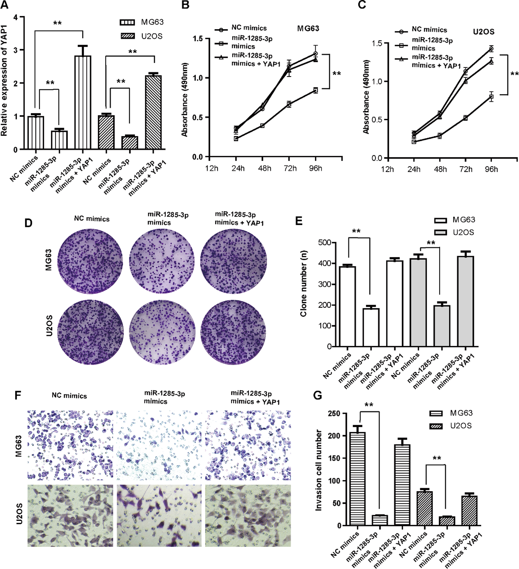
miR-1285-3p expression is down-regulated in osteosarcoma tissues and associated with clinical features
To clarify the miRNAs functions in osteosarcoma, microarray profiling was conducted and bioinformatics analysis suggested that miR-1285-3p was down-regulated in osteosarcoma tissues compared with the normal tissues (Fig. 1A). Consistent with the microarray data, qRT-PCR assay further demonstrated that miR-1285-3p was down-regulation in osteosarcoma tissues (
MiR-1285-3p inhibits the proliferation and invasion of osteosarcoma cells
We next investigated the effects of the altered expression of miR-1285-3p on the cell viability, proliferation and invasion in MG63 and U2OS osteosarcoma cancer cells. Control mimics (NC mimics) and miR-1285-3p mimics were transfected into MG63 and U2OS cells, and the expression levels of miR-1285-3p in both miR-1285-3p mimics transfected MG63 and U2OS cells were dramatically increased compared with that of the control cells (both
YAP1 is the direct downstream target gene of miR-1285-3p
To further investigate the underlying molecular mechanism of miR-1285-3p modulated growth and metastasis suppression in osteosarcoma, we applied bioinformatics tools “miRDB” (
Reintroduction of YAP1 reverses the effects of miR-1285-3p in osteosarcoma cells
To further confirm that the tumor suppressive effects of miR-1285-3p was mediated through directly targeting YAP1 in osteosarcoma cells, the YAP1 overexpression plasmids were employed to transfect the MG63 and U2OS cells. According to our results, the decreased mRNA levels of YAP1 by miR-1285-3p targeting were significantly rescued via the transfection of YAP1 overexpression plasmids in both MG63 and U2OS cells (Fig. 4A). Moreover, MTT assay showed that reintroduction of YAP1 dramatically reversed the inhibitory effects of the miR-1285-3p mimics on the proliferation of MG63 and U2OS osteosarcoma cells (Fig. 4B and C). Additionally, the ectopic expression of miR-1285-3p mimics resulted in a marked decrease in cell colony formation capability while enforcing YAP1 expression significantly rescued cell colony formation capability of MG63 and U2OS cells (Fig. 4D and E). Besides, transwell assays revealed that YAP1 overexpression also abrogated miR-1285-3p mediated suppression of MG63 and U2OS cells invasion (Fig. 4F and G). In summary, the data suggested that the growth and invasion suppressive effects of miR-1285-3p were chiefly via inhibition of YAP1.
Discussion
Osteosarcoma is the most common human primary malignant bone tumor and it has become one of the most promising fields to investigate molecular mechanisms contributing to osteosarcoma development [11]. In recent years, there is accumulating evidence confirming that miRNAs play essential roles in carcinogenesis and miRNAs as well as their targets genes have been proved to represent potential novel therapeutic biomarkers for osteosarcoma [12, 13, 14]. In the present study, miR-1285-3p was found to inhibit the proliferation and invasion of osteosarcoma cells in vitro via direct targeting YAP1.
Previous study had showed that miR-1285-3p could directly repress the expression of JUN oncogene in hepatocellular carcinoma (HCC) which suggested a potential tumor suppressor role of miR-1285-3p acted [15]. Besides, reports had revealed that miR-1285 was significantly down-regulated in plasma of stage I lung squamous cell carcinoma (LSCC) patients and the plasma levels of miR-1285 could serve as LSCC early detection markers [16]. However, the role of miR-1285-3p in osteosarcoma has not been explored. In accordance with the above reported researches, our data in this study also demonstrated that miR-1285-3p was down-regulated in osteosarcoma and the reduction of miR-1285-3p expression predicted a poor overall survival of osteosarcoma patients which suggested a therapeutic and diagnostic potential of miR-1285-3p in osteosarcoma.
MiRNAs function as negative regulators of gene expression at the post transcriptional level by banding to the 3’UTR of the target mRNA which lead to mRNA degradation or suppression of translation [17, 18]. Therefore, identification of the miRNAs targets is pivotal for understanding their roles in tumorigenesis [19, 20]. For example, miRNA-152 attenuated tumor cells growth and induced cancer cells apoptosis though the transcriptional repression of cathepsin L (CTSL) in gastrointestinal stromal tumor and miR-29a suppressed the proliferation of hepatocellular carcinoma cells by directly specifically targeted the 3’UTR of SIRT1 mRNA [21, 22]. In this study, we also confirmed that YAP1 was the direct target gene of miR-1285-3p though performing gain and loss of functions study.
YAP1, acting as one of the most important downstream mediators of Hippo signaling pathway, is known to be involved in cancer cells proliferation and invasion [23, 24]. In the previous study, YAP1 was found overexpressed which was associated with poor prognosis in several cancer types such as gastric cancer (GC), colorectal cancer (CRC), hepatocellular carcinoma (HCC) and non-small cell lung cancer (NSCLC) and inhibition of YAP1 reduced cancer cell proliferation, colony formation and invasiveness [25, 26]. Besides, YAP1 was also discovered in several studies to promote cell proliferation and invasion in osteosarcoma [27, 28, 29]. In our results, YAP1 was demonstrated as the direct target of miR-1285-3p and the Pearson’s correlation analysis suggested that a negative correlation exhibited between the expression of YAP1 and miR-1285-3p in osteosarcoma tissue samples. Furthermore, the inhibitory effects of the miR-1285-3p mimics on the proliferation and invasion of MG63 and U2OS osteosarcoma cells were dramatically reversed by YAP1.
In summary, we certified that the expression of miR-1285-3p was down-regulated in osteosarcoma tissue samples and patients with the reduction of miR-1285-3p expression exhibited shorter overall survival. We further provided evidence that ectopic expression of miR-1285-3p inhibited osteosarcoma growth and metastasis through directly interacting with YAP1. Thus, our data indicated that miR-1285-3p served as a suppressor role in osteosarcoma tumorigenesis as well as metastasis and might be a promising therapeutic targets and biomarker in osteosarcoma.
Footnotes
Acknowledgments
The authors thank all the donors whose names we not included in the author list, but who participated in this program.
Conflict of interest
The authors declare that there are no conflicts of interest.