
Abstract
This article has been retracted, and the online PDF replaced with this retraction notice.
Introduction
Glioma is the most common primary brain tumor in adults, with high malignancy and poor prognosis, accounting for more than 45% of the central nervous system tumors. Like other malignant tumors, the occurrence and development of glioma is a pathological process in which the two major mechanisms of genetics and epigenetics interact together, multiple genes participate, and multiple stages change and accumulate. In the past 20 years, the treatment of glioma has been studied from molecular mechanisms, genetic aspects and pathways, but the mechanism of glioma development is still unclear [1]. At present, surgical resection, radiotherapy and chemotherapy have achieved some progress, but the prognosis of the patients is still not well.
Non coding RNA includes short RNA and long chain noncoding RNA represented by miRNA, siRNA, and piRNA. Long stranded noncoding RNA (lncRNA) is a class of transcripts with a length of more than 200nt RNA, which does not encode proteins per se. Compared with short chain non coding RNA, many functions are still unknown [2]. However, with the development of genome technology, more and more evidences show that non coding long chain RNA has many influences on cell function by regulating the expression of target genes or by modifying the transcription products [3]. Compared with normal tissues, non coding long stranded RNA, which is expressed abnormally in tumor tissues, may affect the function of oncogenes or tumor suppressor genes, thus promoting or inhibiting the formation of tumors [4]. In the past, abnormal expression of molecular markers could be used to determine tumor development or tumor malignancy [5]. Abnormal expression of noncoding long chain RNA is also likely as a new molecular marker to suggest the stage of tumor development. However, in the central nervous system, there are more and more evidence that the expression of long chain non RNA encoding and neural development, neural degeneration and nerve immune disease, is closely related to primary brain tumors, and mental disease, nerve tumor [6]. Abnormal expression of long chain RNA may serve as a cue for glioma types in different types of gliomas [7]. It has also been reported that long chain RNA (anti-NOS2A), an anti NOS2 receptor, plays an important role in the differentiation of glioma, and can affect the effect of chemotherapy in glioma patients [8]. Some previous studies found that lncRNA GHET1 knockdown had effects to suppress cell biological activities in colorectal cancer, gastric cancer and bladder cancer [9, 10, 11, 12, 13, 14, 15, 16]. However, there is no study that the correlation between GHET1 and glioma. This study was firstly showed that the GHET1 knockdown had anti-tumor effects to glioma biological activities, GHET1 might be a potential therapeutic target for glioma cells.
Materials and methods
Clinical sample
The glioma tissues were collected from 30 cases of glioma patients who were treated in our hospital, and the no-tumor tissues were taken from traumatic brain injury patients who were treated in our hospital. The tissues were fixed in the 4% paraformaldehyde until the next experiment.
In situ hybridization (ISH)
Routine dehydration, soaking wax, embedding; sliced with fresh diluted pepsin dilution by 3% citric acid, digestion for 20 min at 37
Immunohistochemistry (IHC)
Tissue samples were taken, paraffin embedded, sliced, and routinely replaced with citric acid solution (pH6. 0), After being heated to boiling, interval 5 min, repeated 1 times, cooled, washed with PBS solution, added goat serum sealing liquid, incubated at room temperature for 20 min, added anti-body working fluid, 4
Cell culture and grouping
The LN229 and U87 cells were purchased from ATCC (USA). The LN229 and U87 cells were respectively divided into 3 groups: NC group, BL group and GHET1-shRNA group. The LN229 and U87 cells were cultured by DMEM medium which contained with 10% fetal bovine serum (FBS) in Incubator (37
Cell transfection
According to the manual operation of liposome 2000, the liposome 2000 and GHET1 shRNA (GHET1-shRNA) and blank control (BL) were respectively mixed. After placing 20 min at room temperature, 48 h and U87 cell culture dishes were dropped into LN229 culture dish respectively. After completion, proceed to the next experiment.
The clinical data. A. The pathology of No-tumor and cancer tissues by HE staining (
The cell proliferation rate of difference groups by MTT assay. A. The cell proliferation rate of difference groups in LN229 
The cell apoptosis rate of difference groups by flow cytometry. A. The cell apoptosis rate of difference groups in LN229 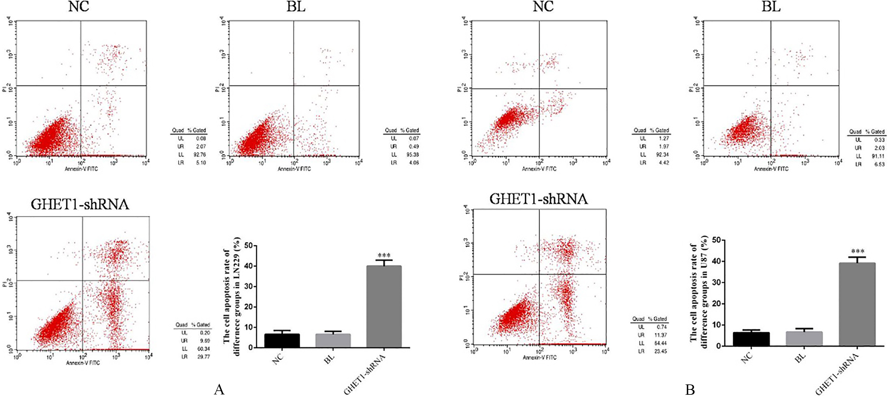
The cell cycle of difference groups by flow cytometry. A. The cell cycle of difference groups in LN229 
The invasion cell number of difference groups by transwell assay. A. The LN229 invasion cell number of difference groups 
The wound healing rate of difference groups by wound healing. A. The wound healing rate of difference groups in LN229 
The relative proteins expressions of difference groups by WB assay. A. The relative proteins expressions of difference groups in LN229 by WB assay 
The nude mice of difference groups. A. The tumor of difference groups in vivo. B. The tumor of difference groups in vitro. C. The tumor volume of difference groups (cm
The positive apoptosis cell of difference groups by TUNEL assay 
The Numb protein expression of difference groups by IHC (
The P53 protein expression of difference groups by IHC (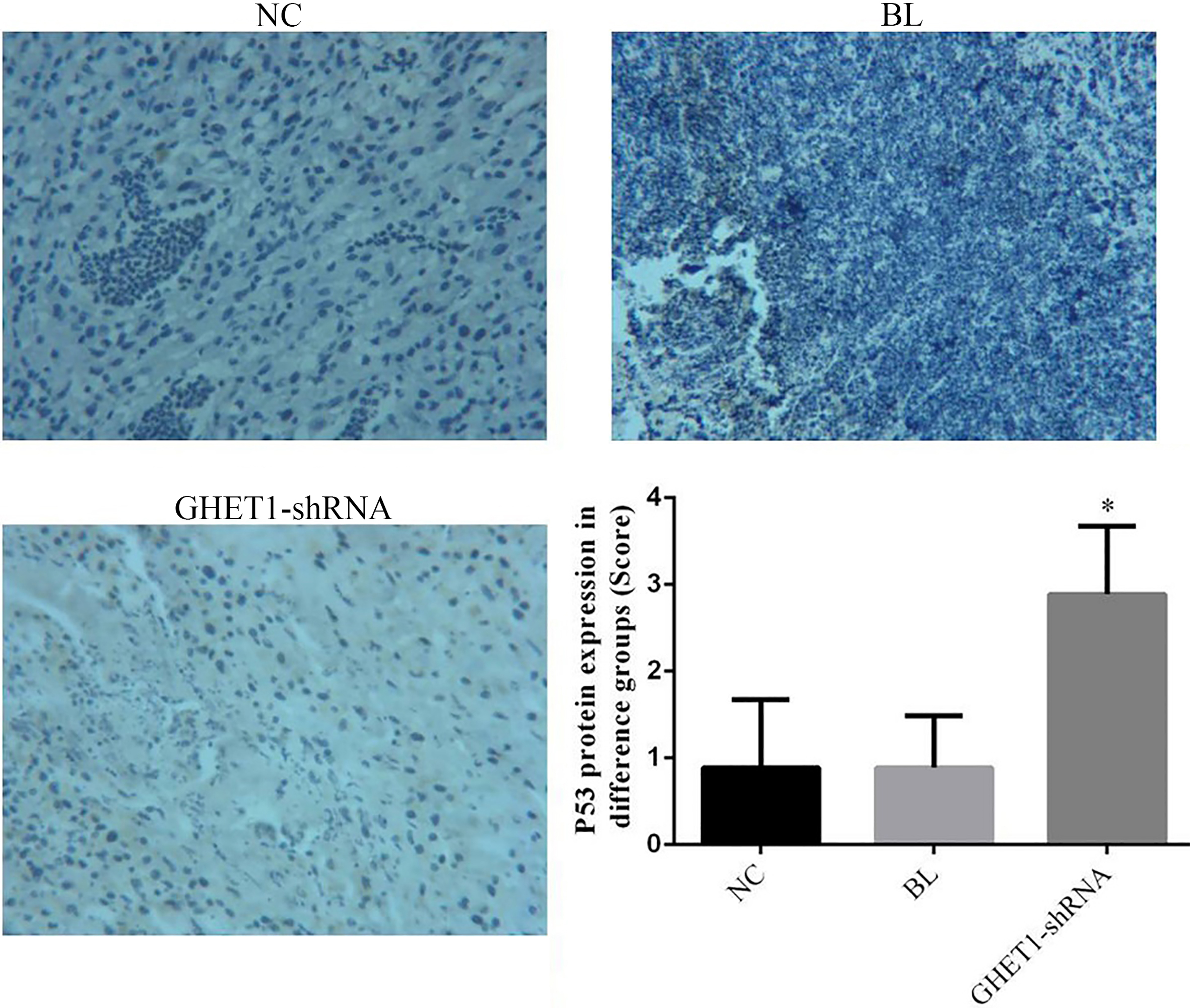
The MMP-2 protein expression of difference groups by IHC (
The MMP-9 protein expression of difference groups by IHC (
The LN229 and U87 cells proliferation rate of difference groups were measured by MTT method, The LN229 or U87 cells of difference groups were inoculated in 96-hole plate as 5
Cell apoptosis by flow cytometry
The LN229 and U87 cells of difference groups were inoculated in the 6-hole plate as 4
Cell cycle by flow cytometry
The cells of difference groups were treated by difference methods for 48 h, the collected cells were washed with PBS and fixed overnight with 75% of the pre cooled ethanol. 50
The cell invasion by transwell assay
One night before inoculation, the cells were diluted with a serum free volume of 1:4 at a volume ratio of Matrigel, and Transwell cells were packed at 50
Wound healing test
The cells of difference groups were inoculated in 6-hole plate as 2
The relative protein expressions in vitro
The total protein was extracted by RIPA reagent 48 hours after transfection, and the protein concentration was determined by BCA kit. Adding the protein sample as 50
Nude mice model
The LN229 cell which were in logarithmic phase were collected, centrifugal as 1000 for 5 min, washing by PBS for once. The cell concentration was adjusted to 10
TUNEL assay
The fresh tumor tissue was fixed with 4% paraformaldehyde, dehydrated, wrapped, sliced, lost to water, washing by PBS, digestion by protease K for 10 min, washing by PBS, inhibition H
Statistical analysis
All of data were analysis by SPSS 19.0 soft ware in our present study. The relative data were explained as mean
Results
Clinical data
Depending on the HE staining, we found that the cell infiltration surrounding normal tissues of cancer tissues was more serious than that of normal tissues (Fig. 1A). By the ISH assay, the GHET1 expression of cancer tissues was significantly up-regulation compared with normal tissues (
GHET1 knock-down suppress cell proliferation in vitro
Compared with NC group, the LN229 and U87 cells proliferation rates of BL groups were no significantly differences (
GHET1 down-regulation improve cell apoptosis in vitro
There were no significantly difference between NC and BL groups in LN-229 and U87 cells apoptosis (
GHET1 knock-down have effects to cell cycle in vitro
Compared with NC group which were treated with normal treatment, the cell cycle rates (G1, S and G2 phases) of BL groups which were transfected with empty vector were similar as those of GHET1-shRNA groups in LN-229 and U87 cells (
GHET1 down-regulation inhibit cell invasion in vitro
There were no significantly differences between NC and BL groups in invasion cell number of LN-229 and U87 cells (
GHET1 knock-down inhibit cell migration in vitro
Compared with NC group, the wound healing rate of GHET1-shRNA groups were significantly suppressed compared with NC groups in LN-229 and U87 cells (
GHET1 down-regulation has effects to relative proteins
Compared with NC group, the Numb and P53 proteins expressions of GHET1 groups were significantly increased (
GHET1 down-regulation inhibit tumor growth in vivo
The tumor volume of difference groups in vivo and vitro were shown in Fig. 8A and B. Compared with NC group, the tumor volume and weight of GHET1-shRNA group were significantly suppressed (
GHET1 knock-down improve cell apoptosis in vivo
The positive apoptosis cell number of GHET1-shRNA group was significantly increase compared with that of NC group (
GHET1 down-regulation have effects to relative proteins expression in vivo
Compared with NC group, the Numb and P53 proteins expressions of GHET1 groups were significantly increased (
Discussion
Glioma is the most common primary brain tumor in adults, with high malignancy and poor prognosis, accounting for more than 45% of the central nervous system tumors. Surgery, radiotherapy and chemotherapy are still the main ways to treat glioma, but the prognosis of patients receiving chemotherapy is still not optimistic [17]. Although progress has been made in the pathogenesis and progression of glioma, the mechanisms underlying the progression of glioma remain unclear because of the complex regulatory network between genes [18]. The past is considered to be the long chain of genome transcription “noise” encoding RNA (long non-coding RNA, lncRNA), but in recent years more and more research found that their participation in the various cell biological functions, including participation in the regulation of tumor occurrence and development mechanism [19]. lncRNA is a class of transcripts with a length of more than 200nt RNA, which itself does not encode proteins. Studies have shown that lncRNA plays an important role in transcriptional level, including transcriptional activation, post transcriptional regulation [20]. In glioma, the study of lcnRNA is still in the exploratory stage, but has made some progress. For example, in gliomas, high expression of lncRNA HOTAIR also increases with tumor grade, and high expression of HOTAIR in glioma patients suggests poor prognosis [21]. The study found that HOTAIR acts on its target gene, PCR2, and ultimately promotes tumor growth [22]. The previous studies found that lncRNA GHET1 expression was significantly up-regulation in the cancer tissues [12, 13, 14, 15, 16]. However, it has been unclear that lncRNA GHET1 expression in the glioma tissues. Our present study was found that the lncRNA GHET1 was high expression in the glioma tissues by ISH. Meanwhile, we also found that the Numb which was an important suppressor gene was significantly down-regulation in the glioma cancer tissues. Depending on these results, we inferred that stimulating lncRNA GHET1 expression may have effects to inhibit Numb to occurrence and development of tumor. In the following studies, we found that GHET1 knock-down might have effects to cell proliferation, invasion and migration in vitro and vivo studies.
In recent years, more and more attentions have been paid to the discovery of Numb protein in tumor formation, invasion and metastasis. Numb has been shown to regulate cellular recognition of differentiation signals. It plays an important role in regulating cell growth, differentiation, tissue regeneration and stabilizing intracellular environment [23, 24, 25]. In our present study, The Numb protein expressions were up-regulation with lncRNA GHET1 knock-down in glioma cancer in vivo and vitro studies. That might the results that the GHET1 knock-down inhibited tumor biological activities (cell apoptosis, cell cycle, cell invasion and migration) in vitro and vivo.
P53 was an important role of Numb down-steam genes [26, 27, 28]. The human p53 gene is an important tumor suppressor gene [29], in cells received external pressure signals, the half-life of p53 can be extended by the accumulation of p53 protein in the cell, at the same time, p53 can play a conformation change, transcription factor function, a series of target genes in regulation leads to cell cycle arrest in G1 phase. P53 can induce cell cycle arrest by transcriptional regulation of p21 and Cyclin B1 genes [30]; P53 also can increase Bax and Puma and apoptosis promoting gene, leading to apoptosis [31]. In our present study, we found that P53 expression was up-regulation with GHET1 down-regulation in vivo and vitro study, that might the results induced cell cycle stayed in the G1 phase induced cell apoptosis enhancing.
Matrix metalloproteinases are a large family, because they require Ca2+, Zn2+ and other metal ions as cofactor. MMPs can degrade almost all kinds of protein components in extracellular matrix, and destroy the barrier of tumor cell invasion, and play a key role in tumor invasion and metastasis [32, 33]. In many MMPs proteins, MMP-2 and MMP-9 are involved in angiogenesis and are directly related to the degradation of basement membranes and are involved in tumor metastasis and prognosis [34, 35]. Meanwhile, MMP-2 and MMP-9 are important roles of Numb downstream genes [36, 37]. In our present study, we found that GHET1 knock-down inhibited the cell invasion and migration via regulation Numb/MMP-2/9 in vitro and vivo study.
Footnotes
Acknowledgments
This research was supported by Yunnan Provincial Science and Technology Department – Kunming Medical University Joint Fund (No. 2015FB068).