
Abstract
BACKGROUND:
Recent findings have identified thousands of long non-coding RNAs (lncRNAs) and reveal that lncRNAs play crucial roles in the regulation of tumor development and progression. However, the clinical significance and potentially functional value of LINC00261 in hepatocellular carcinoma (HCC) remain unknown.
METHODS:
Expression of LINC00261 was detected by qRT-PCR in HCC tissues and adjacent normal tissues. Kaplan-Meier analysis was used to assess the relationship between LINC00261 expression and the overall survival (OS) time. Cell proliferation and invasion were evaluated using MTT assay, cell colony formation assay and transwell assay. The protein expression was determined by western blot analysis.
RESULTS:
In present study, we confirmed that LINC00261 was frequently lower in HCC tissues compared to adjacent normal tissues. Decreased LINC00261 expression associated with lager tumor size, TNM stage (III-IV) and poor overall survival time of HCC patients. The functional assays demonstrated that overexpression of LINC00261 in HCC cells inhibited cell proliferation, cell colony formation, cell invasion and EMT process in vitro. Moreover, we also demonstrated that upregulation of LINC00261 significantly inhibited Notch signaling by downregulating Notch1 and Hes-1 expression in HCC cells.
CONCLUSION:
These results indicated that LINC00261 may be a potential target of HCC treatment.
Introduction
Hepatocellular carcinoma (HCC) is one of the most common malignancies and the second leading cause of cancer-related death worldwide [1]. In the past years, larger advances have improved the prognosis of HCC patients including improvement of early-stage diagnosis, surgery and molecular-targeted therapy for HCC [2]. However, most HCC patients who are diagnosed at advanced stages still have poor prognosis [3]. Thus, identifying novel molecular biomarkers and therapeutic targets for improving the outcome of patients were urgent.
The large scale genome-wide sequencing studies have found a class of abnormal expression of long non-coding RNAs (LncRNAs) in tumors [4, 5]. LncRNAs were well-characterized as tumor suppressors or oncogenes in cancer development and progression [6]. Recent evidence indicates that long non-coding RNA LINC00261 acts as a tumor suppressor to be involved in several tumors, such as, decreased expression of the long non-coding RNA LINC00261 indicates poor prognosis in gastric cancer and suppresses gastric cancer metastasis by affecting the epithelial-mesenchymal transition [7]. Long non-coding RNA linc00261 suppresses gastric cancer progression via promoting Slug degradation [8]. Long non-coding RNA LINC00261 suppresses cell proliferation and invasion and promotes cell apoptosis in human choriocarcinoma [9]. However, the clinical significance and potential value of LINC00261 in hepatocellular carcinoma (HCC) remain unknown.
In the study, we confirmed that LINC00261 was frequently lower in HCC tissues. Decreased LINC00261 was associated with poor overall survival time of HCC patients. LINC00261 overexpression in HCC cells inhibited cell proliferation, cell invasion, and inhibited Notch signaling in HCC. Thus, these results indicated that LINC00261 may be a potential target of HCC treatment.
Materials and methods
Patient tissue samples
Sixty-six cases HCC tissues and adjacent normal tissues were collected from patients undergoing hepatectomy between January 2008 and March 2011 at Sixth People Hospital of Qingdao. All patients were followed up until October 2016. The overall survival (OS) time was defined from the dates of surgery or the last follow up in HCC patients. The study was approved by ethics committee of Sixth People Hospital of Qingdao and informed consent was obtained from all patients.
Cell lines culture, plasmid construction and transfection
Human HCC cell lines (SMCC-7721, MHCC97L and MHCC97H) and human normal liver cell line LO2 were purchased from Shanghai Institute of Cell Biology, Chinese Academy of Sciences (Shanghai, China). All of cell lines were maintained in Dulbecco’s Modified Eagle’s Medium (DMEM) supplemented with 10% fetal bovine serum (FBS) in a 5% CO
Quantitative real-time PCR (QRT-PCR) assay
The RNA derived from tissues and cells samples were extracted using TRIzol (Invitrogen; Thermo Fisher Scientific, Inc., CA, USA) according to the manufacturer’s instruction. The 1
Upregulation of LINC00261 was found in HCC tissues and associated with prognosis of HCC patients. (A) Relative expression of LINC00261 in 66 cases of HCC tissues and adjacent normal tissues were analyzed by qRT-PCR. The mRNA expression was normalized to 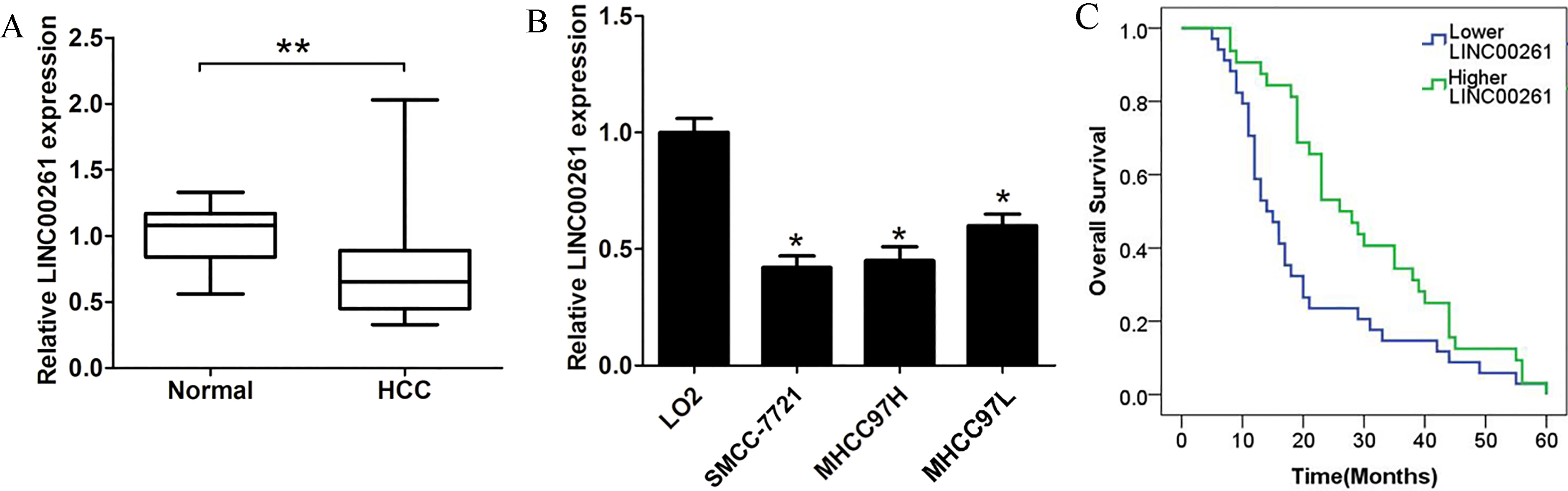
Cell proliferation assay was performed using 3-(4, 5)-dimethyl thiahiazo-(-z-y1)-3, 5-di-phenytetrazoliu mromide (MTT) assay kit (Sigma, USA). After 0, 24, 48 and 72 h of cells transfection, cells were added with 20
Cell colony formation assay
Briefly, the transfected HCC cells were plated in 6-well plates at a cell density (300 cells/well) and incubated at 37
Cell invasion assay
Cell invasion ability was evaluated by Transwell assay according to previous describe [10]. The transwell chambers was used in the study (24-well, 8
Western blot assay
Cells were lysed using RIPA protein extraction reag-ent (Beyotime, Beijing, China) Proteins were separated by sodium dodecyl sulfate-polyacrylamide gel electrophoresis (SDS-PAGE) and transferred on 0.45
Statistical analysis
All statistical analyses were performed by using SPSS 18.0 software (IBM, Chicago, IL, USA). Data are presented as mean
Results
LINC00261 expression is significantly downregulated in HCC tissues and cells
To explore the clinical role of LINC00261 in HCC patients, the expression levels of LINC00261 were determined by qRT-PCR assay. The LINC00261 expression was normalized to GAPDH. The results showed that LINC00261 expression levels were significantly downregulated in HCC tissues when compared to adjacent normal tissues (Fig. 1A). Furthermore, we demonstrated that LINC00261 expression levels were significantly reduced in HCC cells (SMCC-7721, MHCC97H and MHCC97L) when compared to LO2 cells (Fig. 1B). To explore whether LINC00261 expression associated with clinicopathologic factors, the chi-square test was performed. The present results showed that lower LINC00261 expression associated with tumor size and TNM stage in HCC patients (
Correlation between the LINC00261 expression and clinicopathological factors
Correlation between the LINC00261 expression and clinicopathological factors
We further applied siRNA targeting LINC00261 to decrease the expression of LINC00261 in MHCC97L cells, while the pcDNA3.1-LINC00261 plasmid was used to increase the endogenous expression of LINC00 261 in SMCC-7721 and MHCC97H cells, respectively (Fig. 2A). As illustrated by MTT assays, LINC00261 overexpression significantly inhibited the proliferation capability of SMCC-7721 and MHCC97H cells, while LINC00261 knockdown promoted cell proliferation ability in MHCC97L, compared with corresponding control groups (Fig. 2B and C). Furthermore, cell colony formation assays demonstrated that cell colonies number in LINC00261 overexpressed SMCC-7721 and MHCC97H cells was significantly decreased, but was increased in LINC00261 silencing MHCC97L cells, compared with the control groups (Fig. 2D and E). Thus, these results demonstrated that LINC00261 suppressed HCC cell proliferation.
The effects of LINC00261 on HCC cell proliferation ability. (A) Relative expression of LINC00261 was analyzed by transfected with pcDNA3.1 and pcDNA3.1-LINC00261 plasmids into SMCC-7721 and MHCC97H cells or transfected with si-NC and si-LINC00261 oligos into MHCC97L cells. (B)–(C) The cell proliferation ability was detected at 0, 24, 48 and 72 h using MTT assays after cell transfection with pcDNA3.1 and pcDNA3.1-LINC00261 plasmids into SMCC-7721 and MHCC97H cells or transfected with si-NC and si-LINC00261 oligos into MHCC97L cells. (D)–(E) The cell colony number was counted after cell transfection with pcDNA3.1 and pcDNA3.1-LINC00261 into SMCC-7721 and MHCC97H cells or transfected with si-NC and si-LINC00261 oligos in MHCC97L cells at 7 days, Error bars indicate S.D. 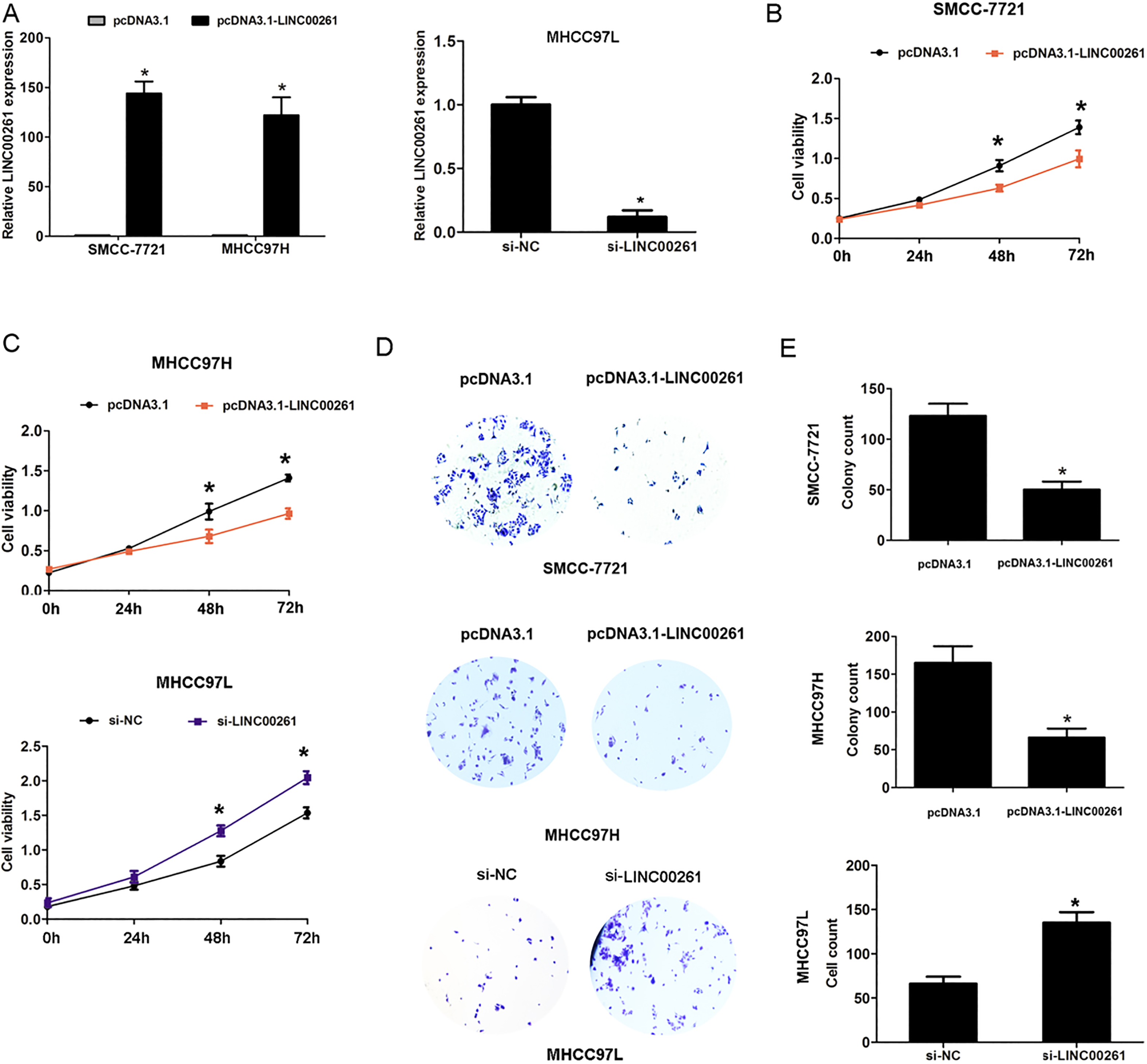
To further explore the LINC00261 expression whether affected cell invasion and EMT process of HCC. The results of transwell assays were present in Fig. 3A, overexpression of LINC00261 markedly suppressed cell invasion ability of SMCC-7721 and MHCC97H cells, compared with the control groups. However, knockdown of LINC00261 inhibited cell invasion ability in MHCC97L, compared with the control group (Fig. 3B). Next, the EMT makers Twist1, E-cadherin and N-cadherin were examined. The results demonstrated that overexpression of LINC00261 markedly upregulated the expression of E-cadherin, but downregulated the Twist and N-cadherin expression in SMCC-7721 and MHCC97H cells compared with the control groups (Fig. 3C and D). Therefore, our results indicated that upregulated LINC00261 expression suppressed HCC cell invasion and EMT process.
The effects of LINC00261 on HCC cell invasion and EMT process. (A)–(B) The cell invasion ability was assessed by performing transwell assay at 48 h after transfecting with pcDNA3.1 and pcDNA3.1-LINC00261 in SMCC-7721 cells and MHCC97H cells or transfected with si-NC and si-LINC00261 oligos into MHCC97L cells. (C)–(D) The relative protein expression of Twist1, E-cadherin and N-cadherin was assessed by transfecting with pcDNA3.1 and pcDNA3.1-LINC00261 plasmids into SMCC-7721 or MHCC97H cells using western blot analysis at 48 h, Error bars indicate S.D. 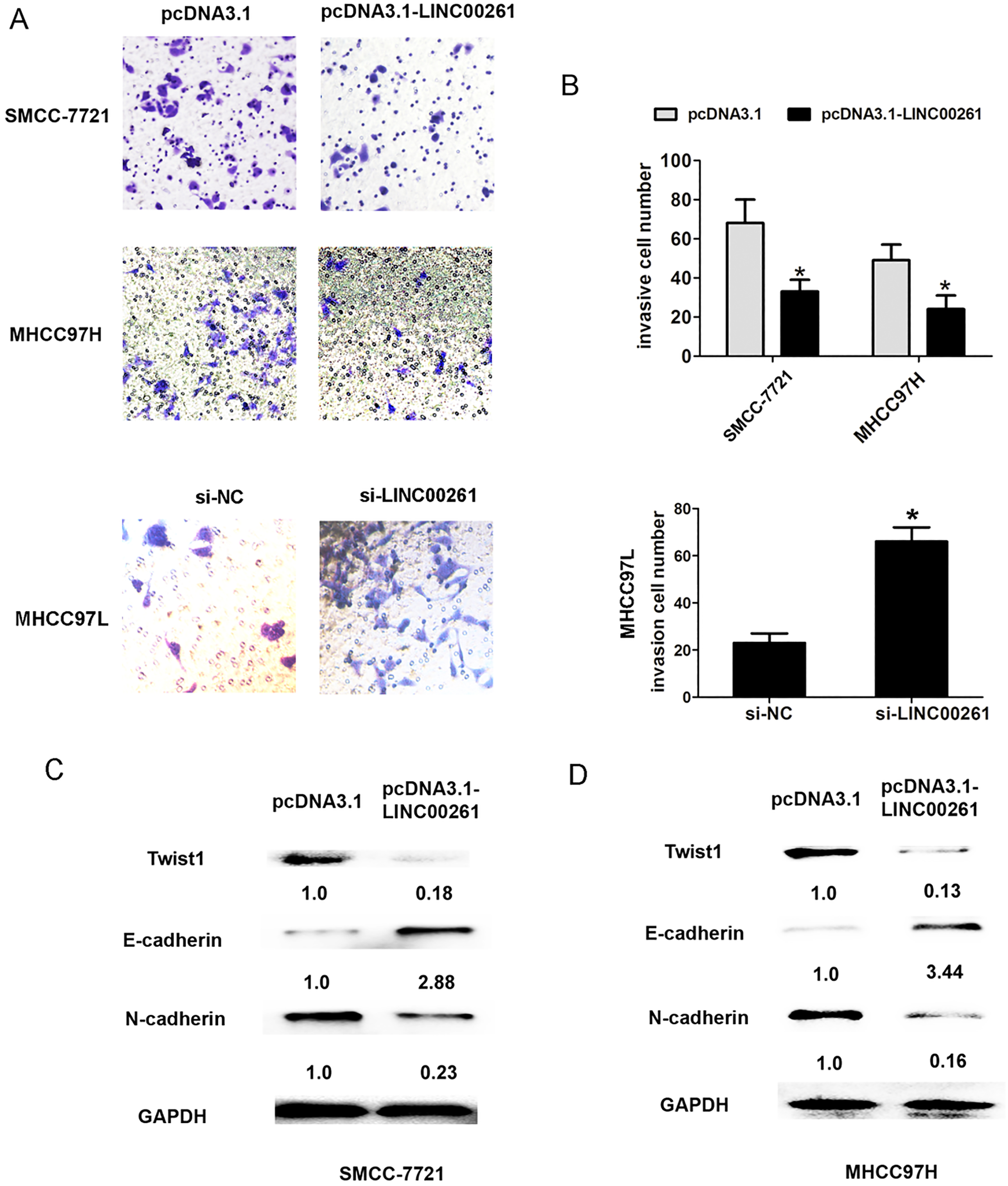
Some signaling pathways have become a major source of targets for novel therapies in hepatocellular carcinoma (HCC). Notch signaling pathway associated with HCC proliferation and invasion [11]. Our results showed that overexpression of LINC00261 markedly downregulated the expression of Notch1 and Hes-1 expression in SMCC-7721 and MHCC97H cells compared with the control groups (Fig. 4A and B). Thus, we demonstrated that increased expression of LINC00261 suppressed Notch signaling pathway in HCC.
The effects of LINC00261 overexpression on Notch signaling pathway in HCC cells. (A) The relative expression of Notch1 and Hes-1 was assessed at 48 h after transfecting with pcDNA3.1 and pcDNA3.1-LINC00261 in SMCC-7721 cells using western blot analysis. (B) The relative expression of Notch1 and Hes-1 assessed at 48h after transfecting with pcDNA3.1 and pcDNA3.1-LINC00261 in MHCC97H cells by western blot analysis. 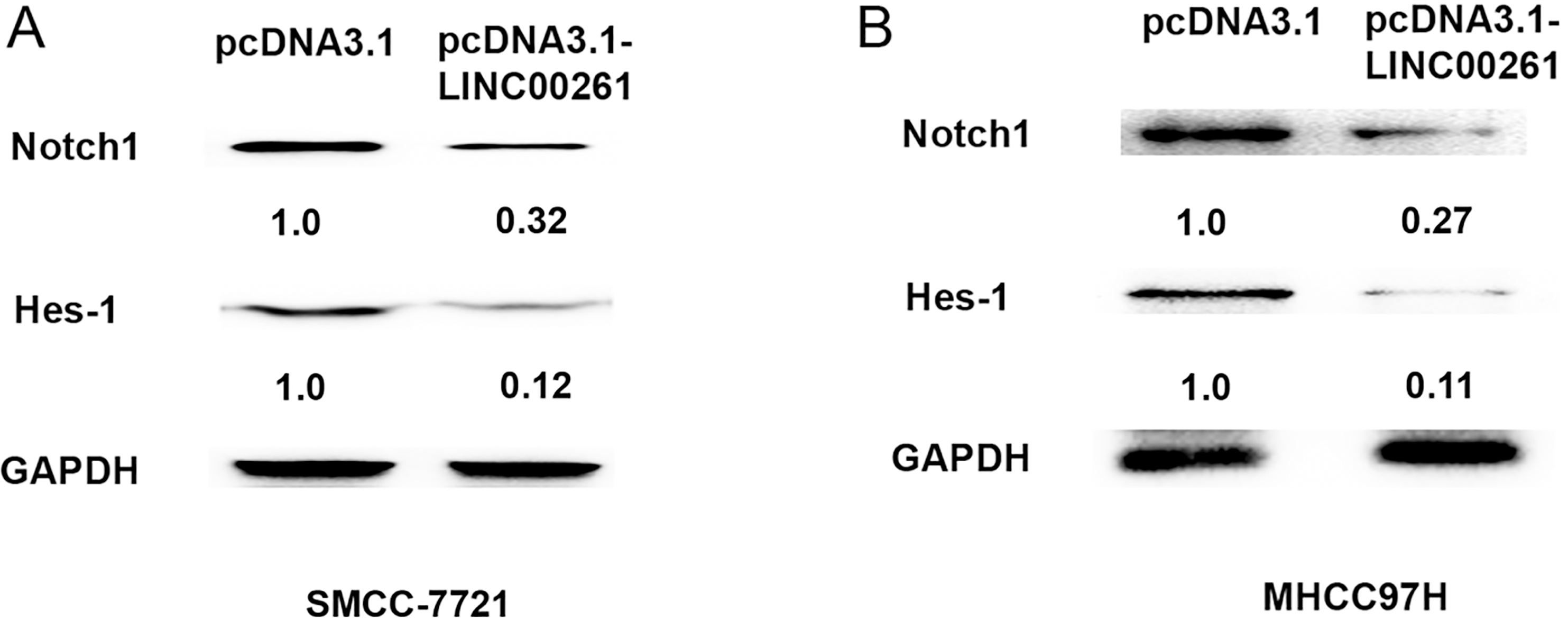
Abnormal expression of lncRNAs has been frequently linked with HCC pathogenesis and lncRNAs are identified as potential biomarkers and prognosis factors in HCC [4]. For example, DANCR acts as a diagnostic biomarker and promotes tumor growth and metastasis in hepatocellular carcinoma [12]. Down-regulation of LncRNA DGCR5 correlates with poor prognosis in hepatocellular carcinoma [13]. LINC00 052 regulates the expression of NTRK3 by miR-128 and miR-485-3p to strengthen HCC cells invasion and migration [14]. In the study, we found that LINC00261 was frequently lower in HCC tissues compared to adjacent normal tissues. Decreased LINC00261 was associated with tumor size, TNM stage and poor overall survival time of HCC patients.
Furthermore, the functional assay showed that upregulated expression of LINC00261 inhibited cell proliferation, colony formation, and cell invasion in HCC, while LINC00261 knockdown had opposite effects in HCC cells. Meanwhile, the results demonstrated that overexpression of LINC00261 suppressed cell EMT process by markedly upregulating the expression of E-cadherin but downregulating the Twist1 and N-cadherin expression in HCC cells. Some lncRNAs have been reported to function as tumor suppressors or oncogenes in HCC progression. Such as, long non-coding RNA uc.338 promotes cell proliferation through association with BMI1 in hepatocellular carcinoma [15]. LINC01225 promotes occurrence and metastasis of hepatocellular carcinoma in an epidermal growth factor receptor-dependent pathway [16]. AFAP1-AS1 indicates a poor prognosis of hepatocellular carcinoma and promotes cell proliferation and invasion by upregulation of the RhoA/Rac2 signaling pathway [17]. Long non-coding RNA FTX inhibits hepatocellular carcinoma proliferation and metastasis by binding MCM2 and miR-374a [18]. In the study, our results showed that LINC00261 functioned as tumor suppressor in HCC.
The Notch pathway is aberrantly activated in tumors and plays the tumor-promoting role in HCC [19]. For instance, LincRNA-p21 inhibits invasion and metastasis of hepatocellular carcinoma through Notch signal-ing-induced epithelial-mesenchymal transition [20]. Our results demonstrated that upregulation of LINC00 261 significantly inhibited Notch signaling pathway through downregulating Hes-1 and Notch1 expression in HCC cells.
In conclusion, our finding demonstrated LINC00261 was lower expression in HCC and increased expression of LINC00261 inhibited cell proliferation and cell invasion in vitro. Moreover, we demonstrated that upregulation of LINC00261 significantly inhibited Notch signaling pathway in HCC cells. Thus, these results indicated that LINC00261 may be a potential target of HCC treatment.
Footnotes
Conflict of interest
The authors declare no conflicts of interest.