
Abstract
BACKGROUND:
Gastric cancer is one of the most common malignancies worldwide. Recent studies reported that Piwil3 was overexpressed in various cancers, including gastric cancer (GC). This study was intended to investigate its function and mechanism in GC progress.
METHODS:
Quantitative real time PCR(RT-PCR) and western blotting assays were utilized to measure mRNA and protein expression levels, respectively. SiRNA transfection was performed to suppress the expression of Piwil3. CCK-8 assay, cell invasion and migration assays were used to determine the cell proliferative, cell invasive and migratory ability.
RESULTS:
The expression of Piwil3 was significantly increased in GC tissues compared with matched normal tissues. The specific siRNA significantly inhibited the protein and mRNA expressions of Piwil3, and effectively inhibited the proliferation and induced G0/G1 phase arrest in GC cells. Downregulation of Piwil3 significantly suppressed the migration and invasion of GC cells. Moreover, the downregulation of Piwil3 also significantly suppressed the tumor volumes in nude mice. Mechanism investigation showed that the downregulation of Piwil3 significantly decreased the mRNA and protein expressions of metastasis-related genes, including RhoC, MTA1, MMP2 and MMP9, and also modulated the phosphorylation levels of JAK2 and STAT3 but not their protein levels.
CONCLUSIONS:
These findings indicate that overexpression of Piwil3 promotes the proliferation, migration and invasion of GC cells partially through JAK2/STAT3 signal pathway.
Introduction
Gastric cancer (GC) is the third leading cause of cancer-related death worldwide (account for 8.8% of total cancers), and is considered to lead the highest mortality rates in Eastern Asia and the lowest in Northern America [1]. Most patients with GC have develop advanced stages when diagnosed and their 5-year survival rates are less than 7% [2, 3].Tumor cell metastasis is major responsible for the high mortality rates but the molecular mechanism of GC metastasis remains unexplained.
PIWI belongs to Argonaute family which includes PIWIL1, PIWIL2, PIWIL3, and PIWIL4. It has been shown that PIWIs are aberrantly expressed in a variety of cancers and play important roles in tumorigenesis and development of cancers. For example, Rosenkranz et al. demonstrated dynamic expression of PIWIL1, PIWIL2 and PIWIL3 during mammalian oncogenesis [4]. The expression of PIWIL1–4 were also increased in the primary tumor and metastatic tissues in epithelial ovarian cancer [5]. PIWILs also regulate the progress of breast cancer [6]. However, the expression and bio-function of Piwil3 in GC development and its underlying mechanism remain largely unclear.
In the present study, we reported that the expression of Piwil3 was significantly increased in GC tissues. Downregulation of Piwil3 induced by specific siRNA significantly suppressed the proliferation and induced G0/G1 phase arrest in GC cells in vitro and in vivo. Moreover, the downregulation of Piwil3 also repressed the migration and invasion of GC cells in vitro. Furthermore, mechanism investigation revealed that Piwil3 regulated the expression of metastasis-related genes in JAK2/STAT3 signaling pathway.
Methods and materials
Patients and samples
Totally, 52 pairs of GC tissues and matched normal tissues were collected from the patients with GC, who were enrolled in Huaihe hospital according to the protocols approved by the Ethics Review Board.
Cell culture
Human GC cell lines (AGS, N87, SGC7901, MKN45 and BGC823) were purchased from Shanghai Cell Bank, Chinese Academy of Sciences (Shanghai, China) and cultured in DMEM (St. Louis, MO, USA) containing 10% FBS and 1% penicillin/streptomycin at 37
SiRNA transfection
MKN45 cells were selected to perform transfection because of its highly expressed Piwil3. The cells were seeded onto 12-well culture plates at a density of 6
Cell proliferation assay
Cell proliferation was evaluated by using Cell Counting Kit 8 (CCK8, Beyotime, Shanghai, China) according to the manufacturer’s protocol. Briefly, MKN45 cells with transfection of the siRNA or control were seeded onto 96-well culture pates at a density of 4
Cell cycle
Flow cytometry was used to analyze cell cycle. After transfection for 48 h, the cells were harvested and fixed in 70% ice-cold ethanol (stored at
Cell invasion and migration assay
Invasion and migration activity of MKN45 cells were analyzed by using 24-well trans-well chambers coated with or without Matrigel (BD Biosciences) on the upper surface of the membrane with a pore size of 8
Quantitative reverse-transcriptase polymerase chain reaction
Total RNA was extracted from the cells with transfection of siRNA or control by using TRIzol (Invitrogen), respectively. Two microgramme of RNA was used to synthesize cDNA by using a ?rst strand cDNA kit (Sigma, Munich, Germany), according to the manufacturer’ protocol. PCR amplification was performed using a SYBR Green PCR kit (Thermo) and run on an ABI 7300 Thermocycler (Applied Biosystems, Foster City, CA, USA). The parameter of PCR reaction was 95
Primers used in FQ-RT-PCR analysis
Primers used in FQ-RT-PCR analysis
The concentrations of total proteins in GC cells were determined by using a BCA Protein Assay Kit (Thermo Scientific). An equal amount of proteins was subjected to SDS polyacrylamide gel electrophoresis, followed by electro-transferring onto PVDF membranes. Five percent skimmed-milk powder in PBS with 0.1% tween-20 was used to block the PVDF membranes for 1 h. The membranes were incubated with diluent antibodies of ICAM, RhoC, MTA1, MMP-2 and MMP-9 for overnight at 4
Cell growth in vivo
MKN45 cells with transfection of siRNA or control were re-suspended in PBS. 2
Statistical analysis
The correlation of Piwil3 expression with the patient’s clinicopathological variables were analyzed by the chi-square test. All data were presented as the mean
Results
Piwil3 is frequently increased in gastric cancer tissues and cell lines
The expression of Piwil3 is showed to be increased in various cancers, but in GC its expression is unclear. We initially detected the expression of Piwil3 in GC and matched normal tissues, and the results showed that the mRNA expression of Piwil3 was significantly higher in the GC tissues than that in the matched normal tissues (
Correlation between Piwil13 expression and clinicopathological parameters in two types of gastric carcinoma
Correlation between Piwil13 expression and clinicopathological parameters in two types of gastric carcinoma
Piwil3 expression in gastric cancer tissues and cell lines. 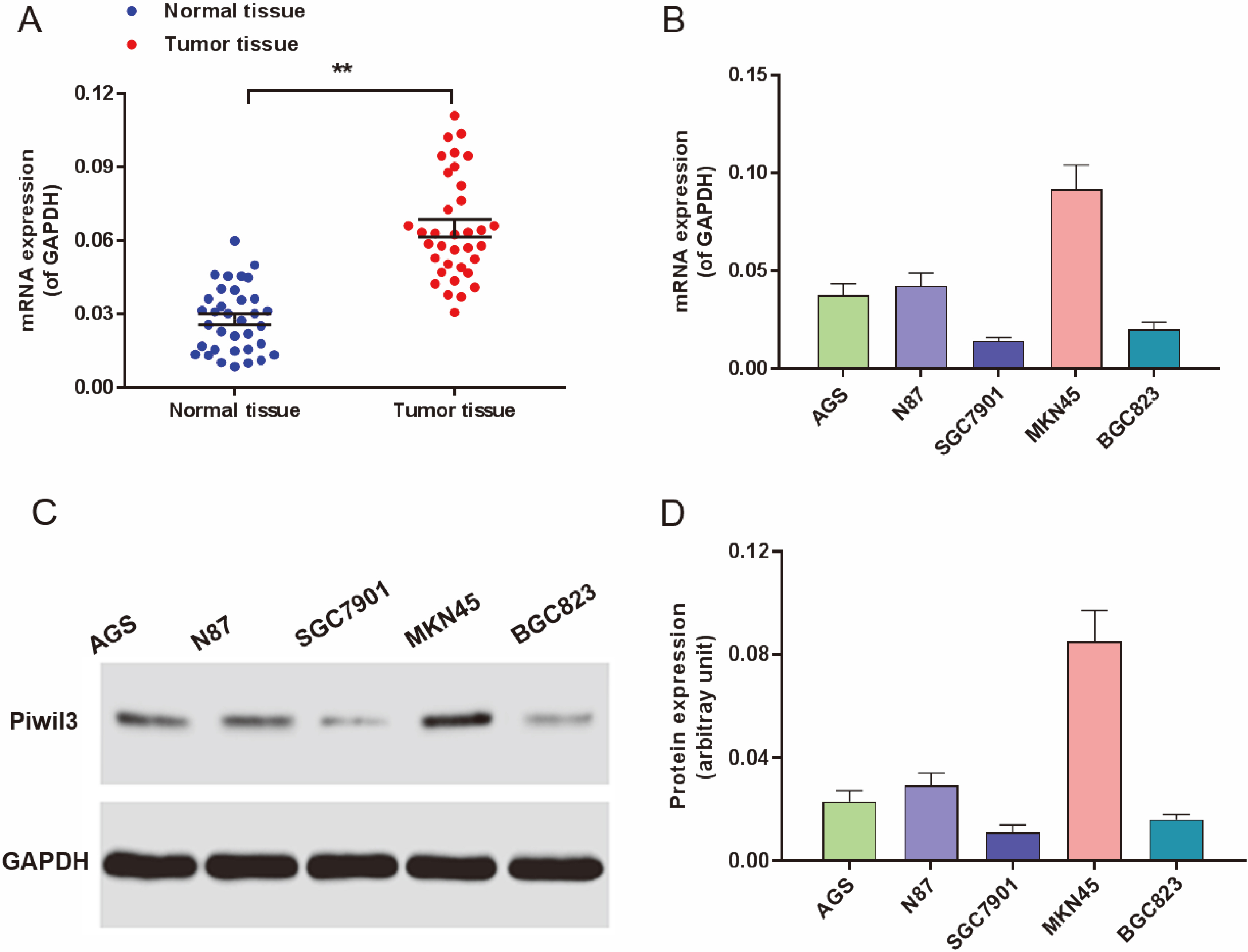
Furthermore, we also detected the expression of Piwil3 in GC cell lines. As shown in Fig. 1D–F, the mRNA and protein expressions of Piwil3 were markedly increased in MKN45 cells compared with the other GC cell lines, including AGS, N87, SGC7901 and BGC823. These results suggested that Piwil3 is overexpressed in GC tissues.
Down-regulation of Piwil3 suppresses the proliferation of MKN45 cells. 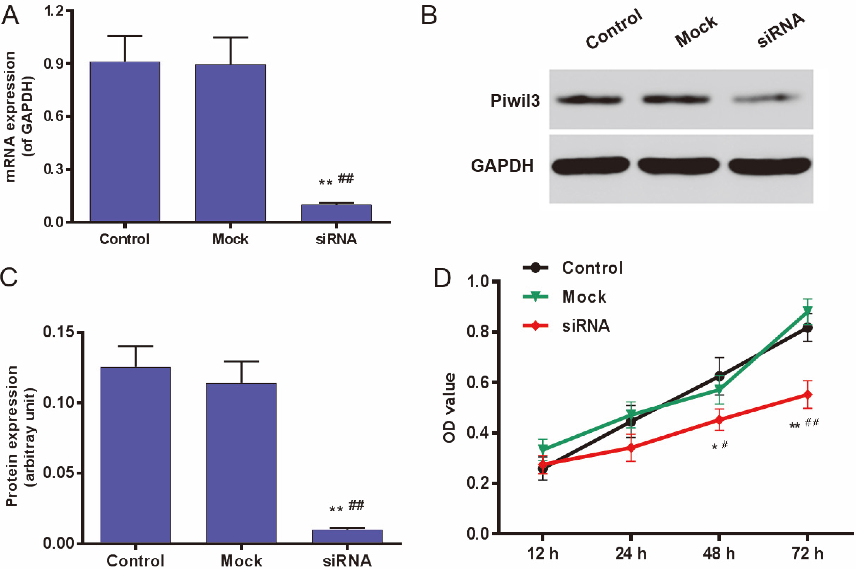
To investigate the bio-function of Piwil3 in GC cells, we selected MKN45 to perform the functional experiments because of its highly expressed Piwil3. The results showed that the specific siRNA significantly inhibited the mRNA and protein expressions of Piwil3 in MKN45 cells (
Down-regulation of Piwil3 induces G0/G1 phase arrest in MKN45 cells. After MKN45 cells were transfected with the specific siRNA for Piwil3 for 48 h, the cell cycle distribution was identified by using flow cytometry. “**” denotes 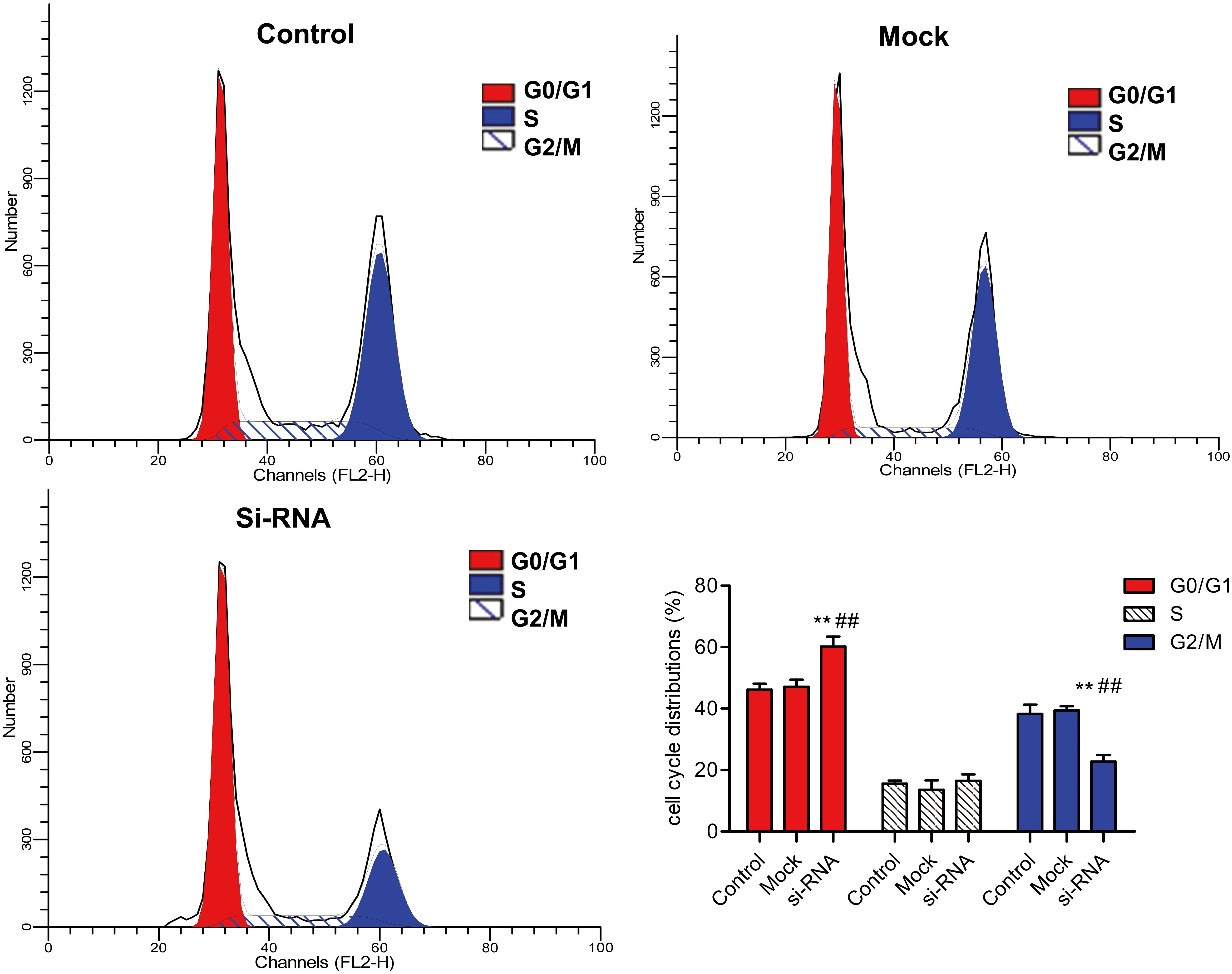
Cell motility is an important factor in regulating cancer metastasis. We investigated whether Piwil3 could affect the abilities of migration and invasion of GC cells. The results showed that the specific siRNA significantly repressed the migration and invasion of MKN45 cells when transfection for 48 h (
Down-regulation of Piwil3 suppresses the migration and invasion of MKN45 cells. After MKN45 cells were transfected with the specific siRNA for Piwil3 for 48 h, cell migration 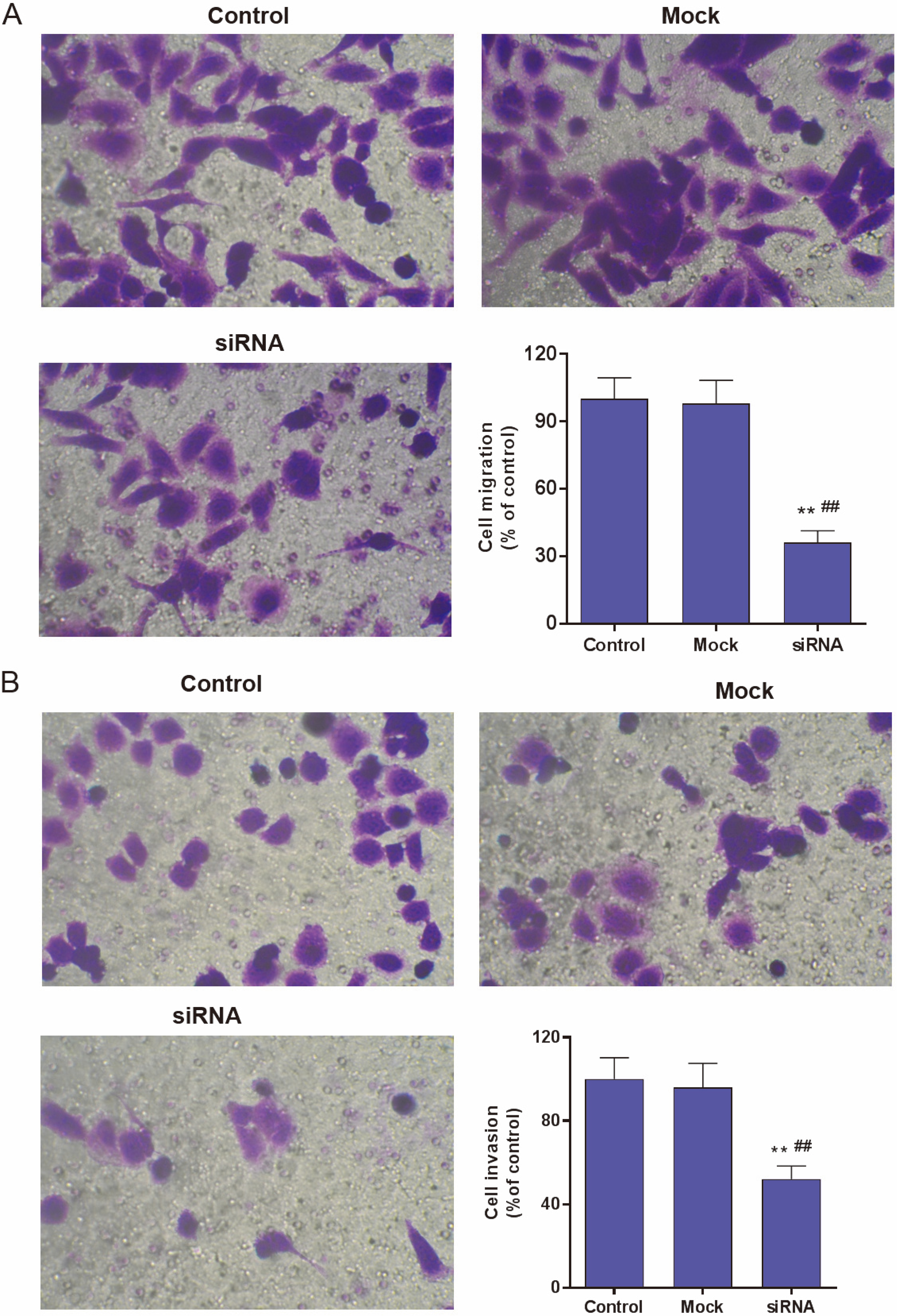
To elucidate the potential mechanism involved in Piwil3-induced the invasion and migration of GC cells, we detected the expressions of cancer metastasis-related genes, including RhoC, MTA1, MMP-2 and MMP-9. The results showed that the specific siRNA for Piwil3 significantly decreased the mRNA and protein expressions of RhoC, MTA1, MMP-2 and MMP-9 in MKN45 cells (
Piwil3 regulates the expressions of multiple cancer metastasis-related genes. After MKN45 cells were transfected with the specific siRNA of Piwil3 for 48 h, the mRNA 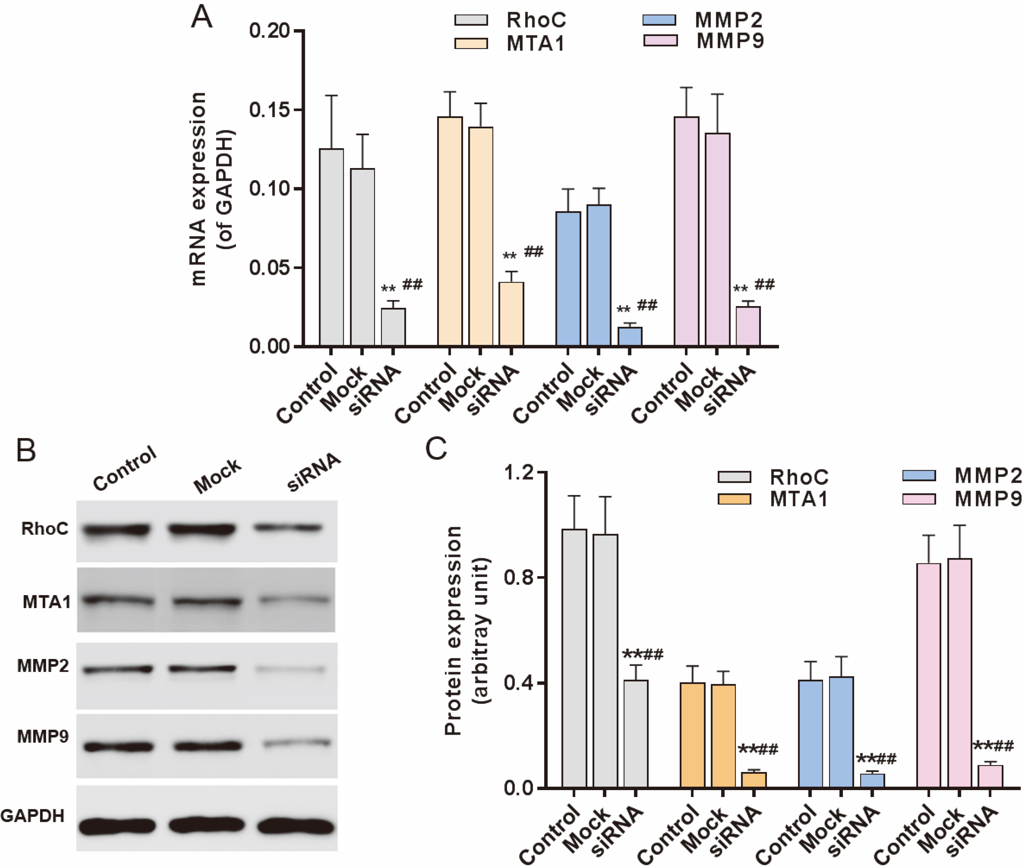
Given the downregulation of Piwil3 significantly decreased the expressions of cancer metastasis-related genes, we investigated whether could its downregulation regulate the activity of JAK2/STAT3 signaling pathway. The results showed that the specific siRNA significantly decreased the phosphorylation levels of STAT3 and JAK2 (
Piwil3 regulates JAK2/STAT3 signaling pathway. After MKN45 were transfected with the specific siRNA of Piwil3 for 48 h, the phosphorylation and protein levels of JAK2 and STAT3 were analyzed by using western blot. “**” denotes 
Although the downregulation of Piwil3 could inhibit the proliferation of GC cells in vitro, its function in vivo also need to be identified. We constructed a lentivirus vector containing a specific shRNA for Piwil3. After MKN45 cells were infected with this lentivirus for 48 h, the mRNA and protein expressions of Piwil3 were significantly decreased (
Downregulation of Piwil3 suppresses cell growth in vivo. MKN45 cells infected with lentivirus containing the specific shRNA for Piwil or negative control were subcutaneously injected in nude mice. The mRNA (A) and protein (B) expressions of Piwil3 were analyzed by qRT-PCR and Western blotting, respectively. GAPDH serves as internal reference. Tumor volumes (C) were evaluated after injection for 6, 12, 18, 24, 30, 36, 42 and 48 days, respectively. The formative tumors (D) were taken out after injection for 48 days and their weight (E) were measured. “**” denotes 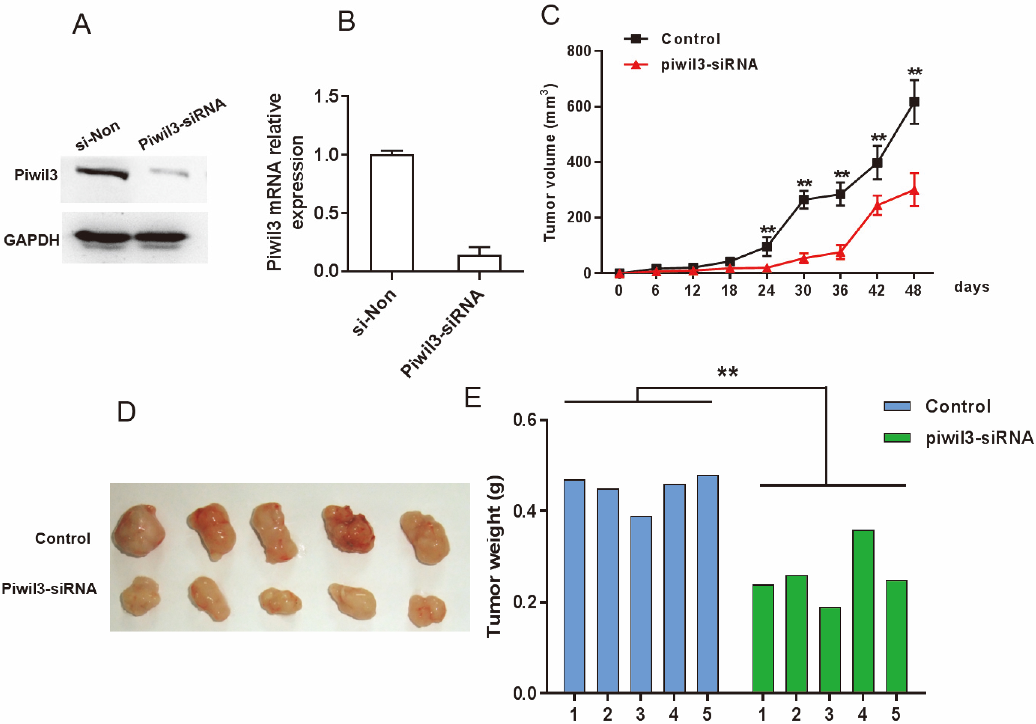
In the present study, we found that the expression of Piwil3 was increased in GC tissues compared with matched normal tissues. Downregulation of Piwil3 significantly inhibited the proliferation and induced G0/G1 phase arrest in GC cells in vitro and in vivo, and decreased the expressions of metastasis-related genes and also regulated JAK2/STAT3 signaling pathway.
PIWI proteins, defined by highly conserved PiWi (P-element-induced wimpy testis) and PAZ (Piwi/Argonaute/Zwille) domains, have been reported to be involve in the development of several human cancers [8, 9]. Our data confirmed that the expression of Piwil3 was increased in GC tissues compared with matched normal tissues and the high level expression of Piwil3 was associated with the depth of invasion, the lymph node metastasis and the clinical stage in the diffuse type of gastric cancer. Wang et al. reported that the expression of PIWIL1–4 was significantly higher in GC tissues, and the expression of PIWIL1 might serve as an independent prognostic factor for GC [10], and these findings supported our results.
The MKN45 cell line is derived from a poorly differentiated adenocarcinoma with highly metastatic potential (PMID: 3962675). In our current study, it showed highly expressed Piwil3 level. This means that MKN45 cells will be a very nice model system to study the loss of function of Piwil3 in GC cells. When the expression of Piwil3 was down-regulated by using the specific siRNA in MKN45 cells, the cell proliferation, migration and invasion were significantly repressed, suggesting that Piwil3 can regulate the progress of GC, and to our knowledge this finding is first reported by us. We also performed the in vivo experiments revealing that the ability of tumor formation of MKN45 cells infected with a lentivirus containing specific shRNA for Piwil3 was significantly alleviated, and this result is consistent with the experiments in vitro. In addition, we tried to overexpress Piwil3 with lenti-Piwil3 in the moderately differentiated SGC-7901 gastric cancer cell line with minimum Piwil3 level. However, cells were killed 24 hr after transduction and this phenomenon repeated in another round of transduction. Transduction condition will be optimized in the future.
Tumor cell invasion is involved in breaking extracellular matrix, which can be degraded by type IV collagen-degrading enzymes, mainly MMP-2 and MMP-9 [11, 12]. MTA1 was reported to regulate the expressions of MMP-2 and MMP-9 in various cancers [13]. RhoC, one of the Rho GTPases, is most convincingly linked with cancer metastasis. Increasing evidences indicated that RhoC participated in the progress of various cancers, including breast cancer [14], non-small cell lung carcinoma [15], colon carcinoma [16], and hepatocellular carcinoma [17]. In order to preliminarily explain the reason for the regulation of migration and invasion by Piwil3 in gastric cancer, we examined the changes of metastasis-related genes mentioned above after the silencing of Piwil3. Our results revealed that Piwil3 regulated the expression of MTA1, MMP-2, MMP-9 and RhoC. These data suggested that Piwil3 promotes the proliferation, migration and invasion of GC cells through regulating the expressions of multiple cancer metastasis-related genes. However, how Piwil3 induced MTA1, MMP-2, MMP-9 and RhoC expression needs further research.
JAK2/STAT3 signaling pathway plays a crucial role in the development of cancer. STAT3 is an important transcription factor, and phosphorylated STAT3 (pSTAT3) directly induced the transcriptions of multiple cancer-related genes in nucleus. Increasing evidences indicated that the activation of pSTAT3 promoted neoplasm metastasis [18, 19]. Our results showed that the downregulation of Piwil3 significantly inhibited the phosphorylation levels of JAK2 and STAT3, suggesting that Piwil3 indeed participated in regulating the JAK2/STAT3 signaling pathway. Although Piwil3 could regulate the expressions of multiple cancer-related genes, the direct target of Piwil3 remains unclear. Therefore, in the future the exact mechanism by which Piwil3 regulates the JAK2/STAT3 signaling pathway needs to be explored.
In conclusion, our study demonstrated that the expression of Piwil3 was increased in GC tissues compared with matched normal tissues. Enhanced Piwil3 expression in the diffuse type of gastric cancer was associated with the depth of invasion, the lymph node metastasis and the clinical stage in the diffuse type of gastric cancer. Downregulation of Piwil3 suppressed the progress of GC through regulating the expressions of multiple cancer metastasis-related genes and JAK2/STAT3 signaling pathway, suggesting that Piwil3 may be a therapeutic target for GC.
Conflict of interest
The authors declare no conflict of interest.
Footnotes
Acknowledgments
This work is supported by Scientific Research Foundation of Henan (092102310090).