
Abstract
Sarcoptic mange was suspected in three of five European albino rabbits (Oryctolagus cuniculus) kept for experimental purposes. Gross examination revealed multifocal areas of alopecia around the eyes, nostrils and lips. Skin snips were processed using PCR for the molecular identification of the suspected mites. Histopathology of the skin snips showed erupted epidermis and stratum corneum with an infiltration of inflammatory cells. Skin scraping examination revealed the presence of adult mites as well as eggs. Microscopic taxonomy identified the adult mites as Sarcoptes (S.) scabiei (Acari: Sarcoptidae). The results of PCR indicated a 311 bp band from all the three cases, which confirmed the S. scabiei infestation in rabbits. Sarcoptes scabiei may be a public health concernthrough direct transmission from rabbits infested with S. scabiei through handling. To the best of our knowledge, this is the first report of S. scabiei infection in rabbits from Pakistan.
Introduction
Sarcoptes scabiei (Acari: Sarcoptidae), commonly known as itch mite, is an ectoparasite that burrows into the skin and causes a disease commonly known as scabies in humans and mange in animals. In mammals, such as wild and domesticated dogs and cats, wild boars, ruminants, wombats, koalas, great apes (Pence and Ueckermann 2002), as well as laboratory animals, including rabbits (Suckow et al. 2002) are affected. The genus Sarcoptes is a part of the larger family of mites collectively known as "scab mites" comprising of one species, S. scabiei with further identification by the variety name indicating the host species e.g S. scabiei var. hominius in humans and S. scabiei var. cuniculi for rabbits. All the life cycle stages of S. scabiei are found on host, and the entire life cycle takes approximately two months (Suckow et al. 2002). The impregnated S. scabiei female about twice the size of the male (Jofre et al. 2009), oviposits into the tunnels made in the stratum corneum of the skin, causing intense itchy skin rashes, hypersensitivity and inflammation. The six-legged larvae hatch with-in three to 10 days and move about on the skin in search of hair follicles, moult into a nymphal stage, and then mature into adult mites (Soulsby 1982). The adult mites live three to four weeks in the host's skin. Mites feed on lymph and sloughed epithelial cells (Hofing and Kraus 1994).
Sarcoptic mange in rabbits is described as an uncommon disease (Scott et al. 2001); however, it has been reported from Israel (Eshar 2010), America (Radi 2004) and India (Soundararajan and Iyue 2005). Wild animals have been reported to transmit sarcoptic mange from dog to dog, dog to rabbits, rabbits to rabbits and rabbit to dogs experimentally as well as naturally (Arlian et al. 1984). In Pakistan, European albino rabbits (Oryctolagus cuniculus) are kept as pets, research laboratory animals and food animals. Rabbit farming is gaining currency in peri-urban and rural areas of Pakistan due to palatable meat quality and the faster rate of reproduction. The present report describes the first study of sarcoptic mange in rabbits from Pakistan diagnosed through clinical signs, gross lesions, taxonomic identification through optical microscopy, histopathology, and polymerase chain reaction (PCR).
Materials and methods
Results
All the three suspected rabbits had clinical signs of pruritis and gross lesions of variable-size alopecia around the nostrils, lips and ears. The affected skin area of ears was covered with tannish yellow, scaly crusts (Fig. 1).
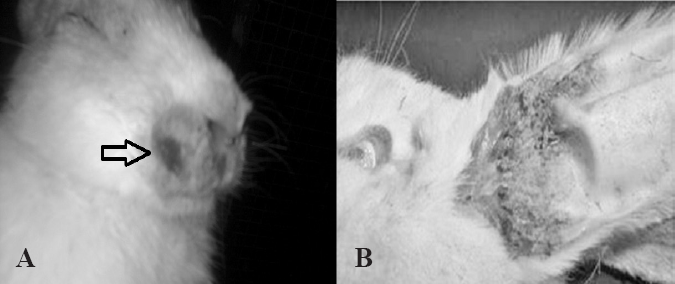
Photographs of gross lesions of sarcoptic mange in a rabbit. A. Notice multifocal areas of alopecia around eyes, nose and lips with scratched wound beside nose (arrows). B. Alopecia and crusting of exudate on the ventral surface of the pinna.
Adult mites were clearly identified in the microscopic examination with round body and shorter legs (Fig. 2A) with the presence of mite eggs attached within the hair follicles (Fig. 2B).
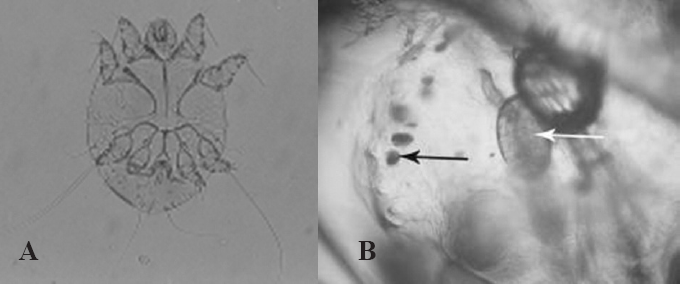
Photomicrographs of sarcoptic mite from the skin scrapping of the rabbit. A. Ventral view of the adult mite with rounded body and shorter legs at 10 x magnification. B. The egg of mite are visible (arrow) at 100 x magnification.
Microscopic examination of the sections revealed the erupted epidermis and stratum corneum with infiltration of inflammatory cells (Fig. 3).
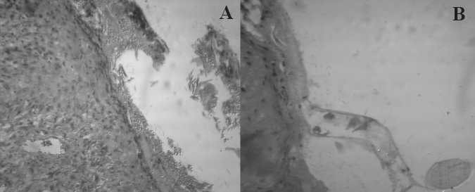
Histological sections of skin scrapping from rabbit with crusted lesion stained with hematoxylin and eosin. A. Note the erupted epidermis and dermis with mild inflammatory infiltrate in the dermis. B. The mite egg beside the hair follicle.
The PCR results of all the three PCR reactions from the three suspected rabbits showed the amplified fragment of 311 bp of actin gene of the S. scabiei var cuniculi. For representation of bands, 3 µL of the reaction mixture was poured in each of the wells 3 and 4 from one of the three positive amplicons for S. scabiei var cuniculi (Fig. 4).
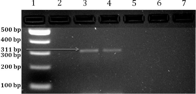
Molecular detection of Sarcoptes scabiei from the gDNA of skin snips of three rabbitssuspected for mite infestation using PCR. Lane 1: DNA 100 bp marker/ladder. Lane 2: Negative control, Lanes 3 and 4: 3 µL of the amplicon was poured into each of the wells which showed band of 311 bp confirming Sarcoptes scabiei var cuniculi. All the three PCR products from three rabbits showed similar bands of 311 bp.
Discussion
In Pakistan, rabbits are mostly kept as pets, experimental lab animals and food animals. These are an excellent source of economical and good quality meat due to their fast rate of reproduction. Sarcoptic mange is a highly contagious disease of the skin transmitted through direct and indirect contact (Arlian et al. 1984).
The present study provides the first report of S. scabiei var cuniculi infestation in rabbits in Pakistan diagnosed through clinical signs, microscopic examination, histopathology and PCR. The clinical signsof acute sarcoptic mange may include severe pruritus, alopecia and seborrhea in result of hypersensitivity reaction (Davis et al. 1991) whereas, crusting and hyperkeratosis occur in chronic infestation (Van-Nesteand Staquet 1986). In the present cases, acute infestation was found with the signs of pruritus and alopecia. Amyloidosis, anemia, and leukopenia have been reported in rabbits with sarcoptic mange (Arlian et al. 1984), however, we did not find evidence for amyloidosis in the study cases. Generally, S. scabiei in rabbits affects the face, nose and external genitalia (Percy and Barthord 2001). In the present cases, affected areas were the face, nose and ears but not the external genitalia.
Diagnosis of sarcoptic mange can be made by identification of the mite by microscopic examination of skin scrapings (Suckow et al. 2002) and histopathology of skin lesions. Sarcoptic mite is round in shape with short legs having a long unjointed stalk with a sucker on front pair of legs. The body wall of Sarcoptic scabiei is thick and chitinous with large spines on the dorsal body surface (Chitwood and Lichtenfels 1972), the anus is terminal and the dorsum possesses scales, cones and bladelike setae. The size of a female sarcoptic mite is 303 to 450 µm x 250 to 350 μm.
Mite infestation is highly specific, however, occasionally; exposure to animals can cause infestation in humans as their aberrant host (Beck 1965). Recently, a 56-years-old human case of scabies infection with a history of contact with his pet dog has been reported (Bandi and Saikumar 2013). Radi (2004) reported that ear canker caused by Psoroptes cuniculi infestation in rabbits is relatively abundant than sarcoptic mange caused by S. scabiei var. cuniculi. The conventional diagnostic tests for mites are having less than 50% accuracy (Shelley and Currie 2007); hence, confirmatory diagnosis of the species of mite infestation through PCR was necessary. Identification of mites at molecular level using PCR is highly sensitive, reliable and specific method.The amplification of specific fragment can confirm the species of the mite (Naz et al. 2013) as confirmed in this report by amplification of specific fragment of the house keeping actin gene of S. scabiei. This report provides the standardized protocol for confirmatory diagnosis of the mite infestation in rabbits which can be helpful for planning future strategies of risk assessment from rabbits as potential reservoirs of infestation.
Footnotes
Acknowledgements
The authors would like to thank Dr. Thomas Nolan, Professor of Parasitology, University of Pennsylvania, Philadelphia, for reviewing the manuscript. Spanish translation of the abstract (resumen) was kindly made by the editorial board of the Revista Colombiana de Entomología. Suggestions of anonymous reviewers of the manuscript are also acknowledged.