
Abstract
Recently, laser capture microdissection (LCM) has become a powerful tool for investigating the genome and protein contents of cells populating normal and pathologically altered tissues. The present work reports a technique for the preparation of tissue specimens for further LCM combined with electron microscopy. In this work, atherosclerotic plaque cells containing oxidized low-density lipoproteins (LDL) were microdissected from human carotid arteries and the intracellular distribution of oxidized LDL was examined, providing novel information about the association of microvesicles containing oxidized LDL with “lipid droplets” in macrophage foam cells.
Keywords
N
LCM was originally designed to capture cells for genetic analysis, but there are instances where the identification of structural peculiarities of a particular cell population is required. Recently, Grant and Jerome (2002) reported a method for preparing microdissected, cultivated cells for further electron microscopic analysis. Although the method described by Grant and Jerome (2002) provides fine structure presentation, it is limited to investigating cultivated cells. Techniques that would utilize LCM for electron microscopic investigation of tissue specimens or techniques that would enable the identification of the location of an antigen of interest within cells are not currently available.
The present work was undertaken to develop a technique suitable for the ultrastructural investigation of cells microdissected from tissue sections. In the present work, human atherosclerotic arterial tissue was used and the intracellular localization of oxidized low-density lipoproteins (ox-LDL) in plaque cells was investigated. The accumulation of ox-LDL in the arterial intima is a key event in atherosclerotic lesion development (Ross 1999; Stocker and Keaney 2004); however, the structural mechanisms of ox-LDL accumulation in human atherosclerosis in situ have had little study and are poorly understood (Kaesberg et al. 1993; Ross 1999). In a previous work by Kaesberg et al. (1993), immunogold-labeled ox-LDL were used to show the presence of the signal within the macrophage-origin cells in human atherosclerotic plaques, but the characteristics of intracellular distribution of ox-LDL are yet to be revealed.
For the present work, atherosclerotic plaques were cut from seven carotid artery specimens obtained at endarterectomy at St. Vincent's Hospital, Sydney, from patients whose ages ranged from 52 to 71 years. The material was collected in accordance with the principles outlined in the Declaration of Helsinki of 1975. The study was approved by the institutional review board of St. Vincent's Hospital, Sydney. Plaques were cut into thin tissue slices (∼2-mm thick) and were fixed for 2 hr at 4C in a solution containing 4% paraformaldehyde and 0.1% glutaraldehyde in phosphate-buffered saline (PBS), pH 7.4.
Consecutive stages (
After washing the tissue slices in PBS (three times for 30 min each) and pretreating them with 0.3% H2O2 and 0.5 μM butylated hydroxytoluene (Sigma; St Louis, MO), immunohistochemical reaction with ox-LDL antibody (2 μg/ml) was carried out on the “floating” tissue slices. The present work utilized ox-LDL antibody (Winyard et al. 1993), a kind gift from Dr. Franz Tatzber. The standard avidin-biotin complex technique (Hsu et al. 1981) was used, as described previously (Bobryshev and Lord 1998), but the periods of the incubations with both the primary and secondary antibodies were increased (12 hr each at 4C). The product reaction was visualized using 3,3′ diaminobenzidine (DAB) (DAKO; Glostrup, Denmark) and, in some cases, with 3-amino 9-ethylcarbazole (AEC) substrate (DAKO). In some cases, for the enhancement of electron density of immunohistochemical reaction product, 6 mg/ml ammonium-nickel sulfate was added to DAB-H2O2 solution according to a method of Tago et al. (1986), modified by Punnonen et al. (1999). Negative controls were carried out as described previously (Bobryshev and Lord 1998).
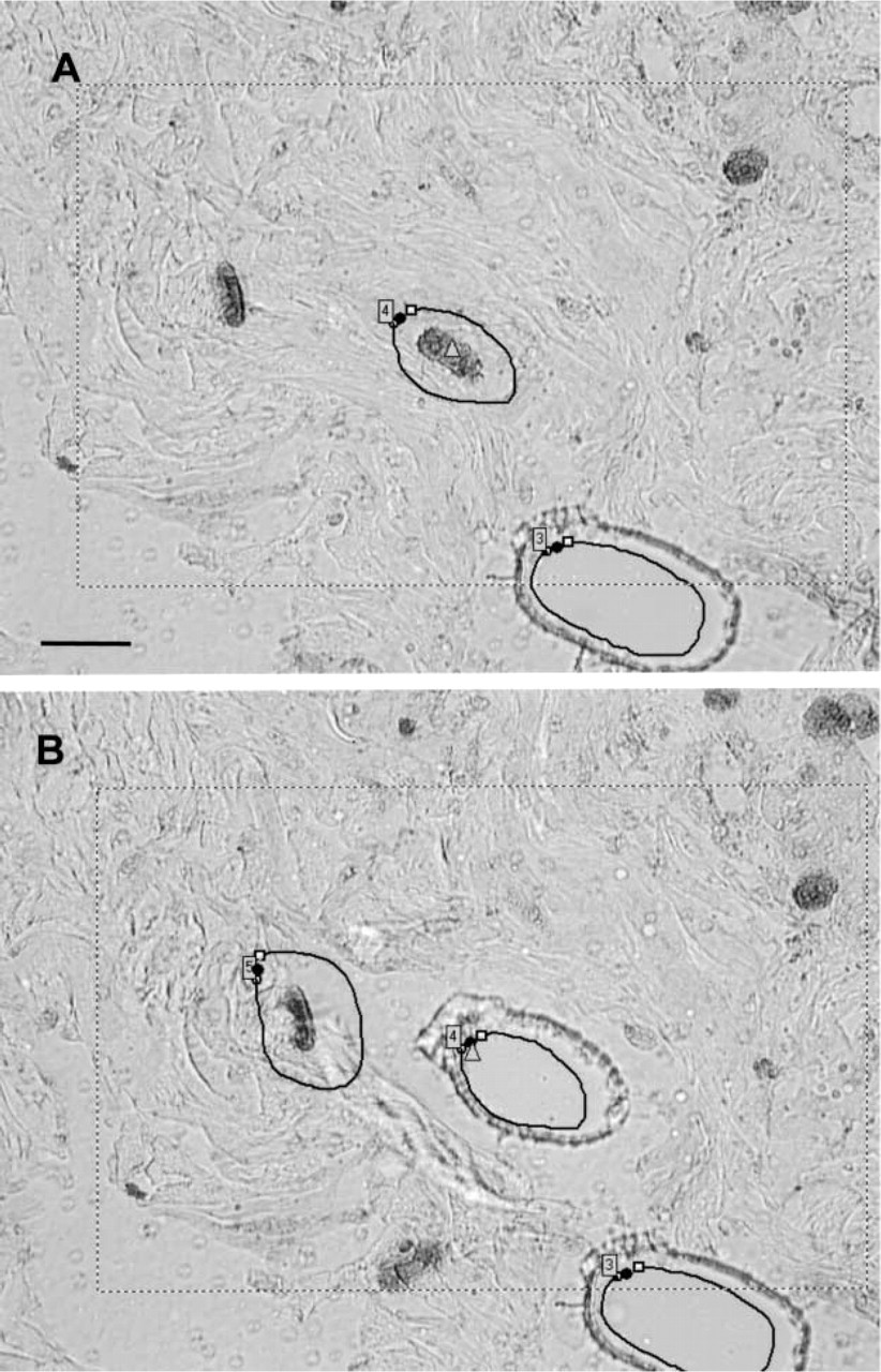
The tissue slices were then washed in PBS, refixed in 1% glutaraldehyde in PBS, orientated and embedded in OCT compound in liquid nitrogen. After cutting in a cryostat, frozen sections were placed onto object slides covered by polyethylene-naphthalate (PEN) membranes (1440–1000; P.A.L.M. Microlaser Technologies, Bernried, Germany). For microdissection, a PALM Laser-MicroBeam System (P.A.L.M. Microlaser Technologies) that enables the contact-free isolation of single cells was used. Cells containing ox-LDL as well as focal extracellular accumulations of ox-LDL were observed in all plaques studied. Cells containing ox-LDL were most frequent in areas surrounding the necrotic cores (Figure 1). Consecutive stages of the selection and capture of cells containing ox-LDL are shown in Figure 1. From several sections obtained from each plaque, up to 40 ox-LDL immunopositive cells were microdissected and catapulted into the lid of a 0.5-ml reaction tube using the laser pressure catapulting technique of the instrument. The reaction tube lid containing microdissected cells was removed from the LCM instrument, fixed in 1% glutaraldehyde in PBS (pH 7.4) for 1 hr, and postfixed in 1% osmium tetroxide. The reaction tube leads with microdissected cells obtained from different plaque specimens were embedded in Araldite resin using a routine technique (Bobryshev and Lord 1996) but with the omission of acetone (or propyleneoxide) to avoid dissolving the reaction tube lids. Consecutive steps in the collection and embedding of ox-LDL immunopositive cells are shown in Figure 2.
Serial ultrathin sectioning at the level of the location of PEN membrane fragments of the embedded reaction tube lids was carried out using a Reichert ultratome (Reichert; Vienna, Austria). Ultrathin sections were placed on formvar-coated nickel grids and then stained with lead citrate for 2 min and with uranyl acetate for 1 min. This short “contrasting” of ultrathin sections enables the visualization of the intracellular structure contours while the product of cytochemical reaction remains more electron dense than any other components of the surrounding cytoplasm. Microdissected cells identified in ultrathin sections were examined with the aid of a Hitachi H7000 electron microscope (Hitachi; Tokyo, Japan) at an accelerating voltage of 100 kV.
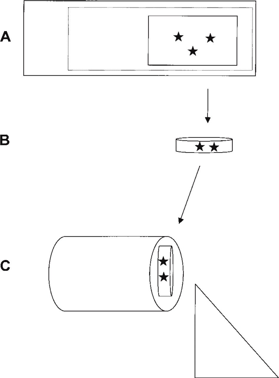
A scheme showing the consecutive steps in the preparation, collection, and embedding of cells of interest (marked by stars) microdissected from an immunostained tissue section (
Electron microscopic analysis demonstrated that the cytoplasm of microdissected cells contained membrane-bound vacuoles and membrane-free lipid vacuoles (“lipid droplets”), enabling the identification of these cells as foam cells (Figure 3A). The cytoplasm of microdissected cells was not only highly vacuolized but was also free of myofilaments, suggesting the macrophage origin of microdissected cells. ox-LDL were observed within vacuoles with the diameters ranging from 200 to 500 nm (Figures 3B and 3C). The association of ox-LDL with microvesicles of 40 to 60 nm in diameter was also observed (Figures 4A and 4B). Some microvesicles containing ox-LDL were located along the border of “lipid droplets” as well as within the peripheral portion of “lipid droplets” (Figure 4B). The study was extended by means of a double immunohistochemistry utilizing ox-LDL antibody and anti-CD68. Double immunostaining included a combination of peroxidase-anti-peroxidase and alkaline phosphatase-anti-alkaline phosphatase techniques, which were carried out as described previously (Bobryshev and Lord 1998). This staining confirmed cellular colocalization of ox-LDL with CD68 antigen (Figure 4C).
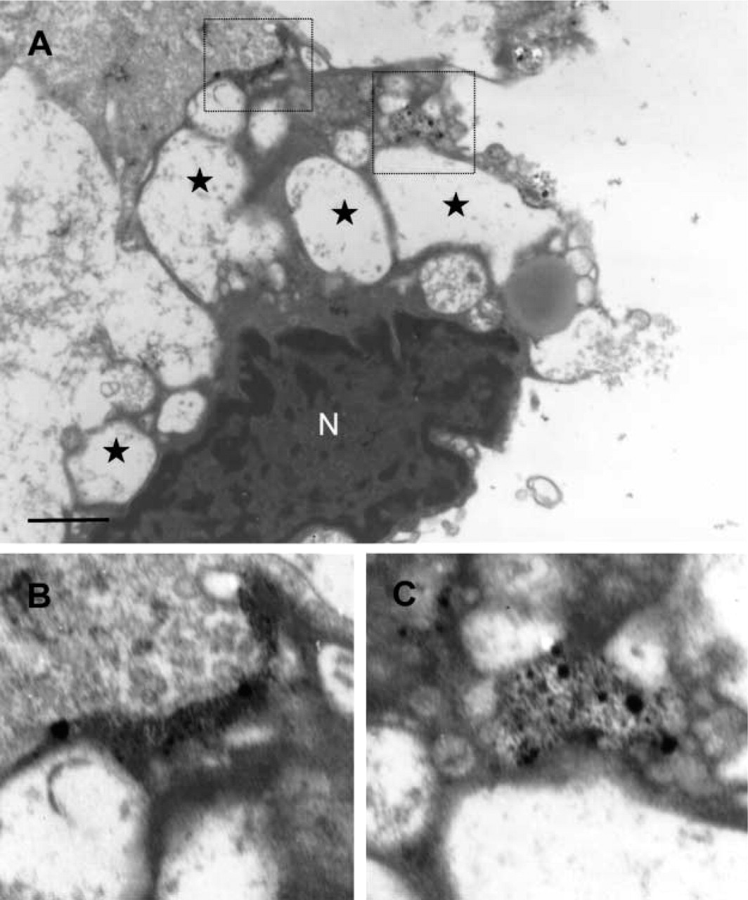
Electron micrographs showing a foam cell containing oxidized low-density lipoproteins LDL (ox-LDL) obtained by means of LCM from a human carotid atherosclerotic plaque (
The present work reports a novel technique for the preparation of immunostained tissue specimens for further LCM, followed by electron microscopic analysis. This technique provides the good preservation of ultrastructural features of microdissected cells and, if DAB is used as a substrate, allows the visualization of the location of the antigen of interest. The “nickel enhancement” of the product of immunohistochemical reaction (Tago et al. 1986) markedly increases electron density. In contrast to experiments utilizing DAB, the use of AEC did not lead to the formation of electron-dense product.
In addition to the procedures described above for the preparation of tissue specimens for further LCM, in some experiments of the present study, arterial tissue slices were immunostained using the labeled streptavidin-biotin complex technique (LSAB; DAKO) according to the manufacturer's instructions, followed by the visualization of the antigen with Fast Red substrate (DAKO). Similarly, as in the experiments utilizing AEC, the visualization of reaction product with Fast Red substrate did not lead to the formation of electron-dense reaction products. If the goal of a study requires the investigation of ultrastructural characteristics of LC-microdissected cells only, the use of Fast Red or AEC substrate may be preferable to the use of DAB. If an antigen of interest is abundant within cells, the use of AEC or Fast Red substrate would prevent an intensive masking of the ultrastructure, which might occur during the visualization of a reaction using DAB.
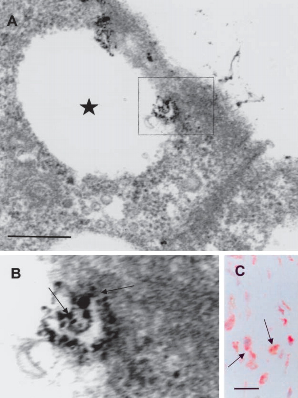
ox-LDL within macrophage foam cells in human atherosclerotic plaques (
Footnotes
Acknowledgements
This research was supported by the St Vincent's Clinic Foundation, Sydney.
I would like to thank Dr. Franz Tatzber, Biomedica, Vienna, Austria, for the kind gift of ox-LDL antibody.