
Abstract
In three cases, banding analysis revealed a normal karyotype except for an enlarged short arm of one chromosome 13 or 15. To clarify whether this enlargement was due to a heteromorphism or to a cryptic chromosomal trisomy, so-called cenM-FISH probe sets containing a microdissection-derived probe specific for the acrocentric human p-arms were applied. The results enabled us to confirm in one case and to exclude in two cases that the enlargement on the suspect chromosome was due to a p-arm polymorphism. M-FISH and/or microdissection were used to resolve the nature of the rearrangements, i.e., partial trisomies 6 and 19.
W
Here we report on three cases with an enlarged p-arm in chromosome 13 (case 2 and 3) or 15 (case 1). Cases 1 and 3 were postnatal cases; case 3 was prenatal. Case 1 was a healthy adult male whose partner had a history of several abortions. Case 2 had a white spot detected in prenatal ultrasound screening. Case 3 was 2 years old and exhibited developmental delay, short stature, and several dysmorphic signs including blepharophimosis, high-arched palate, hypolastic philtrum, low-set ears, and microcephaly.
For cases 1 and 2 we applied the corresponding subcenM-FISH probe sets (Starke et al. 2003), and for case 3 the recently described acrocenM-FISH probe set (Trifonov et al. 2003). Both probe sets contain the microdissection-derived probe midi54 specific for the short arm of all human acrocentric chromosomes (Mrasek et al. 2001), which is essential for the characterization of p-arm heterochromatin presence or absence.
For case 1, the FISH result with the midi54 probe showed that the entire enlarged short arm consisted exclusively of heterochromatic material (Figure 1). Therefore, a balanced translocation was excluded as a reason for abortions in that family, although the large p-arm in one chromosome 15 could not be excluded as connection to that problem (i.e., mitotic problems during embryogenesis). Similar cases with such large p-arm variants and without phenotypic consequences were described previously (Wyandt and Tonk 2004). Especially for chromosome 15 p-arm variants, the possibility of a der(15)t(Y;15)(q12q11.2) must be considered (Alitalo et al. 1988; Wyandt and Tonk 2004).
In cases 2 and 3 the probe midi54 did not stain the entire enlarged short p-arm of the corresponding chromosomes 13 in question (see Figure 2 for case 2; for case 3 see Trifonov et al. 2003). This was a strong clue to the presence of euchromatic material of unknown origin on the derivative chromosomes. For case 2, an M-FISH experiment identified the additional material as being derived from chromosome 8, 19, or 21; a whole chromosome painting probe for chromosome 19 characterized the derivative chromosome 13 as a der(13)t(13;19)(p11.2;p13.1 or q13.1) (Figure 2). The pregnancy was terminated and an autopsy was not performed. For case 3, M-FISH did not lead to informative results. Therefore, glass needle-based chromosome microdissection was performed. The result of the reverse FISH experiment is shown in Figure 3. Apart from the enlarged short arm of chromosome 13 and the short arms of the other acrocentric chromosomes, the end of chromosome 6p was specifically stained. Therefore, and confirmed by other FISH experiments (see Trifonov et al. 2003), it could be deduced that a partial trisomy 6p22.2-pter was present.
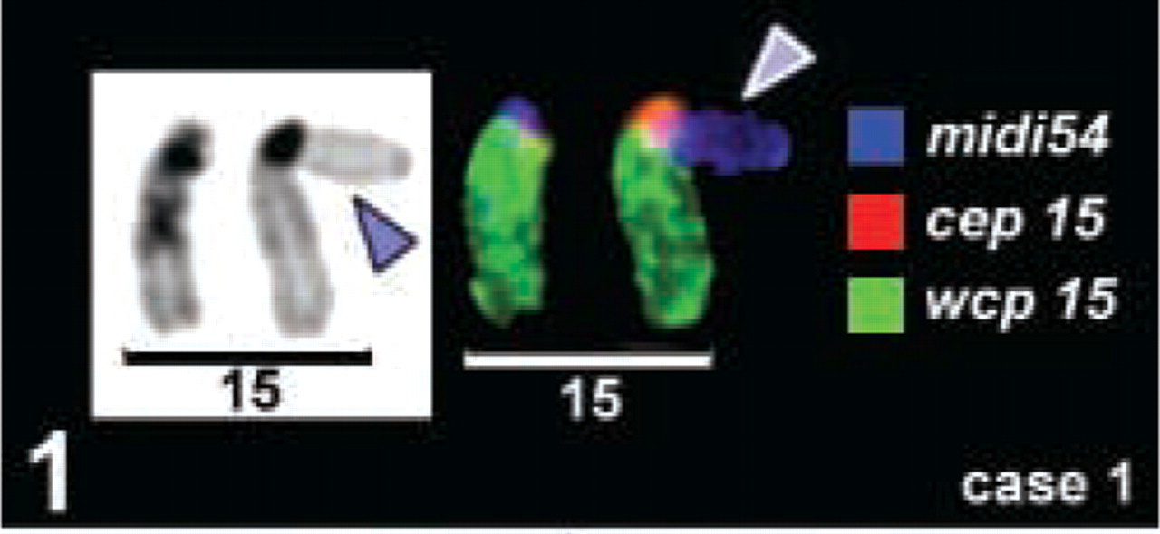
Pair of chromosomes 15 as visible for case 1: an enlarged p-arm was detected after banding cytogenetics (arrowhead), which was completely stained by a probe specific for all acrocentric human p-arms (midi54). A three-color depiction of the subcenM-FISH result (for method see Starke et al. 2003) of the chromosome 15-specific probe set is depicted as inverted DAPI (left) and as truecolor image for the centromere-specific (cep), the whole chromosome painting (wcp), and the midi54 probe (right). The FISH result of the centromere-near probe enclosed in the subcenM-FISH probe set is not shown because it does not provide any additional information.
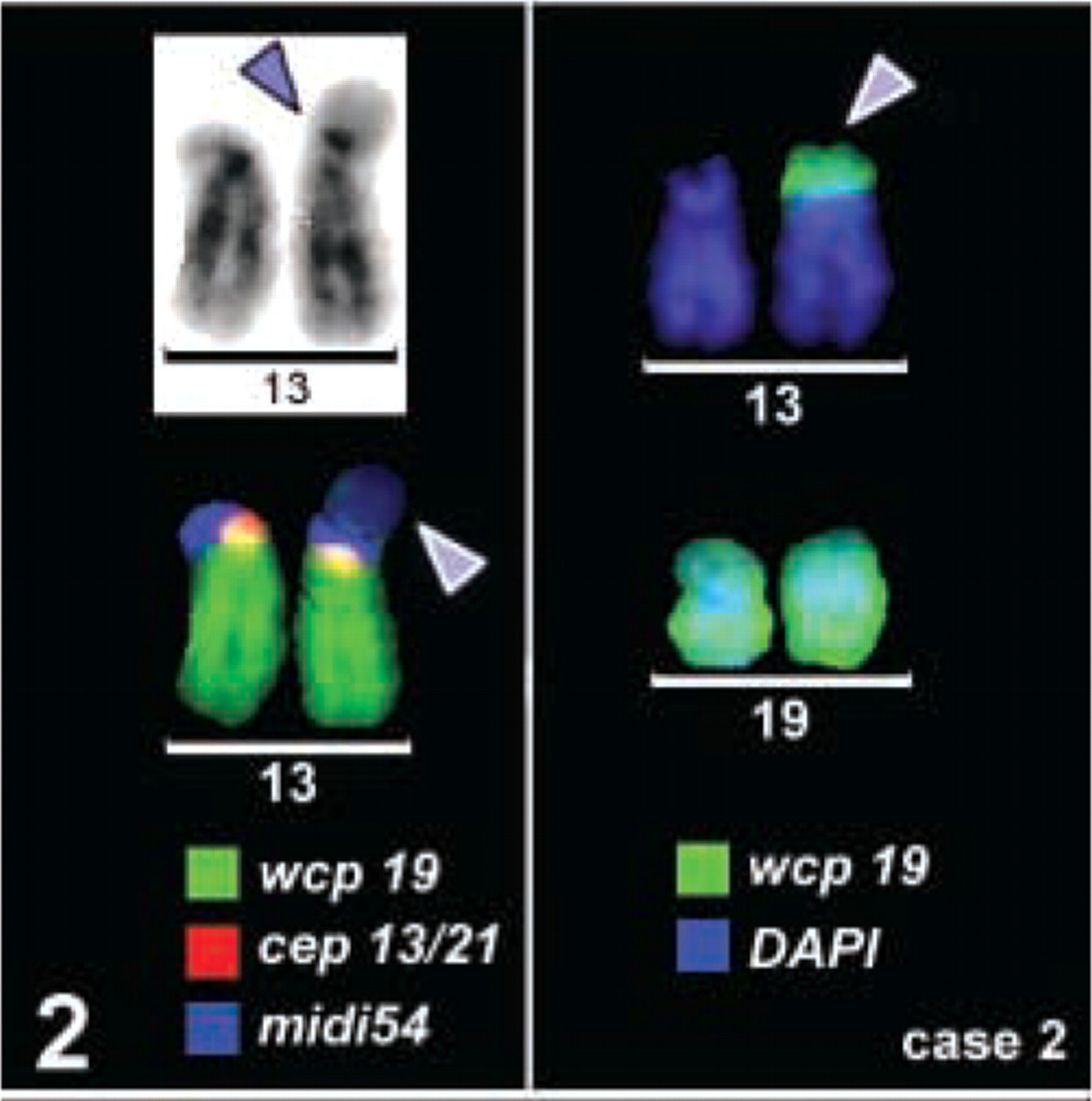
For case 2 a chromosome 13p+ (arrowhead) was found during karyotyping. Inverted DAPI and subcenM-FISH results are depicted on the left part of the figure (as in Figure 1). The arrowhead in the subcenM-FISH figure points to the unstained region of the p-arm in question. The latter was proved to be a part of chromosome 19 by M-FISH (result not shown) and a whole chromosome paint 19 (result shown on the right).
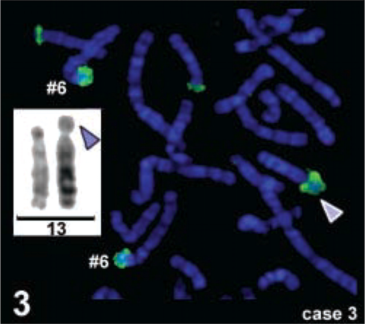
The chromosomes 13p+ of case 3 (arrowhead) was characterized by glass needle-based microdissection as a material derived from chromosome 6p22.2-pter. Inverted DAPI banding of the chromosome 13 pair and a partial metaphase after reverse painting with the microdissected probe are shown.
In summary, it can be assumed that every acrocentric short arm marker of which the size appears abnormally large should be studied in detail by different molecular cytogenetic methods such as M-FISH/SKY, microdissection, and reverse painting or cenM-FISH methods, including a probe such as midi54. In some cases the clinical phenotype may provide clues about the chromosomal region to look for (Benzacken et al. 2001), although in most cases, like those reported here, this advantage will not be present (Morelli et al. 1999; Trifonov et al. 2003).
Footnotes
Acknowledgements
Supported in part by the EU (ICA2-CT-2000–10012) and by the Dr. Robert Pfleger Stiftung.
Cases were kindly provided by Dr Seidel (Jena), Dr Sandig (Weimar), and Dr Wegner (Berlin).