
Abstract
We applied the heat-induced antigen retrieval (HIAR) to aldehyde-fixed fresh frozen sections based on a new approach (i.e., a rapid and complete immobilization of antigen followed by heating). Frozen sections were fixed with 10% formalin in 0.1 M cacodylate buffer (pH 7.4) containing 25 mM CaCl2 for 30 min, or with 0.5% glutaraldehyde in 0.1 M phosphate buffer (pH 7.4) for 1 min at room temperature, and then autoclaved in 20 mM Tris-HCl buffer (pH 9.0) for 10 min at 120C. Both fixatives yielded good tissue structure after autoclaving. In the sections fixed with formalin containing CaCl2, 20 of 22 antigens located in the nucleus, cytoplasm, membranes, and extracellular matrix greatly recovered their antigenicity after autoclaving; only two antigens exhibited stronger immunoreaction in acetone-fixed fresh frozen sections than these sections. Heating also retrieved the immunoreactivity of at least 14 antigens in the sections fixed with glutaraldehyde. We used the similar procedures to localize ligand-free estrogen receptor α (ERα) and glucocorticoid receptors (GR). Mouse uterine cells exhibited almost the same nuclear ERα immunostaining regardless of the hormonal status in glutaraldehyde-fixed fresh frozen sections and unliganded GR was localized mainly in the nucleus of mouse hepatocytes in fresh frozen sections fixed with 20% formalin containing 50 or 75 mM CaCl2 at 40C, after autoclaving. These results demonstrate that HIAR is useful for the immunohistochemistry of many antigens in aldehyde-fixed fresh frozen sections.
Keywords
H
Fresh frozen sections are widely used for immunohistochemical studies, because (1) they preserve antigenecitiy well, (2) they are convenient for comparing antigen expression in the different size of tissues with a constant fixation time, and (3) they allow antigen localization in a short time for pathological diagnosis. In particular, frozen sections fixed with formalin or acetone have been used as a standard to evaluate the efficiency of HIAR in formalin-fixed and paraffin-embedded specimens (Shi et al. 1993; Merz et al. 1995; Mighell et al. 1995), whereas HIAR has not applied to the frozen sections, probably because they are fragile and readily destroyed by heating (Ino 2003). However, if HIAR is based on these mechanisms, it should also be useful to immunohistochemical studies in frozen sections mounted on slide glasses. The main differences between frozen sections and paraffin sections may be as follows: protein denaturation in frozen sections is less intense than in paraffin sections, because proteins are not exposed to organic solvents and heat, and macromolecules may be more extracted from frozen sections during heat-treatment compared with those in paraffin sections.
Fixation is one of the most important factors in immunohistochemistry, and both excess fixation and insufficient fixation weaken immunostaining. Soluble antigens such as ligand-free steroid hormone receptors are readily extracted from fresh frozen sections during fixation. In the present study, we tried to establish a procedure for rapid and complete immobilization of antigens in fresh frozen sections and successive antigen retrieval by heating, and to investigate the mechanisms of HIAR in detail. The procedure was also applied to localize unliganded estrogen receptor (ER) α and glucocorticoid receptor (GR), which are known to be readily extracted from fresh frozen sections during fixation (Gasc et al. 1989; Yamashita and Korach 1989; Pekki et al. 1992).
Materials and Methods
Reagents
The primary antibodies used in this study, their respective sources and dilutions are listed in Table 1. Envision plus/horseradish peroxidase for rabbit immunoglobulin and mouse immunoglobulin were purchased from DakoCytomation (Carpinteria, CA), and anti-goat IgG rabbit antibody was from ICN Biomedicals, Inc (Aurora, OH). A blocking reagent (block ace) was obtained from Dainippon Pharmaceutical Co., Ltd (Osaka, Japan). Formalin and 10% glutaraldehyde were purchased from Wako Pure Chemicals Industries Ltd (Osaka, Japan).
Animals
CD-1 mice and Wistar rats were obtained from Clea Japan, Inc. (Tokyo, Japan). Mice were housed at 21–22C with a 12-hr alternating light-dark cycle at the Keio University Animal Facility, Tokyo, Japan. All animals were maintained and treated according to protocols approved by the Keio University Animal Care Committee. Small pieces of tissues from adult male and female mice (8 weeks old) and male Wistar rats (8 weeks old) were mounted in OCT compound and frozen in dry ice-cooled isopentane.
Primary antibodies used
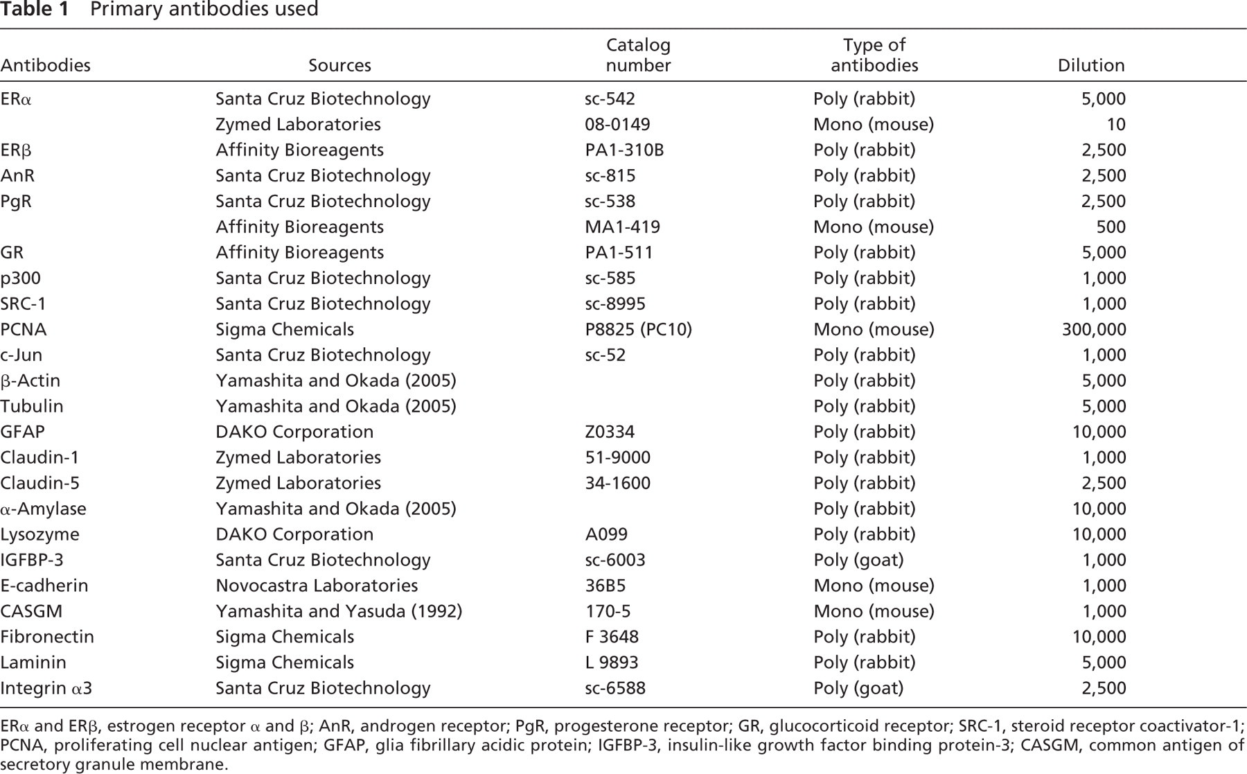
ERα and ERβ, estrogen receptor α and β; AnR, androgen receptor; PgR, progesterone receptor; GR, glucocorticoid receptor; SRC-1, steroid receptor coactivator-1; PCNA, proliferating cell nuclear antigen; GFAP, glia fibrillary acidic protein; IGFBP-3, insulin-like growth factor binding protein-3; CASGM, common antigen of secretory granule membrane.
To detect liganded and unliganded ERα, immature female mice (2 weeks old) were intraperitoneally injected with a single dose of 0.1 ml 17β-estradiol (E2, 20 μg/kg BW) or vehicle solution (1% ethanol and 99% saline) and were killed 1 hr after the injection. To localize hormone-free and hormone-bound GR, female mice (10 weeks old) were adrenalectomized, and 7 days later they were intraperitoneally given 0.2 ml of 70% ethanol containing 25 μg of dexamethasone or vehicle solution, and sacrificed after 1 hr. Four hormone-injected mice and four vehicle-injected control mice were used for each experiment.
Immunohistochemistry
Normal rabbit IgG or mouse IgG was used in place of the primary antibody for control immunostaining. For the control of ERα, ERβ, GR, and androgen receptor (AnR) immunostaining, antibodies absorbed with the respective antigenic peptides were used.
Results
Morphological Effects of Fixatives and Heating
The sections fixed with acetone and immunostained with normal rabbit IgG as a control maintained relatively good morphology, although nuclear staining with hematoxylin was weaker than that of formalin-fixed fresh sections (Figures 1A and 1B). When fresh frozen sections were fixed with 10% formalin in 0.1 M PB or CB for 30–360 min at room temperature or at 40C and then boiled in a microwave oven or autoclaved, the nuclear structure of many cell types, particularly the epithelial cells of the genital tract and intestine, was destroyed, indicating that chromatin had been extracted (Figures 1B and 1C). However, the nuclear structure in the sections fixed with 10% formalin containing CaCl2 was well preserved after heating (Figure 1D), and fixation with 10% formalin containing 25 mM CaCl2 for 30 min yielded much better morphology than fixation with 10% formalin for 5 hr (Figures 1C and 1D). Glutaraldehyde was an excellent fixative for preservation of morphology. Fixation with 0.5% glutaraldehyde even for 1 min at room temperature prevented chromatin extraction after autoclaving (Figure 1E).
HIAR in Fresh Frozen Sections
HIAR was applied to 22 antigens in aldehyde-fixed fresh frozen sections; two antibodies, a monoclonal and a polyclonal antibody, were used for the immunostaining of ERα and progesterone receptor, respectively. Acetone-fixed sections were used a standard for immunostaining of these antigens. Table 2 summarizes the results of HIAR when different fixatives were used for fixation of fresh frozen sections.
In the acetone-fixed tissues, nuclear antigens except for ERα and ERβ were hardly detectable (Figure 2A), although most nuclear antigens were weakly immunostained in the tissues fixed with 10% formalin for 30 min at room temperature (Figure 2B; Figure 4A; Figures 5A, 5B, 5E, and 5F). When the formalin-fixed sections were heated, the staining intensity increased in the cells whose nuclear structure is relatively well preserved, such as the stromal and muscle cells of the genital tract and intestine, but the reaction was weaker in the epithelial cells (Figure 3D and Figure 4B). Both buffers, CB and PB, yielded almost the same staining pattern. Immunostaining for all nuclear antigens was negative when the tissues were fixed with 10% formalin containing 25 mM CaCl2, but the staining was much intensified after heating (Figure 3E and Figure 4D). Autoclaving usually provided stronger immunoreaction than boiling in a microwave oven, and higher concentrations of CaCl2 (75 mM and 100 mM) weakened the staining and heating at high temperature was required for antigen retrieval (data not shown). In glutaraldehyde-fixed fresh sections, autoclaving effectively recovered antigenicity of many nuclear antigens (Figures 3C and 3F).
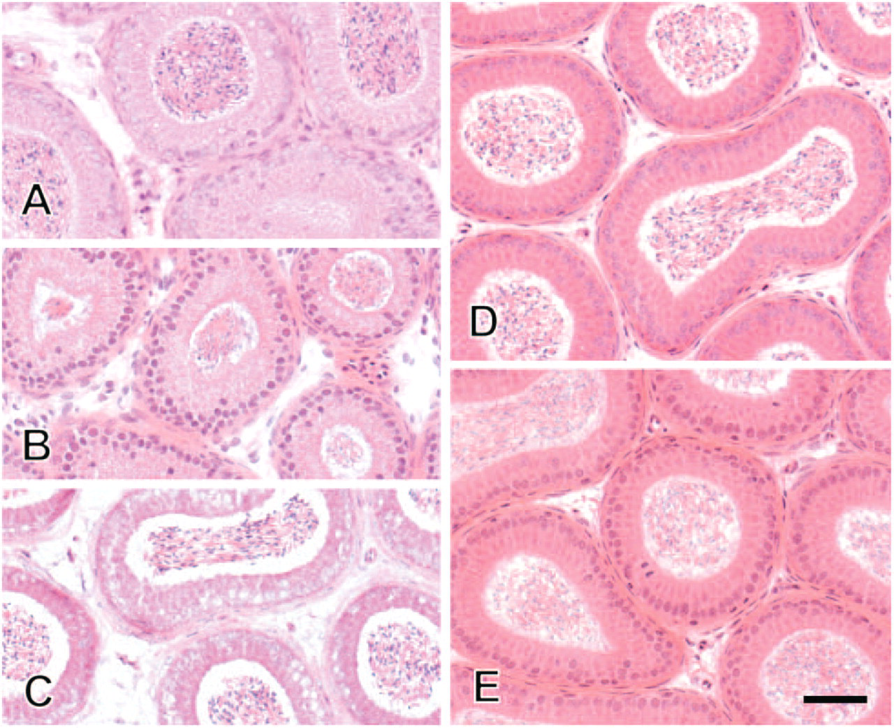
Morphological effect of fixatives and heating. Fresh frozen sections from mouse epididymis were fixed with ice-cold acetone for 30 min, successively dried, and then immunostained with normal rabbit IgG as a control
Most antigens located in the cytoplasm, membranes, and extracellular matrix exhibited similar immunostaining intensity in the tissues fixed with acetone and 10% formalin (Table 2). However, claudin-1 and common antigen of secretory granule membrane were detectable only in acetone-fixed tissues (Figures 2C and 2D). In contrast, insulin-like growth factor binding protein-3 and integrin α3 showed positive immunoreaction in formalin-fixed fresh sections, but not in acetone-fixed sections. In the formalin-fixed fresh frozen sections, most antigens recovered their antigenicity after heating, although heating significantly weakened lysozyme and laminin immunostaining in the tissues fixed with any fixatives. Lysozyme immunostsining was strong in the formalin-fixed fresh sections, but the reaction products were seen not only in the apical cytoplasm but in the nucleus as well. However, formalin with CaCl2 or 0.5% glutaraldehyde yielded almost exclusive apical cytoplasmic staining. α-Amylase immunostaining was diffuse in the cytoplasm of exocrine pancreatic cells when sections were fixed with 10% formalin, but the staining was localized in the apical cytoplasm when fixed with 10% formalin containing CaCl2 (Figures 4E-4H). Heat treatment also activated immunostaining of most antigens localizing in the cytoplasm, membrane, and extracellular matrix in the glutaraldehyde-fixed fresh frozen section (Table 2; Figures 3C and 3F).
Effects of fixatives and heating on antigen retrievala
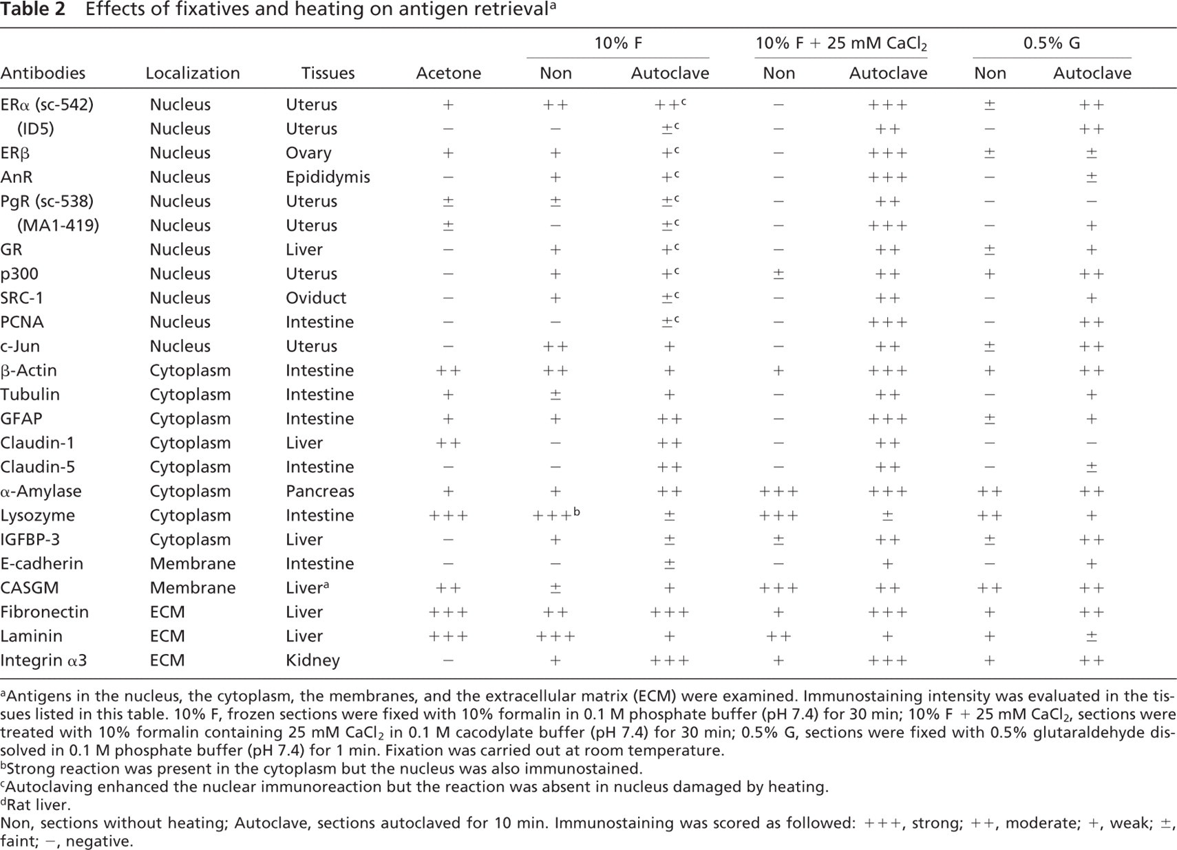
Antigens in the nucleus, the cytoplasm, the membranes, and the extracellular matrix (ECM) were examined. Immunostaining intensity was evaluated in the tissues listed in this table. 10%
Strong reaction was present in the cytoplasm but the nucleus was also immunostained.
Autoclaving enhanced the nuclear immunoreaction but the reaction was absent in nucleus damaged by heating.
Rat liver.
Non, sections without heating; Autoclave, sections autoclaved for 10 min. Immunostaining was scored as followed: + + +, strong; ++, moderate; +, weak; ±, faint; -, negative.
When the aldehyde-fixed frozen sections from mice were immunostained with monoclonal antibodies (mouse IgG), heating completely diminished the immunostaining in the plasma cells, connective tissues, and blood vessels, which corresponds to the endogenous immunoglobulin (Figure 3). All controls for immunohistochemistry exhibited negative staining (Figure 1A; Figures 5D and 5H, insets).
Detection of Unliganded ERα and GR in Aldehyde-fixed Fresh Frozen Sections
When frozen sections were fixed with 10% formalin for 10–30 min at room temperature or 40C, the epithelial cells of the uterus and oviduct of mice injected E2 showed stronger ERα immunostaining than those of vehicle-injected controls (Figures 5A and 5B). When the frozen sections were fixed with 0.5% glutaraldehyde in 0.1 M PB (pH 7.4) for 1 min followed by autoclaving, these cells exhibited almost the same ERα immunostaining intensity regardless of the hormonal status (Figures 5C and 5D), although the reaction was faint or negative without autoclaving. Two antibodies, a polyclonal and a monoclonal antibody, yielded almost the same patterns of HIAR in the glutaraldehyde-fixed fresh frozen sections (data not shown).
When tissues were fixed with 10% formalin for 30 min at room temperature or 40C, strong GR immunostaining was present in the nucleus of various cell-types in the liver, pancreas, and small intestine of adrenalectomized mice treated with dexamethasone (Figure 5F), whereas faint GR immunostaining was observed in the nucleus of vehicle-treated control animals (Figure 5E). In the sections fixed with 20% formalin containing 50 mM or 75 mM CaCl2 at 40C for 30 min and then autoclaved, unliganded GR also showed strong nuclear staining and weak cytoplasmic staining, although the staining intensity in the nucleus was still weaker than that of liganded GR (Figures 5G and 5H).
Effect of EDTA on Immunostaining
To study the effect of calcium ions, fresh frozen sections fixed with formalin containing 25 mM CaCl2 for 30 min were incubated with 20 mM TB (pH 9.0) with or without 1 mM EDTA at room temperature for 2 hr, or autoclaved in the same solutions for 10 min. Treatment with EDTA at room temperature or at 120C had no significant effect on the immunostaining patterns of all antigens (Figure 6), although the immunostaining of some antigens such as ERα, p300, AnR, and integrin α3, decreased slightly when the sections were autoclaved in TB containing 1 mM EDTA.
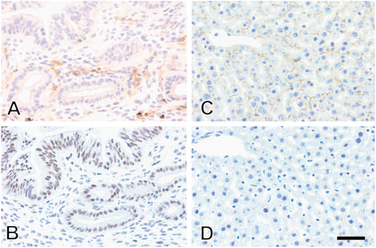
p300 and claudin-1 immunostaining in fresh frozen tissues fixed with acetone or formalin. Fresh frozen sections were fixed with ice-cold acetone for 30 min (
Discussion
The present study demonstrated that immunostaining procedure for fresh frozen sections based on a new strategy is useful for the immunohistochemistry of most antigens located in the nucleus, cytoplasm, membranes, and extracellular matrix. Fresh frozen sections fixed with formalin containing CaCl2 provided excellent antigen localization and strong immunoreaction after heating compared with those fixed with acetone or formalin that have been use as standard for immunostaining: formalin containing CaCl2 may rapidly crosslink and immobilize soluble antigens, and the crosslinks (methylene bridges) are easily cleaved by heating (Rait et al. 2004; Yamashita and Okada 2005). Lysozyme and α-amylase were predominantly localized in the apical cytoplasm without diffusion in the Paneth cells of the small intestine and the acinar cells of exocrine pancreas fixed with formalin containing CaCl2, respectively (Figures 4G and 4H), but they were frequently observed in both the cytoplasm and the nucleus when fixed with CaCl2-free formalin. Glutaraldehyde is also an excellent fixative for a rapid and complete fixation of soluble antigens, and heating may also cleave partially crosslinks formed by glutaraldehyde.
Formaldehyde solutions containing calcium ions, such as Baker's formol calcium, had been used for enzyme histochemistry; they are assumed to stabilize membrane phospholipids and to minimize diffusion of enzymes from cell organelles (Baker 1944; Bancroft 1996). In addition, we have demonstrated that calcium ions accelerate crosslinking of proteins in vitro (Yamashita and Okada 2005). However, they have not been used for immunohistochemical studies, probably because of the severe reduction or loss of immunoreaction. Morgan et al. (1994, 1997) assumed that coordinate bonds between calcium and the methyrol groups of proteins introduced by formaldehyde form a cagelike structure and prevent antigen-antibody interactions. The results of the present study showed that EDTA had no effect on the immunostaining pattern of any of the antigens, with or without heating, and suggested that the cagelike structure is not present in the tissues fixed with formaldehyde as reported by Shi et al. (1999). Our previous study in vitro also suggested that the proteins treated with formaldehyde do not form the tight cagelike structure with calcium ions (Yamashita and Okada 2005). Even if coordinate bonds are present between calcium ions and proteins, the bonds may be unstable than the bonds between calcium ions and EDTA, because polypeptides maintain their secondary and tertiary structure after formaldehyde fixation (Mason and O'Leary 1991) and may be not so flexible enough to form stable coordinate bonds with calcium ions.
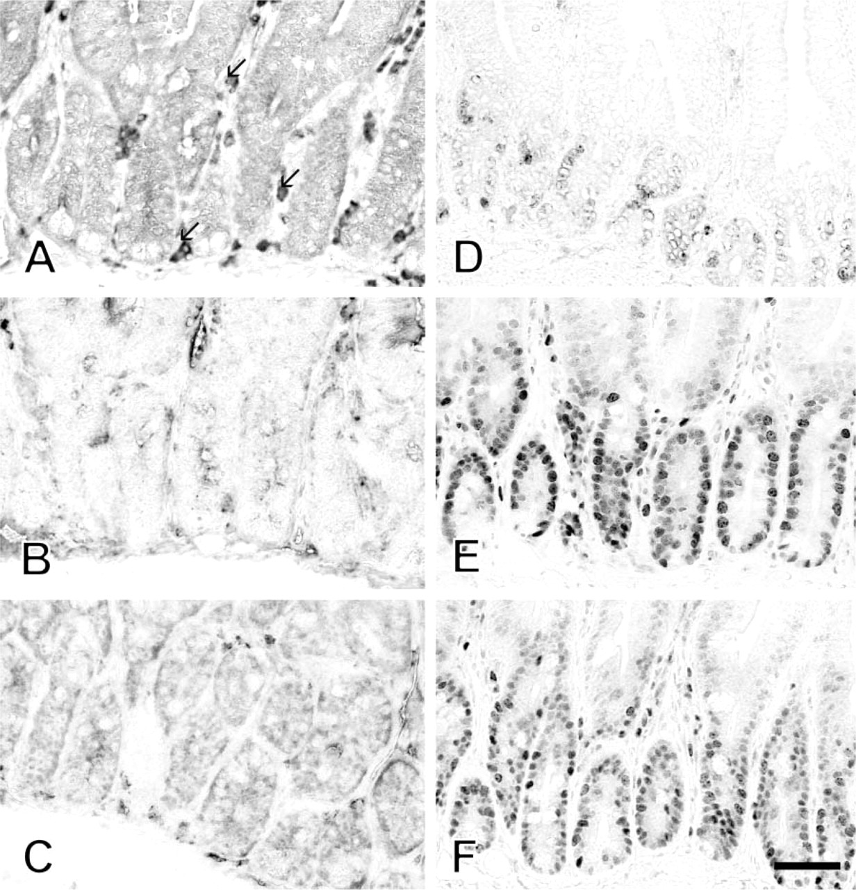
Effect of fixatives and heating on proliferating cell nuclear antigen (PCNA) immunostaining. PCNA was localized in the mouse duodenum. Fresh frozen sections were fixed with 10% formalin in 0.1 M phosphate buffer (PB) (pH 7.4) for 30 min (
It is well known that sex steroid receptors localize in the nucleus, regardless of hormonal status and that unliganded receptors are readily extracted from fresh frozen sections during fixation (Yamashita and Korach 1989; Slayden et al. 1995). Therefore, fixation procedure that yields similar staining intensity of liganded and unliganded ERα is a good model system for evaluating the success of fixation. Liganded and unliganded ERα exhibited almost the same staining intensity in the mouse uterine epithelial and stromal cells when fresh frozen sections were fixed with glutaraldehyde followed by autoclaving. By contrast, the subcellular localization of unliganded GR is still a matter of controversy in a variety of cell types in of tissues and cultured cells (Gasc et al. 1989; McGimsey et al. 1991; Pekki et al. 1992; Yamashita 2001). In the present study, ligand-free GR was barely detected in the nucleus in mouse tissues when fresh frozen sections were fixed with 10% or 20% formalin at room temperature. However, when the sections were fixed with 20% formalin containing 50 or 75 mM CaCl2 at 40C, a strong immunoreaction for ligand-free GR was observed in the nucleus and weak reaction was present in the cytoplasm after HIAR, whereas the reaction of ligand-free GR in the nucleus was weaker than that of liganded GR. The present findings suggest that unliganded GR is present in both the nucleus and the cytoplasm of mouse tissues, and that one of the reasons of conflicting reports concerning localization of ligand-free GR is attributable to differences in fixation protocols.
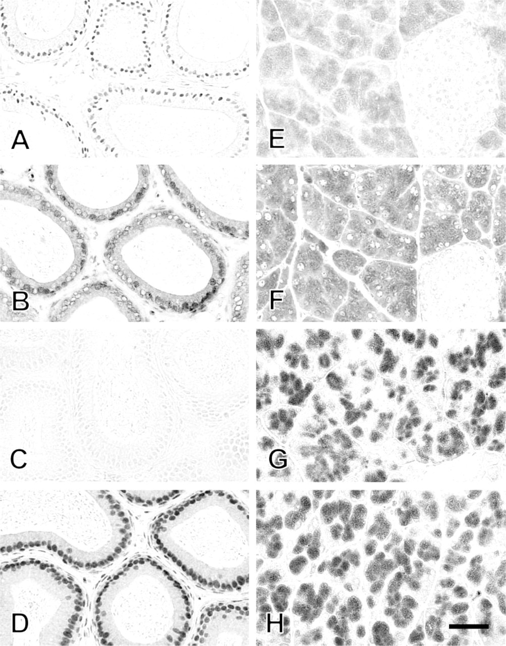
Effect of fixatives and heating on immunostaining of androgen receptor (AnR) and α-amylase. AnR was localized in the head of epididymis (
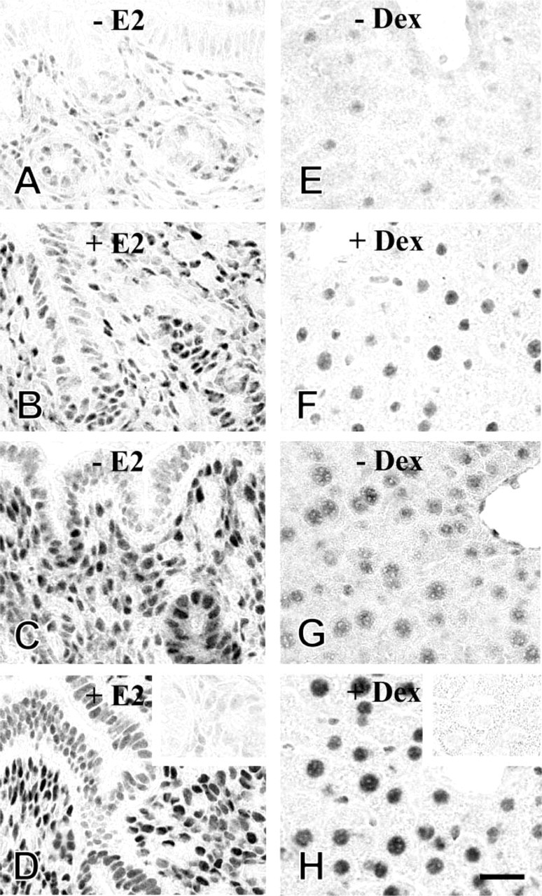
Localization of hormone-occupied and -unoccupied estrogen receptor α (ERα) and glucocorticoid receptor (GR) in frozen sections. To detect liganded and unliganded ERα, immature female mice (2 weeks old) were intraperitoneally injected with 17β-estradiol (20 μg/kg BW) (
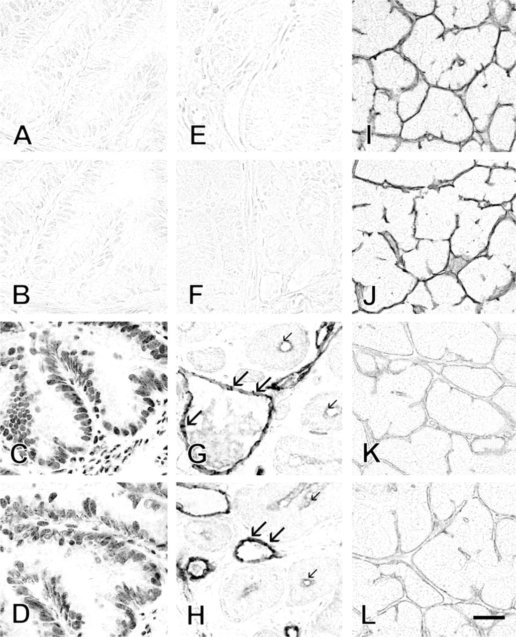
Effects of EDTA on the immunostaining of the tissues fixed with formalin containing CaCl2 Fresh frozen sections were fixed with 10% formaldehyde containing 25 mM CaCl2 dissolved in 0.1 M cacodylate buffer (pH 7.4) for 30 min at room temperature. The sections were then immersed in 20 mM Tris-HCl buffer (TB) (pH 9.0) with (
The results of the present study demonstrate that HIAR is a powerful technique not only for formalinfixed and paraffin-embedded specimens, but also for aldehyde-fixed fresh frozen tissues. The mechanisms of HIAR may be the same in paraffin sections and frozen sections. Heating cleaves intra- and intermolecular crosslinks and extend polypeptide chains and antibodies may be easily penetrate into the tissues. In addition, electrostatic repulsion by negatively charged polypeptides and hydrophobic attraction may balance to prevent intertwining of unfolded polypeptide chains in a retrieval solution at basic pH and antigenic determinants may be exposed to react with antibodies (Yamashita and Okada 2005; Emoto et al. 2005). Rapid and complete fixation procedure that minimizes diffusion artifacts, false localization, and extraction of antigens during fixation and heating is more important for fresh frozen sections than paraffin sections or frozen sections prepared from tissues prefixed with aldehyde. HIAR may also be a useful technique for immunoelectron microscopy of many antigens with the preembedding method, using prefixed frozen unfrozen or frozen sections (Yamashita et al. 1989).
Footnotes
Acknowledgements
This work was supported in part by a Grant-in-Aid (16590154) for Scientific Research from the Ministry of Education, Culture, Sports, Science and Technology, Japan.